Original Article
The effects of interferon-β on neuronal apoptosis
and the expressions of BDNF and TrkB in
rats with spinal cord injuries
Yijia Jia*, Tingsheng Lu*, Shudan Yao, Linsong Ji, Jianwen Yang, Chunshan Luo
Department of Spine Surgery, Guizhou Province Osteological Hospital, Guiyang, China. *Equal contributors and
co-first authors.
Received July 5, 2019; Accepted October 3, 2019; Epub November 15, 2019; Published November 30, 2019
Abstract: Objective: This study aimed to explore the effects of interferon (IFN)-β on neuronal apoptosis, and on brain-derived neurotrophic factor (BDNF), and tropomyosin receptor kinase B (TrkB) expressions in rats with spinal cord injuries. Methods: 40 adult female SD rats were equally and randomly divided into a blank group, a model group, a sham operation group, and an IFN-β group. The spinal cord injury rat model was established in the model and IFN-β groups. Only laminectomies were performed without spinal cord injury in the sham operation group. The rats in the control group were fed routinely. The Basso-Beattie-Bresnahan (BBB) score and the modified Tarlov score were used to evaluate the motor function of each group on the 1st, 3rd, and 7th days after the modeling. The Zea-Longa five-grade score was used to evaluate the rats’ nerve functions. The expressions of the BDNF and TrkB proteins and the apoptosis-related proteins Bax and Bcl-2 in the rat brains were measured on the 7th day after the modeling. The TUNEL method was used to detect the neuronal apoptosis in each group, and the correlations between the expres-sions of the BDNF and TrkB proteins and the neuronal apoptosis rate were analyzed. Results: The BBB score, the modified Tarlov score, and the Zea-Longa score in the blank and sham operation groups were higher than the scores in the model and IFN-β groups (P < 0.05). The blank and the sham operation groups showed higher BDNF, TrkB, and Bcl2 proteins and lower Bax proteins than the model and IFN-β groups (P < 0.05). The IFN-β group exhibited higher BDNF, TrkB, and Bcl2 proteins and lower Bax proteins than the model group (P < 0.05). The expressions of the BDNF and TrkB proteins were negatively correlated with the neuronal apoptosis rate (r = -0.667, P < 0.05; r = -0.791, P < 0.05). Conclusion: The motor and nerve functions were significantly improved, the expressions of the BDNF and TrkB proteins were up-regulated, and the neuronal apoptosis was inhibited by IFN-β in rats with acute spinal cord injury.
Keywords: Interferon-β, spinal cord injury, neurons, BDNF, TrkB
Introduction
Acute spinal cord injury, a serious central ner-vous system injury, can lead to motor and sen-sory dysfunction below the spinal cord injury segment and has a higher disability rate [1, 2]. In recent years, with the development of soci-ety and the rapid growth of the transportation industry, the incidence of acute spinal cord injury is also increasing, and it has brought a very heavy burden to families and society [3]. For a long time, the repair of spinal cord injuries has been one of the key issues in medical research. Most previous studies believed that the mechanism of spinal cord injury was sec-ondary necrosis due to ischemia and cellular hypoxia [4, 5]. However, with the sustained de-
velopment of molecular biology and immunolo-gy in recent years, it has gradually become rec-ognized that neuronal apoptosis exerts an important role in secondary spinal cord injury [6]. Therefore, it is now understood that neuro-nal apoptosis is the main cause of secondary spinal cord injury, so the inhibition of neuronal apoptosis is the key to preventing secondary spinal cord injury [7].
interferon has a more obvious effect [9]. In addition, some studies [10] have indicated that IFN-β could inhibit cell apoptosis under certain conditions. At present, there are few studies on IFN-β as treatment for acute spinal cord injury. However, some studies [11] indicated that IFN-β has a great potential for the treatment of acute spinal cord injury. Therefore, it has been speculated in earlier studies that the immune intervention of IFN-β on the central nervous system could inhibit neuronal apoptosis in acute spinal cord injury.
A spinal cord injury rat model was constructed to explore the effects of IFN-β on neuronal apoptosis in acute spinal cord injury and the expressions of brain-derived neurotrophic fac-tor (BDNF) and tyrosine protein kinase B (TrkB) in spinal cord injury segments, and the thera-peutic effect and mechanism of IFN-β on acute spinal cord injury. This study aimed to provide more possible treatment options for acute spi-nal cord injury.
Materials and methods
Laboratory animals and materials
40 adult female SD rats aged 8 weeks old we- re selected. The body weight of the rats was (173.55 ± 7.34) g. All the rats were purchased from the Shanghai SLAC Laboratory Animal Co., Ltd., with production license SCXK (Shanghai) 2012-0002.
The rats were fed at a temperature of 20-25°C and a relative humidity of 40%-75%. The rats showed normal circadian rhythm alternation, and were free to eat and drank. Every proce-dure was approved by the Animal Care and Use Committee of the Guizhou Province Osteological Hospital.
Modeling and grouping
The rats were randomly divided into a blank group (N = 10), a model group (N = 10), a sham operation group (N = 10), and an IFN-β group (N = 10). The rats in the blank group were fed rou-tinely without any treatment. The rats in the model, sham operation, and IFN-β groups were anesthetized with an intraperitoneal injection of 10% chloral hydrate at a dose of 0.4 mL/100 g. After being anesthetized, the rats were steril-ized in a supine position. The rats’ skin was cut along the midline of the T9-T10 thoracic seg-ment, and their fascia was gradually separated.
The spinous process and vertebral plate were exposed through muscle stripping. The follow-up surgery was not carried out on the rats in the sham operation group after this step. A 20 g weight was freely dropped from a height of 5 cm using an Allen device. The T9-T10 thoracic segment was covered with a 3 mm × 6 mm arc metal sheet in the model and IFN-β groups and was struck by the freefalling weight. Successful modeling was marked by a spastic swing, a retractable flutter of both lower limbs and trunk, and the flaccid paralysis of both hind limbs. Then the incision was stitched. The rats in the IFN-β group were injected with IFN-β of 1*107 IU via the tail vein immediately after suc-cessful modeling, and IFN-β of 0.5*107 IU was injected into the tail vein again 4 hours after the modeling. The drug intervention was not carried out in the blank, sham operation, or model groups.
Outcome measures
(1) The Basso-Beattie-Bresnahan (BBB) score [12] (0-21 points; 0 point as complete paraly-sis; 21 points as perfectly normal) and the modified Tarlov score [13] (0-5 points; 0 points as hind limbs without active motion; 5 points as normal walking) were used to evaluate the motor functions of the rats on the 1st, 3rd, and 7th days after the modeling. (2) The Zea-Longa five-grade score [14] was used to evaluate the nerve functions of the rats on the 1st, 3rd, and 7th days after the modeling. Higher scores rep-resent worse nerve functions. (3) Western blot was used to determine the expressions of the BDNF protein, the TrkB protein, and the apopto-sis-related proteins Bax and Bcl-2 in the rat brains on the 7th day after the modeling. (4) The TUNEL method was used to measure the neuronal apoptosis in each group on the 7th day after the modeling. (5) The correlations between the expressions of the BDNF and TrkB proteins and the neuronal apoptosis rate were analyzed.
The detection of the BDNF protein, the TrkB protein, and the apoptosis-related proteins Bax and Bcl-2 using Western blot
The tissue was mixed on ice and centrifuged at 1300 r/min for 10 min. The protein was sepa-rated by 10% SDS-PAGE and then transferred onto a PVDF After being stored o membrane. 5% skim milk was added, and then the samples were stored overnight at 4°C. vernight, the pri-mary antibodies BDNF (1:1000), TrkB (1:1000), Bax (1:1000), Bcl2 (1:1000) and β-actin were added to incubate overnight at 4°C. Then a goat anti-rabbit antibody labeled with HRP (sec-ondary antibody, 1:1000) was added to incu-bate at room temperature for 1 hour. The pro-tein was rinsed with a PBS solution and colored with an ECL developer. The result was expressed by the gray value of the target band.
Detection of spinal neuronal apoptosis by TUNEL
First, the spinal cord tissue was paraffin-em- bedded, sectioned, dewaxed, and dewatered. Then 20 ug/ml protease K was added. The tis-sue protein was digested for 20 min at 37°C and rinsed with distilled water four times. After the rinsing, PBS containing 2% hydrogen perox-ide was added to the protein and it was incu-bated at room temperature for 5 min. The pro-tein was then rinsed with PBS. After it was rinsed, two drops of TdT enzyme buffer were added to the sections, and the reaction was performed at room temperature for 5 min. Then 50 ul of TUNEL reaction solution was added to and the sections were incubated at 37°C for 30 min. After the sections were washed, the reaction was terminated by adding 0.2 ml of termination solution. Then the protein was stained with hematoxylin again. Finally, the pro-tein was stained with DAB developer, and observed and counted under a lighted micro-scope. Apoptosis rate (%) = Number of apop-totic cells/Number of total cells × 100%.
Statistical analysis
SPSS 19.0 software (Biz Insight (Beijing) In- formation Technology Co., Ltd.) was used in this study. The measurement data were expres- sed as the mean ± standard deviation. One-way ANOVA was used for comparisons among gro- ups, and an LSD/t test was used for compari-sons between two groups. Repeated measure-ment analyses of variance were performed for a multipoint data comparison, and the Pear- son method was used for the correlation analy-sis. P < 0.05 indicates that there was a signifi-cant statistical difference.
Results
Comparisons of the BBB scores at different time points among the four groups
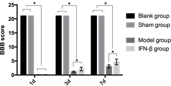
The BBB scores in blank group were 21 points, 21 points and 21 points on the 1st, 3rd, and 7th days after the modeling. The BBB scores in the sham operation group were 21 points, 21 points and 21 points on the 1st, 3rd, and 7th days after the modeling. The BBB scores in the model group were (0.11 ± 0.02) points, (1.13 ± 0.26) points, and (3.05 ± 0.57) points on the 1st, 3rd, and 7th day after the modeling. The BBB scores in the IFN-β group were (0.12 ± 0.02) points, (2.13 ± 0.65) points, and (4.69 ± 1.01) points on the 1st, 3rd, and 7th days after the modeling. The BBB scores of blank and sham operation groups were higher than those of the model and IFN-β groups on the 1st, 3rd, and 7th days after the modeling (P < 0.05). The BBB scores of the model and the IFN-β groups were increased gradually on the 1st, 3rd, and 7th days after the modeling. There was no obvi-ous difference between the model and IFN-β groups on the 1st day after the modeling (P > 0.05). The BBB score of IFN-β group was higher than the score of model group on the 3rd and 7th days after the modeling (P < 0.05) (Figure
1).
A comparison of the modified Tarlov scores at different time points among the four groups
day after the modeling (P > 0.05). The modified Tarlov score of the IFN-β group was higher than the model group score on the 3rd and 7th days after the modeling (P < 0.05) (Figure 2). Comparisons of the Zea-Longa scores at differ-ent time points among the four groups
The Zea-Longa scores in the blank group were (0.17 ± 0.03) points, (0.18 ± 0.02) points, and
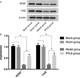
The expressions of BDNF and TrkB in the blank group were (1.03 ± 0.07) and (1.07 ± 0.09) after 7 days of modeling, respectively. The ex- pressions of BDNF and TrkB in the sham opera-tion group were (0.98 ± 0.08) and (1.01 ± 0.07) after 7 days of modeling, respectively. The ex- pressions of BDNF and TrkB in the model group were (0.19 ± 0.03) and (0.20 ± 0.04) after 7 days of modeling, respectively. The expressions of BDNF and TrkB in the IFN-β group were (0.57
Figure 1.Comparison of the BBB scores at different time points among the four groups. The BBB scores of the blank and sham operation groups were higher than those of the model and IFN-β groups on the 1st, 3rd, and
7th days after the modeling (P < 0.05). The BBB scores of the model and
IFN-β groups were increased gradually on the 1st, 3rd, and 7th days after the modeling. There was no observable difference between the model and IFN-β groups on the 1st day after the modeling (P > 0.05). The BBB score of the IFN-β group was higher than the score of the model group on the 3rd
and 7th days after the modeling (P < 0.05). *P < 0.05.
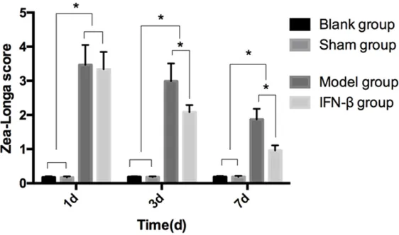
(0.18 ± 0.03) points on the 1st, 3rd and 7th days after the modeling. The Zea-Longa scor- es in the sham operation gro- up were (0.17 ± 0.03) points, (0.18 ± 0.02) points, and (0.19 ± 0.03) points on the 1st, 3rd, and 7th days after the model-ing. The Zea-Longa scores in the model group were (3.47 ± 0.58) points, (2.99 ± 0.52) po- ints, and (1.87 ± 0.31) points on the 1st, 3rd, and 7th days after the modeling. The Zea-Longa scores in the IFN-β gro- up were (3.34 ± 0.51) points, (2.08 ± 0.21) points, and (0.96 ± 0.15) points on the 1st, 3rd, and 7th days after the model-ing. The Zea-Longa scores of the blank and sham operation groups were higher than those of the model and IFN-β groups on the 1st, 3rd, and 7th days after the modeling (P < 0.05). The Zea-Longa scores of the model and IFN-β groups were decreased gradually on the 1st, 3rd, and 7th days after the modeling. There was no obser- vable difference between the model and IFN-β groups on the 1st day after the modeling (P > 0.05). The Zea-Longa score of the IFN-β group was lower than the score of the model group on the 3rd and 7th days after the modeling (P < 0.05) (Figure
3).
[image:4.612.93.371.70.211.2]The expressions of the BDNF and TrkB proteins in the four groups after 7 days of model-ing
Figure 2.Comparison of the modified Tarlov scores at different time points
among the four groups. The Modified Tarlov scores of the blank and sham
operation groups were higher than those of the model and IFN-β groups on
the 1st, 3rd, and 7th days after the modeling (P < 0.05). The modified Tarlov
scores of the model and IFN-β groups were increased gradually on the 1st, 3rd, and 7th days after the modeling. There was no observable difference between the model and IFN-β groups on the 1st day after the modeling (P
> 0.05). The modified Tarlov score of the IFN-β group was higher than the
[image:4.612.92.371.332.492.2]± 0.09) and (0.62 ± 0.10) after 7 days of model-ing, respectively. There were no significant dif-ferences in the expressions of BDNF and TrkB between the blank and sham operation gro- ups (P > 0.05). The expressions of the BDNF and TrkB proteins in the blank sham operation groups were higher than those in the model and IFN-β groups (P < 0.05), and the expres-sions of the BDNF and TrkB proteins in the IFN-β group were higher than those in the model group (P < 0.05) (Table 1 and Figure 4). The expressions of the Bax and Bcl2 proteins in the four groups after 7 days of modeling
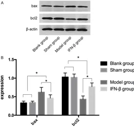
The expressions of Bax and Bcl2 in the blank group were (0.33 ± 0.05) and (1.03 ± 0.11) after 7 days of modeling, respectively. The ex- pressions of Bax and Bcl2 in the sham opera-tion group were (0.34 ± 0.04) and (1.02 ± 0.09) after 7 days of modeling, respectively. The ex- pressions of Bax and Bcl2 in the model group were (0.52 ± 0.13) and (0.43 ± 0.09) after 7 days of modeling, respectively. The expressions of Bax and Bcl2 in the IFN-β group were (0.46 ± 0.09) and (0.77 ± 0.10) after 7 days of model-ing, respectively. There were no significant dif-ferences in the expressions of Bax and Bcl2 between the blank and sham operation groups (P > 0.05). The expression of the Bax protein in the blank and sham operation groups was
1.05)% respectively after 7 days of modeling. There was no remarkable difference in the neu-ronal apoptosis rates between the blank and sham operation groups (P > 0.05). The neuro-nal apoptosis rate in the blank and sham oper-ation groups was lower than it was in the model and IFN-β groups, and the neuronal apoptosis rate in the IFN-β group was lower than it was in the model group (P < 0.05) (Figure 6).
The correlation analysis between the expres-sions of the BDNF and TrkB proteins and the neuronal apoptosis rate
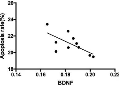
The expressions of the BDNF and TrkB proteins were negatively correlated with the neuronal apoptosis rate (r = -0.667, P < 0.05; r = -0.791, P < 0.05) (Figures 7 and 8).
Discussion
IFN-β is a cytokine with and immunoregulatory effect and can reduce cell apoptosis under cer-tain conditions. It has been applied in MS [15, 16]. Some studies [17-19] have shown that although there are many mechanisms for sec-ondary injury in acute spinal cord injury, the immune system activation exerts a key role. The inflammatory reactions and a series of lipid peroxidation reactions are caused by an aggre-gation of inflammatory cells after the injury,
Figure 3.Comparisons of the Zea-Longa scores at different time points among the four groups. The Zea-Longa scores of the blank and sham op-eration groups were higher than those of the model and IFN-β groups on the
1st, 3rd and 7th days after the modeling (P < 0.05). The Zea-Longa scores
of the model and IFN-β groups were decreased gradually on the 1st, 3rd, and 7th days after the modeling. There was no obvious difference between the model and IFN-β groups on the 1st day after the modeling (P > 0.05). The Zea-Longa score of the IFN-β group was lower than the score of the
model group on the 3rd and 7th days after the modeling (P < 0.05). *P <
0.05.
lower than the expression in the model and IFN-β groups, and the expression of the Bcl2 protein in the blank and sham operation groups was higher than the expression in the mo- del and IFN-β groups (P < 0.05). The expression of the Bax protein in the IFN-β group was lower its was in the model group, and the expression of the Bcl2 protein in the IFN-β group was higher than it was in model group (P < 0.05) (Table 2 and Figure 5).
Comparison of the neuronal apoptosis rate in the four groups
[image:5.612.90.371.72.238.2]which ultimately leads to neuron injury. The- refore, it is speculated that IFN-β might have a certain effect on acute spinal cord injury. In our study, IFN-β intervention was performed on rats with acute spinal cord injuries. It was found that the BBB score, the modified Tarlov score, and the Zea-Longa scores of the blank and sham operation groups were higher than those of the model and IFN-β groups, and the scores of each item in the IFN-β group were markedly improved compared with those in the model group on the 3rd and 7th days after the model-ing. At the same time, it was found that the scores of each item in the model group was gradually improved without drug intervention, suggesting that IFN-β intervention can
[image:6.612.91.515.85.137.2]effec-TrkB proteins in the blank and sham operation groups were higher than those in the model and IFN-β groups, and the expressions of the BDNF and TrkB proteins in the IFN-β group were higher than those in the model group, indicat-ing that the expressions of the BDNF and TrkB proteins in spinal cord tissue could be down-regulated in rats with acute spinal cord injury. It was suggested that IFN-β could promote the recovery of the BDNF and TrkB protein expres-sions in rats with acute spinal cord injury, there-by promoting the recovery of nerve function. It has been reported that the expression of the BDNF protein in the injured spinal cord was down-regulated [23]. As the spinal cord has a partial self-recovery function, the expression of Table 1. The expressions of the BDNF and TrkB proteins in the four groups after 7 days of modeling Protein Blank group n = 10 Sham group n = 10 Model group n = 10 IFN-β group n = 10 F P
BDNF 1.03 ± 0.07* 0.98 ± 0.08* 0.19 ± 0.03 0.57 ± 0.09# 304.8 < 0.001
TrkB 1.07 ± 0.09* 1.01 ± 0.07* 0.20 ± 0.04 0.62 ± 0.10# 263.9 < 0.001
*, # indicates compared with the Model group; P < 0.05.
Figure 4. The expressions of the BDNF and TrkB proteins in the four groups
after 7 days of modeling. There was no significant difference in the expres -sions of BDNF and TrkB between the blank and sham operation groups (P > 0.05). The expressions of the BDNF and TrkB proteins in the blank and sham operation groups were higher than those in the model and IFN-β
groups (P < 0.05), and the expressions of the BDNF and TrkB proteins in the
IFN-β group were higher than those in the model group (P < 0.05).
[image:6.612.92.367.170.436.2]the BDNF protein would be increased to pro-mote the partial recovery of nerve function. However, due to the problem of sample size, it is impossible to monitor the expression of the BDNF protein at multiple time points, which will be further improved on in subsequent experi- ments.
Some studies [24] have shown that cell apopto-sis is an important part of secondary spinal cord injury. The death of nerve cells after spinal cord injury is not caused by external forces, but by neuronal apoptosis. Therefore, the neuronal apoptosis rate and the apoptosis-related
pro-IFN-β on acute spinal cord injury. Therefore, more experiments are needed to confirm our conclusions.
[image:7.612.90.523.85.137.2]In conclusion, the motor and nerve functions are significantly improved, the expressions of the BDNF and TrkB proteins are up-regulated, and neuronal apoptosis is inhibited by IFN-β in rats with acute spinal cord injury. However, there are some deficiencies in this study. First, due to the problem of sample size, a dynamic monitoring of the expressions of the BDNF and TrkB proteins in rat spinal cord tissues was not carried out. Furthermore, the mechanism of Table 2. The expression of the Bax and Bcl2 proteins in the four groups after 7 days of modeling Protein Blank group n = 10 Sham group n = 10 Model group n = 10 IFN-β group n = 10 F P
bax 0.33 ± 0.05* 0.34 ± 0.04* 0.62 ± 0.13 0.46 ± 0.09# 25.14 < 0.001
bcl2 1.03 ± 0.11* 1.02 ± 0.09* 0.43 ± 0.09 0.77 ± 0.10# 83.02 < 0.001
*, # indicates compared with the Model group; P < 0.05.
Figure 5. The Expressions of the Bax and Bcl2 proteins in the four groups
after 7 days of modeling. There were no significant differences in the ex -pressions of Bax and Bcl2 between the blank and sham operation groups (P > 0.05). The expression of the Bax protein in the blank and sham operation groups was lower than it was in the model and IFN-β groups, and the expres-sion of the Bcl2 protein in the blank and sham operation groups was higher than it was in the model and IFN-β groups (P < 0.05). The expression of the
Bax protein in the IFN-β group was lower than it was in the model group, and the expression of the Bcl2 protein in the IFN-β group was higher than it was
in the model group (P < 0.05). Note: *indicates P < 0.05.
[image:7.612.92.367.169.429.2]References
[1] Hayta E and Elden H. Acute spinal cord injury: a review of pathophysiology and potential of non-steroidal anti-inflammatory drugs for phar -macological intervention. J Chem Neuroanat 2018; 87: 25-31.
[2] Li J, Deng J, Yuan J, Fu J, Li X, Tong A, Wang Y, Chen Y and Guo G. Zonisamide-loaded triblock copolymer nanomicelles as a novel drug deliv-ery system for the treatment of acute spinal cord injury. Int J Nanomedicine 2017; 12: 2443-2456.
[3] Martins BC, Torres BBJ, de Oliveira KM, Lavor MS, Osorio CM, Fukushima FB, Rosado IR and de Melo EG. Association of riluzole and dan-trolene improves significant recovery after acute spinal cord injury in rats. Spine J 2018; 18: 532-539.
[4] Li Z, Wang S, Li W and Yuan H. Ferulic acid im-proves functional recovery after acute spinal cord injury in rats by inducing hypoxia to inhibit microRNA-590 and elevate vascular endothe-lial growth factor expressions. Front Mol Neuro-sci 2017; 10: 183.
[5] Zhang Y, Lv Y, Ji W, Zhou R, Gao S and Zhou F. Therapeutic hypothermia effectively reduces elevated extracellular ascorbate concentra-tions caused by acute spinal cord injury. Artif Cells Nanomed Biotechnol 2019; 47: 22-29. [6] Bimbova K, Bacova M, Kisucka A, Pavel J, Galik
[image:8.612.90.286.70.232.2]J, Zavacky P, Marsala M, Stropkovska A, Fe-dorova J, Papcunova S, Jachova J and Lukaco-va N. A single dose of atorLukaco-vastatin applied acutely after spinal cord injury suppresses in-flammation, apoptosis, and promotes axon outgrowth, which might be essential for favor-able functional outcome. Int J Mol Sci 2018; 19.
Figure 6. Comparison of the neuronal apoptosis rates in the four groups. The neuronal apoptosis rate in the blank and sham operation groups was lower than it
was in the model and IFN-β groups, but the neuronal apoptosis rate in the IFN-β group was lower than it
was in the model group (P < 0.05). Note: *Compared with ** and ***P < 0.05. **Compared with ***P < 0.05.
Figure 7. Correlation analysis between the expres-sion of the BDNF protein and the neuronal apoptosis rate. The expression of the BDNF protein was nega-tively correlated with the neuronal apoptosis rate (r = -0.667, P < 0.05).
Figure 8. Correlation analysis between the expres-sion of the TrkB protein and the neuronal apoptosis rate. The expression of the TrkB protein was nega-tively correlated with the neuronal apoptosis rate (r = -0.791, P < 0.05).
IFN-β inhibiting neuronal apoptosis by regulat-ing the expressions of the apoptosis-related proteins Bax and Bcl2 is still unclear, which makes the conclusion inaccurate. In the follow-up experiments, we will further explore these problems.
Disclosure of conflict of interest None.
[image:8.612.323.523.71.210.2] [image:8.612.92.285.347.488.2][7] Xia Y, Xia H, Chen D, Liao Z and Yan Y. Mecha-nisms of autophagy and apoptosis mediated by JAK2 signaling pathway after spinal cord in-jury of rats. Exp Ther Med 2017; 14: 1589-1593.
[8] Mahony R, Gargan S, Roberts KL, Bourke N, Keating SE, Bowie AG, O’Farrelly C and Steven-son NJ. A novel anti-viral role for STAT3 in IFN-alpha signalling responses. Cell Mol Life Sci 2017; 74: 1755-1764.
[9] Hauser SL, Bar-Or A, Comi G, Giovannoni G, Hartung HP, Hemmer B, Lublin F, Montalban X, Rammohan KW, Selmaj K, Traboulsee A, Wo-linsky JS, Arnold DL, Klingelschmitt G, Master-man D, Fontoura P, Belachew S, Chin P, Mairon N, Garren H, Kappos L and Opera I; OPERA I and OPERA II Clinical Investigators. Ocrelizum-ab versus interferon Beta-1a in relapsing mul-tiple sclerosis. N Engl J Med 2017; 376: 221-234.
[10] Guidotti LG, Ishikawa T, Hobbs MV, Matzke B, Schreiber R and Chisari FV. Intracellular inacti-vation of the hepatitis B virus by cytotoxic T lymphocytes. Immunity 1996; 4: 25-36. [11] Gok B, Okutan O, Beskonakli E, Palaoglu S,
Er-damar H and Sargon M. Effect of immunomod-ulation with human interferon-beta on early functional recovery from experimental spinal cord injury. Spine 2007; 32: 873-880.
[12] Muniswami DM, Kanakasabapathy I and Thar-ion G. Globose basal cells for spinal cord re-generation. Neural Regen Res 2017; 12: 1895-1904.
[13] Cheng H, Cao Y and Olson L. Spinal cord repair in adult paraplegic rats: partial restoration of hind limb function. Science 1996; 273: 510-513.
[14] Zheng XW, Yang WT, Chen S, Xu QQ, Shan CS, Zheng GQ and Ruan JC. Neuroprotection of ca-talpol for experimental acute focal ischemic stroke: preclinical evidence and possible me- chanisms of antioxidation, anti-inflammation, and antiapoptosis. Oxid Med Cell Longev 2017; 2017: 5058609.
[15] Singh A, Srivastava RN, Agrahari A, Singh S, Raj S, Chatterji T, Mahdi AA, Garg RK and Roy R. Proton NMR based serum metabolic profile correlates with the neurological recovery in treated acute spinal cord injury (ASCI) sub-jects: a pilot study. Clin Chim Acta 2018; 480: 150-160.
[16] Suárez-Amarán L, Usai C, Di SM, Godoy C, Ni Y, Hommel M, Palomo L, Segura V, Olagüe C, Vales A, Ruiz-Ripa A, Buti M, Salido E, Prieto J, Urban S, Rodríguez-Frias F, Aldabe R and González-Aseguinolaza G. A new HDV mou- se model identifies mitochondrial antiviral signaling protein (MAVS) as a key play- er in IFN-β induction. J Hepatol 2017; 67: S0168827817320469.
[17] Zhang N, Wei G, Ye J, Yang L, Hong Y, Liu G, Zhong H and Cai X. Effect of curcumin on acute spinal cord injury in mice via inhibition of in-flammation and TAK1 pathway. Pharmacol Rep 2017; 69: 1001-1006.
[18] Gong ZM, Tang ZY and Sun XL. miR-411 sup-presses acute spinal cord injury via downregu-lation of Fas ligand in rats. Biochem Biophys Res Commun 2018; 501: 501-506.
[19] Xie W, Yang SY, Zhang Q, Zhou Y, Wang Y, Liu R, Wang W, Shi J, Ning B and Jia T. Knockdown of microRNA-21 promotes neurological recovery after acute spinal cord injury. Neurochem Res 2018; 43: 1641-1649.
[20] Fehlings MG, Tetreault LA, Aarabi B, Anderson P, Arnold PM, Brodke DS, Chiba K, Dettori JR, Furlan JC, Harrop JS, Hawryluk G, Holly LT, Howley S, Jeji T, Kalsi-Ryan S, Kotter M, Kurpad S, Kwon BK, Marino RJ, Martin AR, Massicotte E, Merli G, Middleton JW, Nakashima H, Na-goshi N, Palmieri K, Singh A, Skelly AC, Tsai EC, Vaccaro A, Wilson JR, Yee A and Burns AS. A Clinical practice guideline for the management of patients with acute spinal cord injury: rec-ommendations on the type and timing of reha-bilitation. Global Spine J 2017; 7: 231S-238S. [21] Song Z, Ye Y, Zhang Z, Shen J, Hu Z, Wang Z
and Zheng J. Noninvasive, targeted gene ther-apy for acute spinal cord injury using LIFU-me-diated BDNF-loaded cationic nanobubble de-struction. Biochem Biophys Res Commun 2018; 496: 911-920.
[22] Wang Y, Li W, Wang M, Lin C, Li G, Zhou X, Luo J and Jin D. Quercetin reduces neural tissue damage and promotes astrocyte activation af-ter spinal cord injury in rats. J Cell Biochem 2018; 119: 2298-2306.
[23] Zhou X, Xiao H and Wang H. Developmental changes of TrkB signaling in response to exog-enous brain-derived neurotrophic factor in pri-mary cortical neurons. J Neurochem 2011; 119: 1205-1216.
[24] Xu G, Zhang J, Wang L, Cui Z, Sun X, Liu Z, Zhu Z and Qiu Y. Up-regulation of TRAF2 suppress-es neuronal apoptosis after rat spinal cord in-jury. Tissue Cell 2017; 49: 589-596.