Original Article
The Yi-Qi-Bu-Shen recipe attenuates high
glucose-induced podocyte injury via the inhibition
of IKK-IκBα-NFκB and ERK/P38 MAPK signaling
Rui-Ting Hu1,2, Chang-Ling Li1, Xi Zhang1, Chun-Yao An3, Chun-Mei Ma1, Xiu Li3, Xiao-Hong Liu3, Wen-Jing Li3,
Yun Qiao1, De-Shan Liu1
1Department of Traditional Chinese Medicine, Qilu Hospital of Shandong University, Jinan 250012, P.R. China; 2Hospital of Traditional Chinese Medicine of Linyi City, Linyi 276002, P.R. China; 3Shandong University of Traditional Chinese Medicine, Jinan 250355, P.R. China
Received May 3, 2019; Accepted June 10, 2019; Epub August 15, 2019; Published August 30, 2019
Abstract: The exact mechanisms underlying diabetic nephropathy (DN) remain unknown, but some studies suggest that structural and compositional changes of the slit diaphragms (SD) between podocytes may play an important role in this course. The Yi-Qi-Bu-Shen recipe (YB) is a traditional Chinese herbal formula that is commonly used for the treatment of diabetes mellitus and especially DN. This study investigated the effect of high glucose (HG) on expression of nephrin, podocin, and CD2AP in podocytes and the treatment effect of YB on these molecules. Conditionally immortalized human podocytes were exposed to medium containing normal glucose (NG) or HG for 24 hours. The podocytes cultured in a HG environment had relatively lower expression of nephrin, podocin, and CD2AP both in mRNA and protein synthesis levels compared to NG group. Nephrin, podocin, and CD2AP expression
in the HG group was effectively restored by the treatment of YB. In addition, inflammatory cytokines such as IL-1β, IL-6, TNF-α, and MCP-1 were significantly decreased in HG+YB group compared with the HG group. Moreover, YB suppressed HG-induced activation of IKK-IκBα-NFκB and ERK/P38 MAPK signaling pathways. NFκB, ERK, and P38
inhibitors attenuated HG-induced podocyte injury and increased nephrin, podocin, and CD2AP expression further revealed the healing mechanisms of YB. This study demonstrates that YB treatment attenuated HG-induced
podo-cyte injury through suppressing activation of IKK-IκBα-NFκB and ERK/P38 MAPK signaling pathways and reducing IL-1β, IL-6, TNF-α, and MCP-1 levels, suggesting that YB is a potential therapeutic drug for DN.
Keywords: The Yi-Qi-Bu-Shen recipe, podocyte, nephrin, podocin, CD2AP
Introduction
Diabetic nephropathy (DN) is one of the most serious complications of diabetes mellitus and the main cause of the end-stage renal disease (ESRD) [1, 2]. The characteristics of DN include mesangial cells proliferation, mesangial matrix accumulation, and glomerulosclerosis [3, 4]. Mi- croalbuminuria and later proteinuria are the most important clinical manifestations in the
developing process of DN. The glomerular filtra -tion barrier (GFB), which prevents proteins leak-ing from capillaries to urine, includes endo- thelial cells, glomerular basement membrane (GBM) and podocytes. Any injury of these por-tions may lead to the occurrence of microalbu-minuria and later proteinuria [5, 6].
Podocytes are highly differentiated epithelial cells that interface with the GBM surface and
play a significant role in maintaining the integ -rity of the GFB. Increasing evidence suggests that podocytes prevent protein leakage from plasma to primary urine [7, 8]. Slit diaphragms (SD) between podocytes are the essential determinant of podocyte function [9]. Some
specific proteins in the SD could affect the
structure and function of podocytes, the most important of them are nephrin, podocin, and CD2AP. Nephrin is a transmembrane glycopro-tein and the structural backbone of SD, which encoded by the NPSH1 gene. Podocin is a hair-like structured membrane protein expressed
specifically in SD, which can enhance the signal
in the structure of cytoskeleton. CD2AP is a cohesive protein that connects SD and the cytoskeleton and plays a stabilizing role in podocytes. In DN patients and animal models, the mRNA and protein expressions of these
molecules are significantly reduced and closely
related to the progression of proteinuria
[10-12]. Hence, finding a drug that can protect
these molecules from injury is essential for DN treatment.
Chronic inflammation is closely associated with
permeability changes in the GFB and
protein-uria in DN. Inflammatory cytokines, such as IL-1β, IL-6, and TNF-α, are elevated even in the early stage of DN. Subsequently, TNF-α ampli
-fies the activation of inflammatory cytokines,
thereby leading to DN progression [13-15]. Among several signaling pathways that regulate
chronic inflammation, it has been reported that NFκB and MAPK are associated with podocyte
injury in DN [16, 17].
YB has been used in Qi’lu Hospital for more than 20 years [18, 19], previously clinical data demonstrated that YB reduces proteinuria and decreases blood glucose level in DN patients. However, the exact mechanisms of YB in the treatment of proteinuria remain unknown. Sin- ce podocyte injury has been found to be the pri-mary determinant of proteinuria, the aim of the present study is to investigate the protective effects of YB on HG-induced podocyte injury. Materials and methods
Materials
RPMI-1640 culture medium and D-glucose we- re purchased from Sigma-Aldrich. Fetal bovine serum (FBS), TRIzol reagent, SYBR Green Mas- ter Mix and PierceTM fast Western blot kit were
purchased from Thermo Fisher Scientific.
iSc-ript Advanced cDNA synthesis kit was purch- ased from Bio-Rad. Cytotoxicity detection kit (LDH) and X-tremeGENE HP DNA transfection reagent were purchased from Roche Applied Science. BCA protein assay kit was purchased from Beyotime Biotechnology. The primary anti-bodies were purchased from Cell Signaling
Technology and Affinity Biosciences.
pNFκB-TA-Luc, pRL-TK and a Dual-Luciferase Reporter Assay System were purchased from Promega.
IκBα inhibitor BAY 11-7085 was purchased
from Santa-Cruz. P38 MAPK inhibitor SB203-
580 and ERK MAPK inhibitor U0126 were pur-chased from Cell Signaling Technology. Composition and preparation of the Yi-Qi-Bu-Shen recipe (YB)
The composition and preparation of the YB extract are identical with our previous studies
[18, 19]. Briefly, the YB was composed of crude
Radix Astragali, prepared Rhizome of Rehman- nia, Rhizoma Polygonati, Rhizoma Chuanxiong, Herba Epimedii, Fructus Lycii, Rhizoma Atra- cylodis, Radix Puerariae and Rhizoma Coptidis. All crude drugs were the products of Jinan Jianlian Traditional Chinese Medicine Co. Ltd.
They were chopped finely and extracted with 20
times the amount of distilled water at 100°C for 2 hours, a process that was repeated three times. The extract was evaporated under pres-sure and insoluble substances were removed
via filtration and then dried to obtain YB powder
in a vacuum drying furnace (yield: 28.06%). The YB powder was prepared by the Manufacturing Laboratory of Qilu Hospital, Shandong Univer- sity (one gram was equal to 3.564 g of crude drug).
Study subjects
A total of 160 patients suffering from type 2 diabetes and presence of microalbuminuria, aged 51 to 73 years old, were randomly enrolled between January 2015 and December 2016 from the Qi’lu hospital of Shandong University. Glucose cutoff values were used for diagnosis of DM based on instruction of the World health organization. The urine albumin to creatinine ratio (ACR) ranged from 30 to 300 µg/mg cre-atinine on two of three morning urine collec-tions and was considered as microalbuminuria. Exclusion criteria were history of malignancies,
acute or chronic inflammatory and infectious
NA synthesis kit. qRT-PCR was performed with SYBR Green Ma-
ster Mix. The amplifi -cation protocol used for qRT-PCR analysis was 94°C for 3 min-utes, followed by 35 cycles of 94°C for 30 seconds, 55°C for 30 seconds, 72°C for 30
seconds, and a final
extension at 72°C for 5 minutes. Expression Cell culture
Conditionally immortalized human podocytes were cultured as previously described [20].
Briefly, to induce proliferation, cells were cul -tured in plates containing RPMI-1640 medium supplemented with 10% fetal bovine serum (FBS), D-glucose (5 mM) , Penicillin (100 U/ml) and streptomycin (100 U/ml) at 33°C with 5% CO2. To induce differentiation, podocytes were cultured with RPMI-1640 medium (supplement-ed as above) at 37°C with 5% CO2 for 14 days. All cell experiments were carried out in serum free conditions on well differentiated podo-cytes between passages 10 and 20 after over-night serum starvation.
Cell viability assays
Cell viability was determined by monitoring the lactate dehydrogenase (LDH) levels in the po-
docytes as previously described [21]. Briefly,
1×10⁵ terminally differentiated podocytes in
500 uL culture medium were seeded in each well of the 24-well plates. After overnight serum starvation, the culture medium was replaced with fresh medium containing different concen-trations of glucose or YB. After 24 h, the LDH activity in the cells was determined utilizing a cytotoxicity detection kit. The absorbance was measured using a microplate reader of Thermo
Fisher scientific.
Quantitative real-time polymerase chain reac-tion
Quantitative real-time polymerase chain reac-tion (qRT-PCR) was performed using an
estab-lished procedure [22]. Briefly, total RNA was
extracted utilizing the TRIzol reagent. cDNA was synthesized utilizing the iScript Advanced cD-
levels of target genes were normalized by con-current measurement of GAPDH mRNA levels. All primers used are listed in Table 1.
Western blot analysis
Western blot analysis was performed as previ-ously described [23]. The podocytes were wa- shed three times with a precooled PBS contain-ing sodium vanadate then RIPA lysis buffer con-taining protease inhibitors and phosphatase inhibitors was added to cells. After 30 minutes, the cell lysates collected into 1.5 ml eppendorf tubes and centrifuged at 12000 g for 15 min-utes at 4°C. Total proteins were extracted and determined using a BCA protein assay kit. Protein samples were separated by electropho-resis and transferred to PVDF membranes. After being blocked and incubated with primary antibodies, the PVDF membranes were washed with TBST and subsequently incubated with secondary antibody, then measured by Pie- rceTM fast Western blot kit. The primary
anti-bodies included: anti-Nephrin (AF7951, Affini-ty); anti-Podocin (DF8593, AffiniAffini-ty); anti-CD2AP (DF2298, Affinity); anti-IKKα (AF6012, Affinity Biosciences); anti-IKKβ (AF6009, Affinity sciences); anti-p-IKKα/β (AF3014, Affinity Bio-sciences); anti-IκBα (AF5002, Affinity Biosci-ences); anti-p-IκBα (AF2002, Affinity
Bioscien-ces); anti-ERK1/2 (8867, Cell signaling); anti-p-ERK1/2 (13148, Cell signaling); anti-P38 (14- 451, Cell signaling); anti-p-P38 (4092, Cell sig-naling); anti-JNK (4671, Cell sigsig-naling); anti-p-JNK (3708, Cell signaling); and anti-GAPDH (D16H11, Cell signaling).
Transfection and transcription reporter assay
[image:3.612.89.418.84.215.2]Podocytes were seeded in 24-well plates (1× 10⁵ cells per well) overnight and changed to
Table 1. Primers for qRT-PCR
Primers Forward: 5’-3’ Reverse: 5’-3’ length (bp)Product Nephrin GAGGCTGGTGTGTTTGGCTA GCCAGGATCGTCACGTTAGT 273 Podocin TCTGGTTCTGCGTAAAGGTTGT GCACAAGGAATTGCACAGCTT 294 CD2AP TGGAGATAACAAAAACAGATACCGA TTTGGAGCTGGAGCCTTAGC 271
IL-1β ACAGATGAAGTGCTCCTTCCAG AAGCCCTTGCTGTAGTGGTG 93
IL-6 AATAACCACCCCTGACCCAAC ACATTTGCCGAAGAGCCCT 149
well) and pRL-TK-Luc (10 ng/we- ll) plasmids using ROCHE X-tre- meGENE HP DNA transfection reagent for 18 hours. After treat-ment with drugs, the cells were lysed and transcriptional activi-ties were calculated using a du- al luciferase assay kit and a TD- 20/20 luminometer according to the manufacturer’s instructi- ons.
Statistics
For data analysis, the results are expressed as mean and st- andard deviation in the tables
and figures. The
Kolmogorov-Smirnov test was used to check the normality of the data. Diff- erences between 2 groups were analyzed using the Student’s t
test and differences ≥3 groups
were compared by one ways an- alysis of variance (ANOVA). Wil- coxon test was used to evaluate dependent groups. Mann-Whit- ney test was used for compari-son of the mean differences be- tween the groups. SPSS Statis- tics Version 22 (IBM Corp, Arm-onk, NY) was used for all
analy-ses, and the level of significant
difference was set at P<0.05. Results
The parameters of study sub-jects
Baseline parameters of all the participants in this study are presented in Table 2. There we-
re no significant differences at
baseline in two groups. The pa- rameters after treatment are presented in Tables 3-5. It is notable that the patients’ HbA- 1c, FPG, 2hPG, SBP, DBP, TC, TG, LDL and ACR levels were
sig-nificantly decreased in both
gr-oups after six months’ therapy (P<0.05). Additionally, the HbA- 1c, FPG, 2hPG, and ACR levels serum free medium the next morning. Cells
[image:4.612.91.367.96.373.2]were transfected with pNF-κB-TA-Luc (100 ng/ of the YB treatment group were lower than the control group (P<0.05). Table 2. Baseline characteristics of Control and YB treatment
groups
Index Groups value/xT 2 valueP
Control YB treatment
Gender (M/F) 42/38 39/41 ☆
Age (years) 61.91±5.64 61.18±5.47 0.840 0.402
BMI (kg/m2) 22.38±2.31 22.90±2.21 -1.464 0.145
HbA1c (%) 8.35±0.71 8.44±0.83 -0.759 0.449
FPG (mmol/l) 9.93±0.88 10.12±0.98 -1.248 0.214 2hPG (mmol/l) 13.81±1.02 14.08±1.09 -1.572 0.118 SBP (mmHg) 133.33±9.01 135.55±12.21 -1.304 0.194
DBP (mmHg) 82.16±7.78 83.65±8.24 -1.174 0.242
TC (mmol/l) 4.49±0.59 4.32±0.58 -0.534 0.594
TG (mmol/l) 1.48±0.36 1.51±0.24 1.273 0.205
LDL (mmol/l) 3.24±0.64 3.12±0.60 1.813 0.072
HDL (mmol/l) 1.20±0.16 1.18±0.13 1.152 0.251
Sodium (mmol/l) 140.75±3.75 140.85±2.83 -0.205 0.838 Potassium (mmol/l) 4.33±0.57 4.45±0.48 -1.401 0.163
AST (U/l) 27.34±6.59 28.37±7.03 -0.955 0.341
ALT (U/l) 20.14±7.01 21.19±8.11 -0.882 0.379
Urea (mmol/l) 6.37±2.42 6.89±1.99 -1.484 0.140 Creatinine (µmol/l) 79.22±18.76 77.37±16.43 0.666 0.506 ACR (µg/mg) 185.20±56.65 180.87±56.75 0.483 0.629 ☆Chi square test, others were t test.
Table 3. Parameters of the Control group at the baseline and at end of conventional therapy
Index Control T value P value
Pre treatment Pro treatment
HbA1c (%) 8.35±0.71 6.40±0.35 22.037 <0.001* FPG (mmol/l) 9.93±0.88 6.48±0.41 31.941 <0.001* 2hPG (mmol/l) 13.81±1.02 8.74±0.48 40.303 <0.001* SBP (mmHg) 133.33±9.01 124.80±7.61 6.474 <0.001* DBP (mmHg) 82.16±7.78 76.20±5.78 5.502 <0.001* TC (mmol/l) 4.49±0.59 4.16±0.44 4.072 <0.001* TG (mmol/l) 1.48±0.36 1.29±0.23 3.849 <0.001* LDL (mmol/l) 3.24±0.64 2.85±0.31 4.932 <0.001*
HDL (mmol/l) 1.20±0.16 1.17±0.13 1.681 0.095
Sodium (mmol/l) 140.75±3.75 139.88±3.89 1.436 0.153 Potassium (mmol/l) 4.33±0.57 4.34±0.54 -0.103 0.918
AST (U/l) 27.34±6.59 28.18±6.51 -0.811 0.419
ALT (U/l) 20.14±7.01 18.79±5.61 1.342 0.182
[image:4.612.93.367.432.667.2]to 40 mmol/L for 24 hours) not reveal an effect of HG upon cell viability. To preclude the role of osmosis, we also observed the effect of mannitol (40 mmol/L) at the same time and did not
find mannitol to affect the cell
viability (Figure 1A). Cells were also treated with different con-centrations of YB in normal glu-cose (5.5 mmol/L) and HG (40 mmol/L) medium for 24 hours, respectively. As shown in Figure 1B, YB did not affect cell viability in either condition below the 200 ug/mL YB dose. Thus, sub-sequent experiments employed a 100 ug/mL dose of YB which was demonstrated to have no effect on cell death.
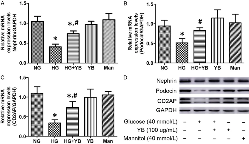
The effects of HG and YB on nephrin, podocin and CD2AP expression
Nephrin, podocin, and CD2AP are all important components of SD. The mRNA and protein expression of these three mark-ers was analyzed to explore the variation of SD under HG condi-tions. First, HG remarkably decr- eased the expression of neph-rin, podocin, and CD2AP at both mRNA and protein levels. How- ever, the same dose of mannitol did not affect their expression (Figure 2). Interestingly, co-tre- atment with YB attenuated the effect of HG evidenced through
significantly restored mRNA and
protein expression of the three markers (Figure 2), suggesting a therapeutic action of YB in HG- induce podocyte injury.
YB suppresses the pro-inflam-matory cytokines in podocytes
The influence of HG and YB on cell viability in podocytes
The viability of podocytes was determined by the LDH assay. Podocytes treated with HG (up
Because chronic inflammation is the main
cause of diabetic nephropathy and podocyte
injury, the mRNA levels of pro-inflammatory cytokines including IL-1β, IL-6, TNF-α, and MCP-1 were first detected in HG-induced podo -Table 4. The parameters of YB therapy group at the baseline and
at end of YB therapy
Index YB treatment T value P value
Pre treatment Pro treatment
HbA1c (%) 8.44±0.83 6.05±0.41 23.083 <0.001* FPG (mmol/l) 10.12±0.98 5.88±0.39 35.773 <0.001* 2hPG (mmol/l) 14.08±1.09 8.17±0.64 41.623 <0.001* SBP (mmHg) 135.55±12.21 125.80±9.67 5.599 <0.001* DBP (mmHg) 83.65±8.24 77.60±7.08 4.981 <0.001*
TC (mmol/l) 4.32±0.58 4.10±0.48 2.725 0.007*
TG (mmol/l) 1.51±0.24 1.30±0.16 6.481 <0.001* LDL (mmol/l) 3.12±0.60 2.82±0.35 3.913 <0.001*
HDL (mmol/l) 1.18±0.13 1.15±0.11 1.354 0.178
Sodium (mmol/l) 140.85±2.83 139.96±3.34 1.808 0.072 Potassium (mmol/l) 4.45±0.48 4.41±0.43 0.524 0.601
AST (U/l) 28.37±7.03 27.55±7.17 0.733 0.465
ALT (U/l) 21.19±8.11 19.42±5.75 1.603 0.111
[image:5.612.91.366.95.333.2]Urea (mmol/l) 6.89±1.99 6.52±1.64 1.256 0.211 Creatinine (µmol/l) 77.37±16.43 75.48±15.71 0.740 0.460 ACR (µg/mg) 180.87±56.75 75.42±24.68 15.243 <0.001* *P<0.05 between pre-treatment and pro-treatment in the YB treatment group.
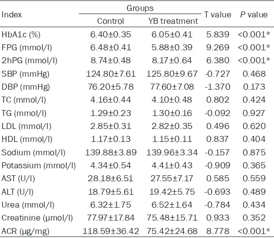
Table 5. The clinical characteristics after conventional therapy and YB treatment
Index Control Groups YB treatment T value P value
HbA1c (%) 6.40±0.35 6.05±0.41 5.839 <0.001* FPG (mmol/l) 6.48±0.41 5.88±0.39 9.269 <0.001* 2hPG (mmol/l) 8.74±0.48 8.17±0.64 6.380 <0.001* SBP (mmHg) 124.80±7.61 125.80±9.67 -0.727 0.468 DBP (mmHg) 76.20±5.78 77.60±7.08 -1.370 0.173
TC (mmol/l) 4.16±0.44 4.10±0.48 0.802 0.424
TG (mmol/l) 1.29±0.23 1.30±0.16 -0.092 0.927
LDL (mmol/l) 2.85±0.31 2.82±0.35 0.496 0.620
HDL (mmol/l) 1.17±0.13 1.15±0.11 0.837 0.404
Sodium (mmol/l) 139.88±3.89 139.96±3.34 -0.157 0.875 Potassium (mmol/l) 4.34±0.54 4.41±0.43 -0.909 0.365
AST (U/l) 28.18±6.51 27.55±7.17 0.585 0.559
ALT (U/l) 18.79±5.61 19.42±5.75 -0.693 0.489
[image:5.612.91.366.392.631.2]cytes. Interestingly, qRT-PCR revealed that the mRNA expressions of nephrin, podocin and CD2AP were all up-regulated in the HG group compared with the control group. Treatment
with YB could significantly inhibit the transcrip
-tional activity of these inflammatory cytokines
induced by HG (Figure 3). These results re-
vealed a potent anti-inflammatory profile of YB
in podocytes.
Molecular mechanism by which YB attenuated HG-induced inflammation
Because these pro-inflammatory cytokines are
mainly regulated at the transcriptional level
by the activation of NFκB and MAPKs, the
ef-fect of YB on HG-induced activation of the- se signaling pathways in podocytes was an- alyzed.
[image:6.612.89.522.74.216.2]Figure 1. Cell viability assays. A. Podocytes were cultured in serum-free medium with normal glucose (NG, 5.5 mmol/L), high glucose (HG, 40 mmol/L) or mannitol (Man, 40 mmol/L) for 24 hours. Cell viability was assessed by a cell death kit as described in method and materials. B. Podocytes were cultured in serum-free medium with or without the Yi-Qi-Bu-Shen recipe (YB) in NG or HG medium for 24 hours. Cell viability was assessed by a cell death kit as described in method and materials. (*P<0.05 vs NG control; #P<0.05 vs HG control).
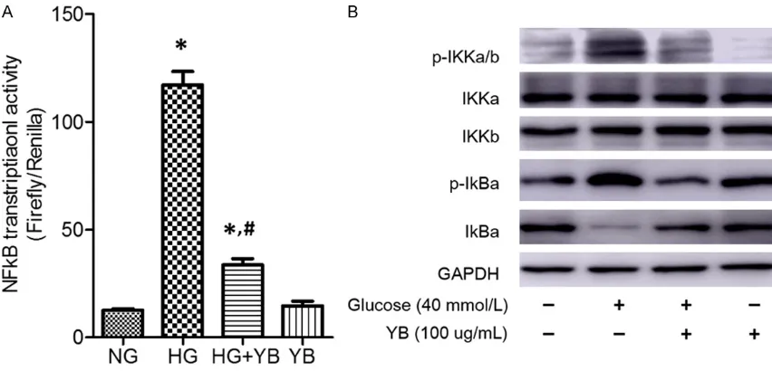
[image:6.612.92.521.299.547.2]Effect of YB on NFκB signaling pathway
As shown in Figure 4A, podocytes treated with HG alone show increased transcriptional
activ-ity of NFκB compared with the control group. Moreover, the phosphorylation of IKKα/β and IκBα were also up-regulated in the HG group
(Figure 4B), suggesting activation of NFκB sig
-Figure 3. Effect of YB on HG-induced expression of IL-1β, IL-6, TNF-α and MCP-1 in podocytes. A-D. Podocytes were treated with HG and YB for 24 hours and then mRNA levels of IL-1β, IL-6, TNF-α and MCP-1 were determined by
[image:7.612.94.521.69.327.2]qRT-PCR. (*P<0.05 vs control; #P<0.05 vs HG group).
Figure 4. Effect of YB on NFκB signaling pathway in podocytes. A. Podocytes were transfected with NFκB reporter
constructs as well as internal control plasmids of pRL-TK, stimulated for 6 hours, and then followed with the assay
[image:7.612.92.518.388.593.2]Figure 6. Effect of signaling pathway inhibitors and YB on nephrin, podocin and CD2AP expression in podocytes. Podocytes were treated with HG, Bay
11-7085 (IκBα inhibitor), U0126 (ERK inhibitor),
SB203580 (P38 inhibitor) and YB for 24 hours and then protein levels of nephrin, podocin and CD2AP were determined by Western blot.
naling pathway. However, the protein levels of
p-IKKα/β and p-IκBα were decreased after YB
treatment (Figure 4B). Furthermore, the
tran-scriptional level of NFκB was also down-regulat -ed by YB (Figure 4A), which demonstrates the
inhibitory effects of YB on NFκB signaling
pa-thway.
Effect of YB on MAPK signaling pathway
As shown in Figure 5, HG significantly activated
the phosphorylation of ERK and P38, but only slightly increased the protein level of p-JNK. After co-treatment of YB, the phosphorylation
level of ERK and P38 can be significantly
re-duced, suggesting that YB may exert its
anti-inflammatory effect by inhibiting the ERK and
P38 signaling pathway.
Effect of inhibitors on nephrin, podocin and CD2AP expression
Based on the above results, YB may attenuate
podocyte injury by inhibiting the IKK-IκBα-NFκB
and ERK/P38 MAPK signaling pathways. In
order to confirm this hypothesis, the IκBα inhib -itor (BAY 11-7085), ERK inhib-itor (U0126) and P38 inhibitor (SB203580) were utilized in co-treatment with HG in podocytes. Interestingly, each inhibitor could partly reverse the reduced expression of nephrin, podocin, and CD2AP observed in HG conditions (Figure 6), which fur-ther suggests that YB attenuated the podocyte
injury induced by HG through the IKK-IκBα-NFκB and ERK/P38 MAPK signaling pathways.
Discussion and conclusion
Recent studies have reported that the tradition-al Chinese medicine prescriptions and constitu-ents exhibit a variety of biological effects such as decreasing blood glucose levels, improving
the glomerular filtration rate (GFR) and reduc -ing urine proteins in diabetic patients and ani-mal models [24-27]. The Yi-Qi-Bu-Shen recipe (YB) is a traditional Chinese herbal formula has been used in Qi’lu Hospital of China to treat DN for more than 20 years, the active ingredients of the YB including nine herbs: Astragalus mem-branaceus (Fisch.) Bge., Rehmannia glutinosa Libosch., Polygonatum sibiricum Red., Ligustic- um chuanxiong Hort., Lycium barbarum L., Epi- medium brevicornu Maxim., Atractylodes lan-cea (Thunb.) DC., Pueraria lobata (Willd.) Ohwi, Coptis chinensis Franch. Previous studies have shown that the YB extract can enhance the antioxidant activity of neurons, inhibit hippo-campal neuronal apoptosis and promote neuro-nal survival under HG and hypoxia conditions [18, 19]. In the present study, the function and mechanism of YB in HG-induced podocyte inju-ry were investigated.
Nephrin, podocin, and CD2AP are all important membrane proteins of podocytes, when their expression down-regulated, the SD and the actin cytoskeleton will be altered. Previous research has revealed that HD could reduce the expression of these membrane proteins through different molecular mechanisms in-
cluding the MAPK and NFκB signaling pathways
[image:8.612.325.522.73.209.2][28, 29]. However, there are scarcely any drugs able to interfere this process. As shown in Figure 1, HG reduced the mRNA and protein expression levels of these molecules, but YB
significantly reversed the reduction. These
re-sults suggest that YB may be an effective ther-apeutic drug for the restoration of SD.
Previous studies have indicated that chronic
inflammation plays a key role in the pathogen
-esis of DN. Many inflammatory factors such as pro-inflammatory cytokines, chemokines, and
cell adhesion factors are highly expressed in the renal tissue of DN patients. In addition, cytokines and chemokines were also detected in the blood and urine of DN patients [30, 31]. Interleukin is a group of cytokines produced by a variety of cells, which are divided into
anti-inflammatory and pro-anti-inflammatory factors
ac-cording to their physiological functions. Among
them, IL-1β and IL-6 are increased in DN pa-tients. As pro-inflammatory factors, they
incr-ease cell permeability, enhance extracellular matrix, and promote the production of other
inflammatory cytokines, thereby participating in the pathogenesis of DN. TNF-α has a direct
cytotoxic effect in podocytes. It can activate the determinants of DN, including various sec-ond messengers, transcription factors, growth factors, ICAM and cytokine expression or syn-thesis. MCP-1 is involved in inducing macro-phages moved to the diabetic kidney and the
level of MCP-1 in DN patients is significantly
increased. The level of urine MCP-1 can, inde-pendently or with proteinuria, predict the rate of renal function decline [32, 33]. These results indicate that YB can inhibit the transcription of
these inflammatory factors, suggesting that YB
may ameliorate podocyte injury by inhibiting
chronic inflammatory reactions.
Although several signaling pathways contribute
to HG-induced IL-1β, IL-6, TNF-α, and MCP-1
expression as aforementioned, YB might
inter-fere with specific cascades, thereby regulating
these molecules’ expression in podocytes. Pre-
vious studies have shown that MAPK and NFκB signaling pathways could mediate these
infla-mmatory factors expression [34]. Accordingly,
first it was explored whether YB could suppress NFκB in podocytes. As shown in Figure 4, YB dramatically suppressed HG-induced
phos-phorylation of IKKα/β and IκBα as well as sub
-sequent NFκB transcriptional activity. In the
present study, HG activated the ERK, JNK, and P38 MAPK cascades but YB mainly inhibited ERK and P38 cascades in HG conditions (Figure 3B). In addition, it was confirmed that the IκBα
inhibitor BAY 11-7085, ERK inhibitor U0126, and P38 inhibitor SB203580 all partly improved nephrin, podocin and CD2AP expression in HG- induced podocytes.
In conclusion, YB significantly ameliorated the
decrease of nephrin, podocin, and CD2AP ex- pression induced by HG, reduce the
intracellu-Figure 7. Schematic diagram illustrating the signal-ing pathways involved in Yi-Qi-Bu-Shen Recipe at-tenuates HG-induced podocytes injury through the
inhibition of ERK/P38 and IKK-IκBa-NFκB signaling
cascade. YB acts via direct down-regulation of ERK/
P38 MAPK phosphorylation and indirect influence of IKKa/β phosphorylation, which subsequently re
-sults in suppression of HG-induced inflammation
[image:9.612.89.277.80.450.2]lar mRNA and protein expression of IL-1β, IL-6, TNF-α, and MCP-1 in podocytes, supporting its
protective effect in podocytes against injury
induced by chronic inflammation. The mecha
-nism underlying the anti-inflammatory effect
may be that YB can inhibit the activation of
IKK-IκBα-NFκB and ERK/P38 MAPK signaling path -ways in podocytes (Figure 7). Therefore, this study might offer mechanistic insights concern-ing a potentially useful therapy for preventconcern-ing podocyte injury induced by HG.
Acknowledgements
We thank Hettinghouse Aubryanna of New York University, School of Medicine for checking the manuscript. This work was partly supported by the National Natural Science Foundation of China (Grant No. 81173250) and the grants from the Project of Natural Foundation of Sh- andong Province, China (Project No.ZR2017- MH039).
Disclosure of conflict of interest
None.
Address correspondence to: Dr. De-Shan Liu, De- partment of Traditional Chinese Medicine, Qilu Hospital of Shandong University, 107#, Wenhua Xi
Road, Jinan 250012, P.R. China. Tel: +86 531 82166329; Fax: +86 531 86927544; E-mail: liude -shan@sdu.edu.cn
References
[1] Barrett EJ, Liu Z, Khamaisi M, King GL, Klein R, Klein BEK, Hughes TM, Craft S, Freedman BI, Bowden DW, Vinik AI and Casellini CM. Diabetic microvascular disease: an endocrine society
scientific statement. J Clin Endocrinol Metab
2017; 102: 4343-4410.
[2] Tsai WC, Wu HY, Peng YS, Ko MJ, Wu MS, Hung KY, Wu KD, Chu TS and Chien KL. Risk factors for development and progression of chronic kidney disease: a systematic review and ex-ploratory meta-analysis. Medicine (Baltimore) 2016; 95: e3013.
[3] Kolset SO, Reinholt FP and Jenssen T. Diabetic nephropathy and extracellular matrix. J His- tochem Cytochem 2012; 60: 976-986. [4] Hu C, Sun L, Xiao L, Han Y, Fu X, Xiong X, Xu X,
Liu Y, Yang S, Liu F and Kanwar YS. Insights into the mechanisms involved in the expres-sion and regulation of extracellular matrix pro-teins in diabetic nephropathy. Curr Med Chem 2015; 22: 2858-2870.
[5] Satchell S. The role of the glomerular endothe-lium in albumin handling. Nat Rev Nephrol 2013; 9: 717-725.
[6] Jarad G and Miner JH. Update on the
glomeru-lar filtration barrier. Curr Opin Nephrol
Hyper-tens 2009; 18: 226-232.
[7] Leeuwis JW, Nguyen TQ, Dendooven A, Kok RJ and Goldschmeding R. Targeting podocyte-as-sociated diseases. Adv Drug Deliv Rev 2010; 62: 1325-1336.
[8] Altintas MM, Moriwaki K, Wei C, Moller CC, Fle- sche J, Li J, Yaddanapudi S, Faridi MH, Godel M, Huber TB, Preston RA, Jiang JX, Kerjaschki D, Sever S and Reiser J. Reduction of protein-uria through podocyte alkalinization. J Biol Chem 2014; 289: 17454-17467.
[9] Lennon R, Randles MJ and Humphries MJ. The importance of podocyte adhesion for a heal- thy glomerulus. Front Endocrinol (Lausanne) 2014; 5: 160.
[10] Hauser PV, Collino F, Bussolati B and Camussi G. Nephrin and endothelial injury. Curr Opin Nephrol Hypertens 2009; 18: 3-8.
[11] Ying Q and Wu G. Molecular mechanisms in-volved in podocyte EMT and concomitant dia-betic kidney diseases: an update. Ren Fail 2017; 39: 474-483.
[12] Hyvonen ME, Ihalmo P, Sandholm N, Stavarachi M, Forsblom C, McKnight AJ, Lajer M, Maestroni A, Lewis G, Tarnow L, Maestroni S, Zerbini G, Parving HH, Maxwell AP, Groop PH and Leh- tonen S. CD2AP is associated with end-stage renal disease in patients with type 1 diabetes. Acta Diabetol 2013; 50: 887-897.
[13] Chen YL, Qiao YC, Xu Y, Ling W, Pan YH, Huang YC, Geng LJ, Zhao HL and Zhang XX. Serum TNF-alpha concentrations in type 2 diabetes mellitus patients and diabetic nephropathy pa-tients: a systematic review and meta-analysis. Immunol Lett 2017; 186: 52-58.
[14] Chung CH, Fan J, Lee EY, Kang JS, Lee SJ, Pyagay PE, Khoury CC, Yeo TK, Khayat MF, Wang A and Chen S. Effects of tumor necrosis factor-alpha on podocyte expression of mono-cyte chemoattractant protein-1 and in diabetic nephropathy. Nephron Extra 2015; 5: 1-18. [15] Navarro-Gonzalez JF, Mora-Fernandez C, Mu-
ros de Fuentes M and Garcia-Perez J.
Inflam-matory molecules and pathways in the patho-genesis of diabetic nephropathy. Nat Rev Ne- phrol 2011; 7: 327-340.
[16] Zhang L, Zhang Q, Liu S, Chen Y, Li R, Lin T, Yu C, Zhang H, Huang Z, Zhao X, Tan X, Li Z, Ye Z, Ma J, Zhang B, Wang W, Shi W and Liang X. DNA methyltransferase 1 may be a therapy tar-get for attenuating diabetic nephropathy and podocyte injury. Kidney Int 2017; 92: 140-153. [17] Wang Y, Deb DK, Zhang Z, Sun T, Liu W, Yoon D,
receptor signaling in podocytes protects ag- ainst diabetic nephropathy. J Am Soc Nephrol 2012; 23: 1977-1986.
[18] Liu DS, Gao W, Lin WW, Hao YY, Zhong LH, Li W, Inoguchi T and Takayanagi R. Effects of the Chinese Yi-Qi-Bu-Shen recipe extract on brain-stem auditory evoked potential in rats with dia-betes. J Ethnopharmacol 2011; 137: 414-420. [19] Liu DS, Zhou YH, Liang ES, Li W, Lin WW, Chen
FF and Gao W. Neuroprotective effects of the Chinese Yi-Qi-Bu-Shen recipe extract on injury of rat hippocampal neurons induced by hypox-ia/reoxygenation. J Ethnopharmacol 2013; 145: 168-174.
[20] Badshah II, Baines DL and Dockrell ME. Erk5 is a mediator to TGFbeta1-induced loss of pheno-type and function in human podocytes. Front Pharmacol 2014; 5: 71.
[21] Mullick Chowdhury S, Lalwani G, Zhang K, Ya-
ng JY, Neville K and Sitharaman B. Cell specific
cytotoxicity and uptake of graphene nanorib-bons. Biomaterials 2013; 34: 283-293. [22] Wu F, Li Y, Song H, Zhang Y, Jiang M, Wang F,
Mu Q, Zhang W, Li L and Tang D. Preventive effect of dihydromyricetin against cisplatin-in-duced nephrotoxicity in vitro and in vivo. Evid Based Complement Alternat Med 2016; 2016: 7937385.
[23] Bao L, Li J, Zha D, Zhang L, Gao P, Yao T and Wu X. Chlorogenic acid prevents diabetic ne-phropathy by inhibiting oxidative stress and
in-flammation through modulation of the Nrf2/
HO-1 and NF-kB pathways. Int Immunophar- macol 2018; 54: 245-253.
[24] Han H, Cao A, Wang L, Guo H, Zang Y, Li Z, Zhang X and Peng W. Huangqi decoction am- eliorates streptozotocin-induced rat diabetic nephropathy through antioxidant and regula-tion of the TGF-beta/MAPK/PPAR-gamma sig-naling. Cell Physiol Biochem 2017; 42: 1934-1944.
[25] Guo H, Wang Y, Zhang X, Zang Y, Zhang Y, Wa- ng L, Wang H, Cao A and Peng W. Astragalosi- de IV protects against podocyte injury via SERCA2-dependent ER stress reduction and AMPKalpha-regulated autophagy induction in streptozotocin-induced diabetic nephropathy. Sci Rep 2017; 7: 6852.
[26] Chen H, Guo J, Zhao X, He X, He Z, Zhao L and Tong X. Retrospective analysis of the overt pro-teinuria diabetic kidney disease in the
treat-ment of modified Shenzhuo formula for 2
years. Medicine (Baltimore) 2017; 96: e6349. [27] He L, Wang H, Gu C, He X, Zhao L and Tong X.
Administration of traditional Chinese blood cir-culation activating drugs for microvascular complications in patients with type 2 diabetes mellitus. J Diabetes Res 2016; 2016: 1081657. [28] Lu HJ, Tzeng TF, Liou SS, Da Lin S, Wu MC and
Liu IM. Polysaccharides from liriopes radix ameliorate streptozotocin-induced type I dia-betic nephropathy via regulating NF-kappaB and p38 MAPK signaling pathways. BMC Co- mplement Altern Med 2014; 14: 156.
[29] Chai WX, Yu WM, Li RS, Xue JJ and Zhao X.
[Effects of leflunomide on high glucose-in -duced podocyte cytoskeleton and its mecha-nism]. Zhonghua Yi Xue Za Zhi 2013; 93: 780-784.
[30] Donate-Correa J, Martin-Nunez E, Muros-de-Fuentes M, Mora-Fernandez C and
Navarro-Gonzalez JF. Inflammatory cytokines in diabet -ic nephropathy. J Diabetes Res 2015; 2015: 948417.
[31] Barutta F, Bruno G, Grimaldi S and Gruden G.
Inflammation in diabetic nephropathy: moving
toward clinical biomarkers and targets for treatment. Endocrine 2015; 48: 730-742. [32] Navarro-Gonzalez JF and Mora-Fernandez C.
The role of inflammatory cytokines in diabetic
nephropathy. J Am Soc Nephrol 2008; 19: 433-442.
[33] Haller H, Bertram A, Nadrowitz F and Menne J. Monocyte chemoattractant protein-1 and the kidney. Curr Opin Nephrol Hypertens 2016; 25: 42-49.