ISSN Online: 2164-3032 ISSN Print: 2164-3024
DOI: 10.4236/ojrad.2017.73018 Aug. 24, 2017 164 Open Journal of Radiology
Chilaiditi Syndrome
Jean Marie Ovungu
1*, Pierlesky Elion Ossibi
2, Franck Mvumbi
1, Ismael Dandakoye Soumana
2,
Badre Alami
1, Meryem Boubbou
1, Mustapha Maaroufi
1, Khalid Mazaz
2, Khalid Ait Taleb
2,
Youssef Lamrani
11Department of Radiology, Hassan II University Hospital, Fez, Morocco 2Department of Visceral Surgery, Hassan II University Hospital, Fez, Morocco
Abstract
The interposition of the colon or the small intestine between the liver and the diaphragm otherwise called Chilaiditi syndrome remains a rare condition. Its incidence varies between 0.025% and 0.28% according to recent literature and is only found incidentally on diagnostic imaging. Hence, it constitutes a clas-sic pitfall in the diagnosis of false right pneumoperitoneum. We deem inter-esting to report a case of Chilaiditi syndrome in a 44-year-old patient with no significant history who was admitted at emergency department for abdominal trauma following a road accident.
Keywords
Chilaiditi, Interposition, Colon, Small Intestine, Diaphragm, Liver Imaging
1. Introduction
Chilaiditi syndrome is a condition characterized by the interposition of colon or small intestine between the liver and the diaphragm. It is a condition first de-scribed in 1865 by Cantini. However, it was only in 1910 that Demetrius Chilai-diti reported 3 cases of patients with radiological evidence of colonic
interposi-tion between the diaphragm and the liver [1]. Its discovery is most often
inci-dental during chest or abdominal imaging. Chilaiditi sign is typical pitfall in the misdiagnosis of right false pneumoperitoneum. Global incidence of the
malposi-tion varies from 0.025% to 0.28% with a clear male predominance [2]. The
affec-tion is more common in the elderly. We hereby report the case of Chilaiditi syn-drome in a 44-year-old patient with no significant history who presented at the emergency department with suspected abdominal trauma following a road acci-dent.
How to cite this paper: Ovungu, J.M., Ossibi, P.E., Mvumbi, F., Soumana, I.D., Alami, B., Boubbou, M., Maaroufi, M., Mazaz, K., Taleb, K.A. and Lamrani, Y. (2017) Chilaiditi Syndrome. Open Journal of Radiology, 7, 164-169.
https://doi.org/10.4236/ojrad.2017.73018
Received: February 26, 2017 Accepted: August 21, 2017 Published: August 24, 2017
Copyright © 2017 by authors and Scientific Research Publishing Inc. This work is licensed under the Creative Commons Attribution International License (CC BY 4.0).
DOI: 10.4236/ojrad.2017.73018 165 Open Journal of Radiology
2. Case Report
Patient, 44 years old with no significant clinical history admitted to emergency ward for abdominal trauma following a road accident. Symptoms dated back a few hours prior to his admission after patient was involved in a motorcycle colli-sion with right-side impact on fall.
Physical examination found a conscious patient, stable vitals with abdominal examination revealing right upper quadrant tenderness.
Lab tests came back unremarkable.
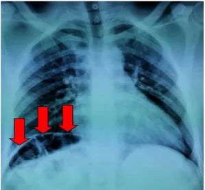
Plain chest radiograph centered on the diaphragmatic dome revealed a gas
shadow in the epigastric region (Figure 1).
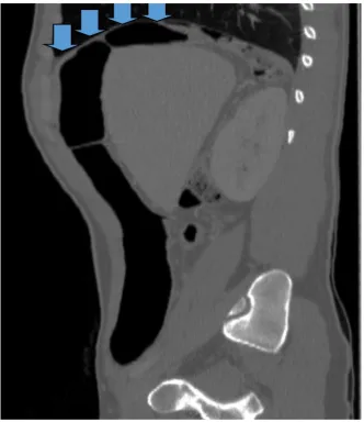
Abdominal CT showed a colonic interposition between the liver and the
di-aphragm (Figures 2-4) without pneumatosis in the cavity and segment VI liver
contusion.
Management entailed administration of intra venous analgesics and pro-ton-pump inhibitors with close monitoring of vitals and biological markers.
Clinical course was uneventful with favorable outcome after conservative management.
3. Discussion
Chilaiditi syndrome is a rare condition. Globally, its incidence is estimated be-tween 0.025% to 0.28% with the affection more common in adult males as was
[image:2.595.225.525.426.703.2]the case in our patient [2].
DOI: 10.4236/ojrad.2017.73018 166 Open Journal of Radiology
Figure 2. Abdominal CT showing colic interposition between liver and anterior abdominal
wall.
Figure 3. Abdominal CT coronal section showing colic interposition between liver and
[image:3.595.209.541.336.693.2]DOI: 10.4236/ojrad.2017.73018 167 Open Journal of Radiology
Figure 4. Abdominal CT sagittal section showing interposition of colic interposition
be-tween liver and diaphragm.
As per its pathogenesis, it is well established that normal embryological de-velopment of the liver as well as means of fixation of the intestine prevents in-terposition of the colon between the diaphragm and the liver. However, in cer-tain rare anatomical variations, notably hypotrophy of the liver or agenesis of the right lobe of the liver, or in the event of elongation of the suspensory liga-ment of the liver, elongation of the colon (dolichocolon), poor fixation or poor position of the colon, congenital pathologies of the small intestine or diaphragm with elevation of the right hemidiaphragm (eventration), relaxation or agenesis
of the suspensory ligament of the mesentery [3][4] [5] the latter could occur.
These factors may be present in 6% of patients at birth. On the other hand, in
adults other factors including cirrhosis [6], chronic constipation, increased
ab-dominal pressure (pregnancy), obesity [7], enlargement of the lower rib cage due
to chronic obstructive pneumonitis with a large space in which the interposition
of the colon may occur [1] are also believed to be contributory factors.
DOI: 10.4236/ojrad.2017.73018 168 Open Journal of Radiology between the liver and the diaphragm or the abdominal wall are mostly the transverse colon, followed by the right colic flexure even though cases involving
small intestine interposition have also been reported [7].
Colonic interposition is often asymptomatic despite relative higher incidence
of radiological evidence [8].
As far as its diagnosis is concerned, interposition of the colon (Chilaiditi sign) is defined as the presence of a gas shadow below the right hemidiaphragm on plain abdominal X-ray. Positive diagnosis of Chilaiditi syndrome based on im-aging requires the presence of the following criteria: elevation of the right di-aphragmatic hemicoupola by the intestine, gas distension of the colon beneath the diaphragm, lowering of the upper margin of the liver than the level of the left
diaphragmatic hemicoupola with no other positional anomalies [9].
Differential diagnosis of the syndrome involves mainly pneumoperitoneum
[10][11]. In addition, changes in patient positions do not change the location of
radiological evidence unlike in a patient with free air in the peritoneal cavity.
4. Conclusion
Chilaiditi syndrome is a rare pathological entity, discovered incidentally during diagnostic imaging and constitutes a major pitfall in the diagnosis of right false pneumoperitoneum.
References
[1] Nitin, T., Sameer, S., Priyanka, S., Dinesh, M., Sachin, B., Neeraj, B., Sulbha, S. and Puneet, S. (2014) Coexistence of Pneumothorax and Chilaiditi Sign: A Case Report.
Asian Pacific Journal of Tropical Biomedicine, 4, 75-77.
[2] Cedrick, S.M., Maruis, K.F., Mireille, K.Z., Nelly, M.S., Patience, M.P., Shem, M., Chamy, C.L. and Josephine, M.K. (2014) Chilaiditi Syndrome in a Newborn, in Case Report. Pan African Medical Journal, 19, 239.
[3] White, J.J., Chavez, E.P. and Souza, J. (2002) Internal Hernia of the Transverse Co-lon-Chilaiditi Syndrome in a Child. Journal of Pediatric Surgery, 37, 802-804. https://doi.org/10.1053/jpsu.2002.32293
[4] Orangio, Fazio, V.W., Winkelman, E. and McGonag, B.A. (1986) The Chilaiditi’s Syndrome and Associated Volvulus of the Transverse Colon: An Indication for Sur-gical Therapy. Diseases of the Colon and Rectum, 29, 653-656.
https://doi.org/10.1007/BF02560330
[5] Plorde, J.J. and Raker, E.J. (1996) Transverse Colon Volvulus and Associated Chi-laiditi’s Syndrome: Case Report and Literature Review. The American Journal of
Gastroenterology, 91, 2613-2616.
[6] Haddad, C.J. and Laclé, J. (1998) Chilaiditi’s Syndrome: A Diagnostic Challenge.
Postgraduate Medical Journal, 9, 249-250.
[7] Murphy, J.M., Maibaum, A., Alexander, G. and Dixon, A.K. (2000) Chilaiditi’s Syndrome and Obesity. Clinical Anatomy, 13, 181-184.
https://doi.org/10.1002/(SICI)1098-2353(2000)13:3<181::AID-CA4>3.0.CO;2-7 [8] Cetin, D., Unubol, M., Soyder, A., Guney, E., Coskun, A., Ozbas, S., et al. (2012)
DOI: 10.4236/ojrad.2017.73018 169 Open Journal of Radiology [9] Gupta, P.P. and Agarwal, D. (2011) Medical Image: Chilaiditi Syndrome. The New
Zealandmedical Journal, 124, 81-83.
[10] Farinella, E., Nazzaro, C., Rossetti, B., Giuliani, D., Giustozzi, G.M. and Sciannameo, F. (2006) Chilaiditi’s Syndrome: A Rare Cause of Abdominal Pain in the Differential Diagnosis of the Abdominal Perforation. Case Report Giornale di
Chirurgia, 27, 417-421.
[11] Moaven, O. and Hodin, R.A. (2012) Chilaiditi Syndrome: A Rare Entity with Im-portant Differential Diagnoses. Gastroenterology and Hepatology, 8, 276-278.
Submit or recommend next manuscript to SCIRP and we will provide best service for you:
Accepting pre-submission inquiries through Email, Facebook, LinkedIn, Twitter, etc. A wide selection of journals (inclusive of 9 subjects, more than 200 journals)
Providing 24-hour high-quality service User-friendly online submission system Fair and swift peer-review system
Efficient typesetting and proofreading procedure
Display of the result of downloads and visits, as well as the number of cited articles Maximum dissemination of your research work
Submit your manuscript at: http://papersubmission.scirp.org/