Eff ect of early immunocastration on testicular histology
in pigs
Z. Sladek
1*
, M. Prudikova
1, A. Knoll
1, P. Kulich
2, I. Steinhauserova
3,
G. Borilova
31Mendel university, Brno, Czech Republic
2Veterinary Research Institute, Brno, Czech Republic
3University of Veterinary and Pharmaceutical Sciences Brno, Brno, Czech Republic
*Corresponding author: [email protected]
ABSTRACT: The objective of this study was to investigate short- and long-term effects of early immunocastra-tion with Improvac® vaccine, administered in two doses, at ages eight and 14 weeks, on testicular histology in pigs
slaughtered eight or 15 weeks after the second dose. We hypothesised that the effectiveness of early vaccination could be diminished by late application of the booster dose and/or delayed time of slaughter. Thirty non-castrated male pigs of a commercial hybrid breed were used in this study. Pigs (n = 15) in the control group (NOCA) remained intact throughout the study. Pigs (n = 15) in the experimental group (IMCA) were administered Improvac in two doses: a priming dose at eight weeks and a booster dose at 14 weeks. Subsequently, nine of the IMCA pigs were slaughtered at eight weeks and the remaining six at 15 weeks after the second dose. In NOCA pigs, we observed normal spermatogenesis in the tubuli seminiferi and many prominent interstitial endocrine (Leydig) cells. In IMCA8 pigs, there was a noticeable decrease in the diameter and area of seminiferous tubules and spermatogenesis was absent. Interstitial endocrine cells appeared atrophied with pyknotic nuclei. In IMCA15 pigs, we observed a larger diameter of tubuli, thickened germinal epithelium and larger and more numerous interstitial endocrine cells when compared to IMCA8. In conclusion, these results demonstrate that early immunocastration with Improvac disrupts spermatogenesis and reduces the number and size of interstitial endocrine cells. This indicates that vaccination at an age of eight weeks and again at 14 weeks in pigs causes disruption of testicular histology and spermatogenesis at least through the subsequent 15 weeks.
Keywords: testis; structure; microscopy; pig; Improvac
In pork, meat taint is a common and unpleas-ant odour caused by the accumulation of 16-an-drostenone and skatole in the fat of male pigs. The androstenone steroids (5α-androst-16-en-3-one) are synthesised primarily in interstitial endocrine (Leydig) cells of pig testes and transported to fat tissue (Patterson 1968). Skatole is produced in the large intestine by microbial breakdown of the amino acid tryptophan originating from dietary or
endogenous protein (Wilkins 1990). The presence of these compounds significantly affects carcass and meat quality, traits that commonly play a role in rejection by pork consumers.
In Europe and world-wide, surgical castration of male piglets is the most common method for pre-venting pig meat taint. Besides castration, breeding and genetic selection have been used as alternative solutions to boar taint (Strathe et al. 2014; Drag
et al. 2017). However, surgical castration of male piglets is traditionally practiced in the pork indus-try, for reasons beyond prevention of meat taint. Indeed, a primary goal of this type of castration is the reduction of undesirable sexual and aggressive behaviour in pigs and the calming of the herd.
Castrations have traditionally been performed without the use of anaesthesia or analgesia, and in recent years many consumers have called for pigs to be managed with less stressful, painful and inva-sive practices. Consequently, the European Union has introduced a Council Directive 2008/120/EC from December 2008, which specifi es that surgi-cal castration of pigs shall only be performed with prolonged analgesia and/or anaesthesia. In 2010, the ‘European Declaration on alternatives to surgical castration of pigs’ was adopted. Th e Declaration stipulates that, as of 2018, surgical castration of pigs should be phased out altogether. However, some EU member states (including the Czech Republic) have objected to this requirement although coun-tries using analgesia and/or anaesthesia (Norway, Sweden, Switzerland, Th e Netherlands) have found this method to be practical and eff ective. Only a few countries seem to be aiming to meet the deadline for phasing out surgical castration (De Briyne et al. 2016).
Immunocastration constitutes an alternative method for the control of pig meat taint. This method involves the vaccination of all male pigs with a gonadotropin-releasing hormone analogue so that antibodies are produced against gonado-tropin-releasing hormone. The hypothalamic-pitu-itary-gonadal axis is thereby blocked and testicular steroid hormone production ceases (Pauly et al. 2010; Fabrega et al. 2011). For effective immunisa-tion, two doses of the vaccine are administered. The first (primer) dose and second (booster) dose are administered at least four weeks apart. Generally, the second dose should be administered not later than four weeks prior to slaughter (Einarsson et al. 2011).
Immunocastration significantly affects the mor-phological characteristics of the testes. Past obser-vations show that the size and weight of testes are generally reduced after vaccination (Einarsson et al. 2011; Lealiifano et al. 2011). Histological changes in the seminiferous tubules and interstitium of the testes are also objective indicators of successful cas-tration. These changes include a reduced diam-eter of the seminiferous tubules, a reduced number
of interstitial endocrine cells and a reduction of germinal epithelium (Brunius et al. 2011). These immunocastration effects persist for 22–26 weeks after the second/booster vaccination, as manifested in androstenone and skatole levels, the size of re-productive organs (Einarsson et al. 2011; Han et al. 2017) and histomorphological traits observed using light and/or electron microscopy (Kubale et al. 2013; Li et al. 2015; Han et al. 2017).
It is clear from studies to date that earlier vac-cination leads to more dramatic and irreversible changes in the testes than vaccination at later ages. Specifically, there is a decline in testosterone lev-els, a reduction in testicular and bulbourethral size as well as a reduction in spermatogenesis and in the size and number of interstitial endocrine cells. These dramatic effects are causally linked to the de-creased levels of testosterone and other hormones produced by the interstitial endocrine cells, which are below those levels required for spermatogenesis (Ge et al. 2008).
However, a decreased efficacy of the Improvac vaccine has also been observed in the case of an early vaccination scheme. While Wicks et al. (2013) used the primary dose of Improvac at eight weeks, their second dose was administered as late as 20 weeks. In this study, testicular function was observed to be restored in some pigs at about 10 weeks after Improvac immunisation.
Therefore, the effectiveness of early vaccina-tion could be diminished by late applicavaccina-tion of the booster dose and/or delayed time of slaughter. The question arises regarding the optimal time in an early vaccination scheme for the administration of the booster dose of Improvac and the timing of slaughter.
The objective of the present study was to inves-tigate short- and long-term effects of early immu-nocastration with Improvac administered at eight and 14 weeks of age on testicular structure in pigs slaughtered eight and 15 weeks after the second/ booster dose of Improvac. In this study, we inves-tigated the possibilities of following a more flexible vaccination schedule.
MATERIAL AND METHODS
scopic fields from each microscopic section were digitised with a camera using different objectives (Keyence VH-Z100R 100–1000 × and VH-Z20R 20–200 ×) and then assessed using Keyence soft-ware ver. 1.04 (Keyence).
The testis samples were evaluated for the follow-ing parameters: seminiferous duct diameter (μm); seminiferous duct area (μm2); height of the germinal epithelium (μm); and, in interstitial endocrine cells, the diameter (μm), n uclear volume (μm3), nuclear area (μm2) and cytoplasm area (μm2). Inasmuch as the nuclei of interstitial endocrine cells in pigs are spherical, the nuclear volume of interstitial endo-crine cells was calculated from the mean nuclear diameter, following Avelar et al. (2010). One to five measurements were made for seminiferous tubule parameters and more than five measurements were made for interstitial endocrine cell parameters in one microscopic field.
Transmission electron microscopy. Samples for electron microscopy were obtained from two IMCA and two NOCA pigs.
Ultrathin sections of the samples were fixed in 3% glutaraldehyde in cacodylate buffer, post-fixed in 1% OsO4 solution in cacodylate buffer, dehydrated in 50%, 70%, 90% and 100% acetone and embed-ded in a mixture of Epon 812 (Serva, Heidelberg, Germany) and Durcupan ACM (Fluka, a subsidiary of Sigma-Aldrich). Sections were stained with 2% uranyl acetate and 2% lead citrate and observed at 80 kV using a Philips EM 208 transmission electron microscope (Philips, The Netherlands).
Statistical analysis. Results were evaluated using multifactorial analysis of variance (ANOVA) for determining significant sources of variability. The significance of differences between evaluated pa-rameters was tested by Scheffé’s method. P-values were considered statistically significant at the levels of P < 0.05 (*), P < 0.01 (**) or P < 0.001 (***). The data were processed using STATISTICA 7.1 soft-ware (StatSoft CR Ltd, Prague, Czech Republic).
RESULTS
Immunocastration significantly affected the tes-tes macroscopically, microscopically and ultrami-croscopically.
Macroscopically, the testes of IMCA8 and IMCA15 pigs were significantly smaller and lighter than tes-tes from NOCA pigs (Table 1 and Figure 1). study. Piglets were weaned at 10.5 kg and kept in
identical boxes (five animals) in the accredited experimental barns of the Veterinary Research Institute, Brno, Czech Republic. Pigs were fed twice daily with a standard commercial diet A1– A3 according to the weight category. Animal care conformed to the Institute’s good care practices protocol. All experimental procedures were ap-proved by the Central Commission for Animal Welfare of the Czech Republic.
The 30 male piglets were randomly divided into two groups. Pigs (n = 15) in the control group (des-ignated NOCA, not castrated) remained intact throughout the study. Pigs (n = 15) in the experi-mental group (designated IMCA, immunocastrat-ed) were administered Improvac® vaccine (Pfizer
Animal Health, S.A., Belgium) by subcutaneous injection in the neck, immediately behind the ear. Improvac was administered in two doses of 2 ml each. The first (priming) dose was administered at an age of eight weeks and the second (booster) dose at 14 weeks. The timing of the first Improvac vaccination was consistent with the manufactur-er’s recommendation for the timing of vaccination. Subsequently, a portion of the IMCA pigs (n = 9) were slaughtered eight weeks (IMCA8)and the remainder (IMCA15; n = 6) at 15 weeks after the second dose of Improvac. All animals were slaugh-tered under controlled conditions. Live weight at slaughter was 110.7 ± 9.2 kg for IMCA8, 127.4 ± 6.1 kg for IMCA15 and 106.1 ± 8.8 kg for NOCA.
Tissue sampling. Testicles (without epididymis) were removed at slaughter. Testes were measured individually and were then weighed in pairs at the slaughterhouse.
The microscopic structure of testes was evaluated for all pigs from IMCA and NOCA experimental groups. Testes were cut in half and on the trans-verse section and tissue samples were collected from the centre, above the mediastinum (Kubale et al. 2013). Two tissue samples were obtained from each animal for light microscopy.
10 micro-Light and transmission electron microscopy
Although there was individual variation between pigs in terms of testicular morphology, the most important factor was length of time between boost-er vaccination and slaughtboost-er.
In testicular tissue of NOCA, spermatogenesis was fully developed, with all stages of spermatogen-esis from spermatogonia to spermatozoa evident. The testicular histology in IMCA pigs, by contrast, was clearly negatively affected. Seminiferous tu-bules did not contain all developmental stages of spermatogenic cells. Significant changes were also noted in the interstitial space and the morphology of interstitial endocrine cells.
Germinal epithelium
In NOCA pigs, we observed normal spermatogen-esis in the tubuli seminiferi. As is evident in Figure 2,
[image:4.595.317.518.94.357.2]spermatogenesis was fully developed with all stages from spermatogonia to spermatozoa evident in TEM (Figure 3). Th e relatively thick germinal epithelium
Table 1. Testes parameters in immunocastrated and non-castrated pigs. Data are presented as mean ± SD meas-ured in immunocastrated (IMCA) and non-castrated (NOCA) pigs
Group Testis length (cm) Testes weight (g) IMCA8 4.5*** ± 0.5 34.6*** ± 8.2 IMCA15 4.9*** ± 0.3 42.8*** ± 11.9 NOCA 11.2 ± 0.9 587.6 ± 130.7
[image:4.595.64.290.152.209.2]Th e analysis is between NOCA samples and IMCA samples ***P < 0.001; Scheff é’s method
[image:4.595.305.533.508.675.2]Figure 1. Example of the signifi cantly diff erent testes sizes obtained from non-castrated (NOCA) and immu-nocastrated (IMCA8) pigs
Figure 2. Testicular histology of intact pigs (NOCA) with normal tubuli seminiferi. (A) Spermatogenesis products ranging from spermatogonia to spermatozoa are present in tubuli seminiferi (scale bar 100 μm). (B) Detail of a large intestitial space with numerous interstitial endo-crine cells (*, scale bar 50 μm)
Figure 3. Transmission electron microscopy of non-castrated (NOCA) pigs. Fully developed epithelium with spermatogonia (sg), spermatocytes (sc), spermatids (st) and head of a spermatozoon (arrow). Th e large space near the base of the germinal epithelium with visible myofi brocytes (bm) is characteristic of NOCA pigs
(A)
(B)
NOCA
[image:4.595.64.292.538.713.2]extended nearly to the centre of the tubuli, and only small lumina remained. Th e mean diameter of tubuli in NOCA pigs was nearly 200 μm (Table 2).
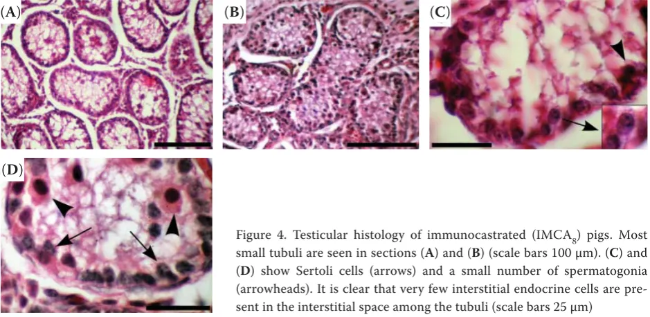
In IMCA8 pigs, there was a noticeable decrease in the diameter and area of seminiferous tubules (Figure 4). Samples from IMCA8 had approximately half the tubule diameter and one-third the area, compared to NOCA pigs (Table 2); these differ-ences were highly significant (P < 0.001). There was no spermatogenesis, as evidenced by the absence of spermatogenic cells at any stage of spermiation in the seminiferous tubules. The de facto mon-olayer epithelium contained only spermatogonia
and sustentacular (Sertoli) cells attached to the basal lamina (Figures 4 and 5). In some samples, spermatogonia were sparce. Sustentacular cells had round and elongated nuclei with spotted hetero-chromatin (Figure 5). In some samples, the tubule lumen was obscured by apical projections of sus-tentacular cells (Figure 4). The height of the lining epithelium was approximately 8–15 μm.
In IMCA15 pigs, we observed only minor changes in the histological structure of tubuli compared to IMCA8 pigs. Notably, these pigs exhibited a larger diameter of the tubuli and thicker germinal
epithe-Table 2. Testes parameters (seminiferous duct diam-eter (SDD; μm), seminiferous duct area (SDA; μm2) and
germinal epithelium height (GEH; μm)) in immunocas-trated and non-casimmunocas-trated pigs. Data are presented as mean ± SD measured in immunocastrated (IMCA) and non-castrated (NOCA) pigs
Group Tubuli seminiferi
SDD SDA GEH
IMCA8 91.2*** ± 17.9 9803*** ± 93.3 33.8*** ± 7.3 IMCA15 116.3*** ± 27.5 13 803*** ± 218.3 46.6 ± 16.6 NOCA 191.4 ± 9.9 31 772 ± 76.9 64.6 ± 4.2
The analysis is between NOCA and IMCA samples. Ten microscopic sections were made from one sample of testis and ten microscopic fi elds were examined from one micro-scopic section. One to fi ve measurements were made for each parameter in one microscopic fi eld
[image:5.595.305.532.94.269.2]***P < 0.001; Scheff é’s method
Figure 4. Testicular histology of immunocastrated (IMCA8) pigs. Most small tubuli are seen in sections (A) and (B) (scale bars 100 μm). (C) and (D) show Sertoli cells (arrows) and a small number of spermatogonia (arrowheads). It is clear that very few interstitial endocrine cells are pre-sent in the interstitial space among the tubuli (scale bars 25 μm)
Figure 5. Transmission electron microscopy of immuno-castrated pigs. Epithelium of tubuli seminiferi with Ser-toli cells with an elongated nucleus (*), vacuoles in the cytoplasm and less numerous spermatogonia (+) can be distinguished. Myofi brocytes (bm) are also present near the base of the germinal epithelium
(A) (B)
(D)
[image:5.595.64.291.179.252.2] [image:5.595.63.533.531.760.2]lium (Table 2). Compared to IMCA8 pigs, a “double layer” epithelium was observed containing susten-tacular cells and a large number of spermatogonia (Figure 6).
Interestingly, the return of spermatogenesis was noted in one IMCA15 pig whose testes were simi-lar in size to those of NOCA pigs: length 9.7 cm and weight 369.2 g. In this case, the diameters of tubuli were 150 μm ± 30 μm and the histological structure of the epithelium was identical to NOCA pigs (Figure 7).
Interstitial space
In NOCA pigs, the interstitial space contained a large number of prominent interstitial endocrine cells responsible for production of testosterone and performing the main function of triggering the formation of spermatozoa (Figures 2 and 8). Morphometric parameters of these cells are pre-sented in Table 3.
In IMCA8 pigs, immunocastration clearly dis-rupted the number and morphology of the inter-stitial endocrine cells (Figures 4 and 9). Interinter-stitial endocrine cells appeared as shrunken and small cells with pyknotic nuclei compared to NOCA pigs (Figures 9 and 10). It was difficult to distinguish
terstitial endocrine cells from other cells of the in-terstitium, e.g., fibrocytes. As noted in Table 3, the nucleocytoplasmic index was noticeably changed and the cell diameter and area of cytoplasm was significantly decreased compared to NOCA pigs (P < 0.001).
In IMCA15 pigs, we observed higher numbers of interstitial endocrine cells in comparison to IMCA8. Moreover, the interstitial endocrine cells were greater in size and the interstitial space was similar to that seen in NOCA pigs (Figure 6). As shown in Table 3, the area of cytoplasm was nearly twice as large and the nucleocytoplasmic index only half that of NOCA pigs.
DISCUSSION
[image:6.595.64.389.97.222.2]In this study, we demonstrated the short- and long-term effects of early immunocastration on testicular structure in comparison to untreated ani-mals. We used Improvac vaccine, administered at eight and 14 weeks, and pigs were slaughtered eight and 15 weeks following the booster dose. Improvac is the fi rst commercial product for the immuno-castration of male pigs and is frequently used. Th e vaccine is the fi rst anti-GnRF product approved for general use in the swine industry. According to
Figure 6. Testicular histology of immunocastrated (IMCA15) pigs. (A) Tubuli seminiferi with Ser-toli cells and some spermatogonia (scale bar 100 μm). (B) Interstitial endocrine cells (*) are clearly vis-ible in higher magnifi cation of the interstitium (scale bar 50 μm)
Figure 7. Testicular histology of immunocastrated (IMCA15) pigs with recovery of spermatogen-esis. (A) Note the fully developed epithelium in the tubuli (scale bar 100 μm). (B) Moreover, large numbers of interstitial endocrine cells (*) are evident within the interstitium (scale bar 50 μm)
(A) (B)
[image:6.595.64.384.611.758.2]the manufacturer’s instructions, the vaccine’s pri-mary dose can be administered no earlier than at eight weeks. In fact, researchers have used various vaccination schedules, with the primer dose being administered at 11–16 weeks and the booster dose at 18–21 weeks (Einarsson et al. 2009; Batorek et al. 2012; Kubale et al. 2013; Li et al. 2015; Shi et al. 2016; Needham et al. 2017). In these cases, the eff ect of vaccination may persist for as long as 22 weeks (Zamaratskaia et al. 2008; Einarsson et al. 2009).
In addition to the standard vaccination proto-col, some investigators have used early (pre-pu-bertal or early pu(pre-pu-bertal) vaccination with Improvac (Brunius et al. 2011; Andersson et al. 2012; Lugar et al. 2017). This early vaccination can be used as an alternative to the recommended schedule in order to minimise problematic aggressive behaviour of
the males, while having no effect on profitability. Einarsson et al. (2011) administered Improvac at 10 and 14 weeks and observed a long-lasting negative effect on fertility, with none of the animals recov-ering testicular function, at least up to 25 weeks.
[image:7.595.305.533.94.270.2]The first sign of immunocastration was a reduc-tion in testes size. It has been reported that im-munocastration decreases the size of reproductive organs (Batorek et al. 2012) and we observed very small testes in IMCA8 and IMCA15 pigs compared to NOCA pigs. Brunius et al. (2011) reported that a more pronounced reduction in reproductive organ size occurred in pigs vaccinated earlier. Einarsson et al. (2011) stated that the reduction in testis size was most likely the combined result of arrested testicular development at an earlier age and a pro-longed period without spermatogenesis. Brunius et al. (2011) and Einarsson et al. (2011) used early
Figure 8. Transmission electron microscopy of non-cas-trated pigs. Th e interstitial space with prominent inter-stitial endocrin e cells (*), myofi brocytes (fi ) and blood vessels (+). A vacuole is present in the Sertoli cell in the tubuli seminiferi (v)
[image:7.595.64.292.97.269.2]Figure 9. Transmission electron microscopy of immuno-castrated pigs. Interstitial endocrine cells (*) with a small amount of cytoplasm and myofi brocytes (bm) are evi-dent in the interstitial space
Table 3. Testes parameters in immunocastrated (IMCA) and non-castrated (NOCA) pigs. Data are presented as mean ± SD measured in IMCA and NOCA pigs
Group
Interstitial endocrine cells
diameter (μm)
nuclear volume
(μm3) nuclear area(μm2) cytoplasm area(μm2) (nuclear area/cytoplasm area)nucleocytoplasmic index
IMCA8 7.1*** ± 0.6 27.6 ± 1.5 11.1 ± 0.3 26.4*** ± 5.4 0.42 IMCA15 8.2 ± 2.2 24.4 ± 6.3 10.6 ± 2.7 42.4* ± 13.4 0.25
NOCA 11.3 ± 0.9 32.5 ± 1.6 12.3 ± 0.6 88.2 ± 16.3 0.14
Th e analysis is between NOCA samples and IMCA samples. Ten microscopic sections were made from one sample of testis and ten microscopic fi elds were examined from one microscopic section. At least fi ve measurements were made for each parameter in one microscopic fi eld
[image:7.595.64.531.617.700.2]Improvac immunocastration, at 10 and 14 weeks, and the paired testes weights reported were 74 g and 109 g. In our study, we used early immunocas-tration at eight and 14 weeks and the paired testes weight was approximately 35 g. It is evident that early vaccination dramatically reduces testicular maturation. Improvac was administered at eight weeks, when testicles start to undergo significant differentiation and growth (Avelar et al. 2010). When the second dose of Improvac is administered as late as 20 weeks, paired testes weight can exceed 250 g (Wicks et al. 2013). Interestingly, Batorek et al. (2012) and Han et al. (2017) observed testes weights of about 300 g and 147 g, respectively, for a standard vaccination scheme of 10–12 weeks and booster dose at 18 weeks.
In addition to macroscopically detectable chang-es, Improvac markedly affected the histology of testes in IMCA8 and IMCA15 pigs. Histological changes are related to inhibition of hypothalamic synthesis and secretion of gonadotropin-releasing hormone, which normally stimulates adenohypo-physeal production and release of follicle-stimulat-ing hormone and luteinisfollicle-stimulat-ing hormone. Luteinisfollicle-stimulat-ing hormone is responsible for initiating testosterone production by the interstitial endocrine cells. Follicle-stimulating hormone and testosterone stimulation of the sustentacular (Sertoli) cells ini-tiates spermatogenesis and spermiation (Basu et al. 2017). In the absence of these hormones, normal stimulation of the testes is compromised; gonadal regression and infertility follow (Miller et al. 1997).
In accordance with earlier studies, Improvac vac-cination significantly disrupted spermatogenesis and decreased the number and size of interstitial endocrine cells (Hilbe et al. 2006; Einarsson et al. 2009, Einarsson et al. 2011). We also observed these changes in our study, but our study differed in relation to the length of time from booster dose administration to slaughter. The most noticeable changes were observed in IMCA8 pigs as opposed to in IMCA15 animals. In this case, we observed smaller seminiferous duct perimeter and height of the germinal epithelium along with inhibited spermatogenesis. In IMCA8 samples, only a few spermatogonia were present alongside susten-tacular cells, suggesting that spermatogenesis was disrupted reversibly. Others have observed tubuli with only sustentacular cells, indicating an irrevers-ibility of spermatogenesis (Einarsson et al. 2011). In our IMCA15 samples, we observed large num-bers of spermatogonia, suggesting that these cells were mitotically active and could potentially act to restore spermatogenesis. Overall, the histology of germinal epithelium in IMCA pigs was similar to that seen prior to puberty. The structure of susten-tacular cells also suggests this. In IMCA samples, sustencacular cells had round and elongated nuclei, a pattern which is characteristic for pre-pubertal ages (Avelar et al. 2010).
Alterations in the morphology and functionality of the tubuli seminiferi coincide with changes ob-served in interstitial endocrine cells (Kubale et al. 2013). In our study we observed significant changes in interstitial endocrine cells in immunised pigs. Similar to changes seen in sustentacular cells and spermatogonia, the extent of changes seems pro-portional to the time remaining until slaughter. In IMCA8 pigs, immunocastration caused a reduction in the number and size of interstitial endocrine cells. In some cases, these cells were nearly indis-tinguishable from other cells of the interstitium. Interstitial endocrine cells were shrunken and had both a small nucleus and limited cytoplasmic area. Similar results have been reported by other studies (Einarsson et al. 2011; Kubale et al. 2013; Li et al. 2015). These changes in the morphology of inter-stitial endocrine cells were consistent with a loss of function and the decrease in testosterone and androstenone (Kubale et al. 2013).
[image:8.595.63.290.96.267.2]In IMCA15 samples of our study, the size of in-terstitial endocrine cells and their numbers were greater; the histology of the interstitial space was
similar to that of NOCA pigs. These changes – ap-parent 15 weeks after the second dose of Improvac – strongly suggest progression toward a return of spermatogenesis. Moreover, in a single IMCA15 pig there was a return to spermatogenesis. The histology was nearly the same as in NOCA pigs. This is not surprising inasmuch as a return to spermatogenesis after Improvac vaccination has also been observed in other studies. Kubale et al. (2013) reported that the testicles from animals ad-ministered vaccine four months prior to slaugh-ter showed recovery of spermatogenic function. Similarly, Wicks et al. (2013) reported that tes-ticular function in boars treated with Improvac began to recover 10 weeks after immunisation. It should be noted that these laboratories used differ-ent vaccination protocols. Kubale et al. (2013) did not use an early vaccination scheme, and the first dose was administered at 12 weeks. Wicks et al. (2013) used an early vaccination protocol, but the booster dose of Improvac was administered only 12 weeks after the first dose. In contrast, Einarsson et al. (2011) reported a long-lasting disruption of spermatogenesis in pigs vaccinated at as early as 10 and 14 weeks. Moreover, these investigators noted a persisting effect of immunocastration up to 25 weeks. Again, it is evident that the histologi-cal changes in testes are more prominent in pigs vaccinated early. This is consistent with the results of our study.
In conclusion, our hypothesis that the effective-ness of early vaccination could be diminished by late application of the booster dose and/or de-layed time of slaughter was not confirmed. The results reported in this study demonstrate that early immunocastration using Improvac leads to a reduction in the size of testes, disruption of sper-matogenesis as well as a decrease in the number and size of interstitial endocrine cells. This indicates that vaccination at ages of eight and 14 weeks in pigs causes disruption of testicular structure and function through the subsequent 15 weeks. This time-related effect allows the use of a more flexible vaccination schedule.
REFERENCES
Andersson K, Brunius C, Zamaratskaia G, Lundstrom K (2012): Early vaccination with Improvac®: effects on per-formance and behaviour of male pigs. Animal 6, 87–95.
Avelar GF, Oliveira CF, Soares JM, Silva IJ, Dobrinski I, Hess RA, Franca LR (2010): Postnatal somatic cell proliferation and seminiferous tubule maturation in pigs: a non-ran-dom event. Theriogenology 1, 11–23.
Basu S, Arya SA, Usmani A, Pradhan BS, Sarkar RK, Gan-guli N, Shulla M, Mandal K, Singh S, Sarda K, Majumdar SS (2017): Defective Wnt3 expression by testicular Sertoli cells compromise male fertility. Cell Tissue Research, doi: 10.1007/s00441-017-2698-5.
Batorek N, Candek-Potokar M, Bonneau M, Van Milgen J (2012): Meta-analysis of the effect of immunocastration on production performance, reproductive organs and boar taint compounds in pigs. Animal 6, 1330–1338. Brunius C, Zamaratskaia G, Andersson K, Chen G, Norrby
M, Madej A, Lundstrom K (2011): Early immunocastration of male pigs with Improvac(®) – eff ect on boar taint, hor-mones and reproductive organs. Vaccine 28, 9514–9520. De Briyne N, Berg C, Blaha T, Temple D (2016): Pig castra-tion: will the EU manage to ban pig castration by 2018? Porcine Health Management 20, 29.
Drag M, Skinkyte-Juskiene R, Do DN, Kogelman LJA, Ka-darmideen HN (2017): Differential expression and coex-pression gene networks reveal candidate biomarkers of boar taint in non-castrated pigs. Scientific Reports 7, 1–18.
Einarsson S, Andersson K, Wallgren M, Lundstrom K, Rodriguez-Martinez H (2009): Short- and long-term ef-fects of immunization against gonadotropin-releasing hormone, using Improvac, on sexual maturity, reproduc-tive organs and sperm morphology in male pigs. Theri-ogenology 15, 302–310.
Einarsson S, Brunius C, Wallgren M, Lundstrom K, Anders-son K, Zamaratskaia G, Rodriguez-Martinez H (2011): Effects of early vaccination with Improvac(®) on the de-velopment and function of reproductive organs of male pigs. Animal Reproduction Science 127, 50–55.
Fabrega A, Guyonnet B, Dacheux JL, Gatti JL, Puigmule M, Bonet S, Pinart E (2011): Expression, immunolocalization and processing of fertilins ADAM-1 and ADAM-2 in the boar (Sus domesticus) spermatozoa during epididymal maturation. Reproduction Biology and Endocrinology 30, 96.
Ge SQ, Kang XJ, Liu GR, Mu SM (2008): Genes involved in spermatogenesis. Yi Chuan 30, 3–12.
Han X, Zhou Y, Zeng Y, Sui F, Liu Y, Tan Y, Cao X, Du X, Meng F, Zeng X (2017): Effects of active immunization against GnRH versus surgical castration on hypotha-lamic-pituitary function in boars. Theriogenology 15, 89–97.
immuno-histochemical findings in testes, bulbourethral glands and brain of immunologically castrated male piglets. Schweizer Archiv fuer Tierheilkunde 148, 599–608. Lealiifano AK, Pluske JR, Nicholls RR, Dunshea FR,
Camp-bell RG, Hennessy DP, Miller DW, Hansen CF, Mullan BP (2011): Reducing the length of time between slaughter and the secondary gonadotropin-releasing factor immu-nization improves growth performance and clears boar taint compounds in male finishing pigs. Journal of Animal Science 89, 2782–2792.
Li Y, Liu Y, Su S, Pu Y, Zhang X, Fang F (2015): Immuniza-tion against recombinant GnRH-I alters testicular struc-ture in an experimental boar model. Zygote 23, 125–135. Lugar DW, Rhoads ML, Clark-Deener SG, Callahan SR,
Revercomb AK, Prusa KJ, Estienne MJ (2017): Immuno-logical castration temporarily reduces testis size and function without long-term effects on libido and sperm quality in boars. Animal 11, 643–649.
Kubale V, Batorek N, Skrlep M, Prunier A, Bonneau M, Fazarinc G, Candek-Potokar M (2013): Steroid hormones, boar taint compounds, and reproductive organs in pigs according to the delay between immunocastration and slaughter. Theriogenology 1, 69–80.
Miller LA, Johns BE, Elias DJ, Crane KA (1997): Compara-tive efficacy of two immunocontracepCompara-tive vaccines. Vac-cine 15, 1858–1862.
Needham T, Hoffman LC, Gous RM (2017): Growth re-sponses of entire and immunocastrated male pigs to di-etary protein with and without ractopamine hydro-chloride. Animal 20, 1–6.
Patterson (1968): 5α-drost-16-ene-3-one: Compound re-sponsible for taint in boar fat. Journal of the Science of Food and Agriculture 19, 31–37.
Pauly C, Spring-Staehli P, O’Doherty JV, Kragten SA, Dubois S, Messadene J, Bee G (2010): The effects of method of castration, rearing condition and diet on sensory quality of pork assessed by a trained panel. Meat Science 86, 498–504.
Shi X, Li C, Cao M, Xu X, Zhou G, Xiong YL (2016): Com-parative proteomic analysis of longissimus dorsi muscle in immuno- and surgically castrated male pigs. Food Chemistry 15, 885–892.
Strathe AB, Velander IH, Mark T, Kadarmideen HN (2014): Genetic parameters for androstenone and skatole as in-dicators of boar taint and their relationship to production and litter size traits in Danish Landrace. Journal of Ani-mal Science 91, 2587–2595.
Wicks N, Crouch S, Pearl CA (2013): Effects of Improvac and Bopriva on the testicular function of boars ten weeks after immunization. Animal Reproduction Science 30, 149–159.
Wilkins CK (1990): Analysis of indole and skatole in porcine gut contents. International Journal of Food Science and Technology 25, 313–317.
Zamaratskaia G, Andersson HK, Chen G, Andersson K, Madej A, Lundstrom K (2008): Eff ect of a gonadotropin-releasing hormone vaccine (Improvac) on steroid hor-mones, boar taint compounds and performance in entire male pigs. Reproduction of Domestic Animals 43, 351–359.