Use of castor bean polymer in developing
a new technique for tibial tuberosity advancement
for cranial cruciate ligament rupture correction in dogs
R.M. Medeiros
1, M.A.M. Silva
2, P.P.M. Teixeira
3, L.G.G.G. Dias
1,
D.G. Chung
1, C.C. Zani
1, M.A.R. Feliciano
1, M.E.B.A.M. Da Conceicao
1,
M.R.F. Machado
1, A.G. Rocha
1, G.O. Chierice
4, L.N. Coutinho
5,
J.G. Padilha Filho
11Sao Paulo State University, Jaboticabal Campus, Jaboticabal, Sao Paulo, Brazil
2College of Agronomy and Veterinary Medicine, University of Passo Fundo, Passo Fundo,
Rio Grande do Sul, Brazil
3Veterinary Hospital, Para Federal University, Castanhal, Para, Brazil 4Sao Carlos Chemistry Institute, Sao Paulo University, Sao Paulo, Brazil
5Animal Health and Production Institute, Amazonia Rural Federal University, Belem, Para, Brazil
ABSTRACT: The purpose of the current study was to develop a new tibial tuberosity advancement (TTA) tech-nique, by replacing the original titanium cage with a Ricinus communis polyurethane resin-made wedge polymer. The implants were manufactured using the same size and angles of the original titanium cages, though larger distally. The modified TTA technique (TTAm) was performed in 42 knees of 35 dogs diagnosed with rupture of the cranial cruciate ligament (RCCL). Animals were submitted to radiographic and gait assessments preoperatively, early postoperatively and following 30, 60, 90 and 120 days. All animals exhibited good clinical outcome. There were no cases of impaired healing or bone resorption until 120 days postoperatively and there was no patient with patellar distress after TTAm. Scores of gait evaluation revealed differences between time points. There were also differences between the evaluations of control and pre- and post-operative times. However, there was no difference among the assessment of 30 days and the following time points. The use of the polyurethane polymer for TTAm was advantageous, not only due to biocompatibility and osseointegration, but also for providing easy handling; it can be moulded intra-operatively if necessary. Moreover, it allows precise adaptation to the osteotomy site, as opposed to the original TTA metallic implants, which cannot be moulded. It is suggested that incision lengths for TTAm are slightly shorter than those required for the conventional TTA as this requires the distal fixation of the plate at the beginning of the middle third of the body of the tibia. The TTAm does not require the use of fixation plates and it is performed only at the cranial aspect of the tibia. The method of attachment of the tibial tuberos-ity in the craniocaudal direction was effective. The setting associated with the use of the polyurethane polymer allowed simplification of the technique for easier implementation, and the amount of implant material required to perform TTAm was reduced in comparison to the conventional TTA. This technique can be used for treating the knees of dogs with RCCL, and provides for easy execution, less invasiveness to the tissues of the knee joint and more versatility in comparison to conventional TTA.
Keywords: orthopaedic implant; corrective osteotomy; knee; ligament tear; canine
Several techniques have been developed for the repair of cranial cruciate ligament (CCL) disease, including arthroscopic techniques (Kudnig 2000;
extra-articular factors, including anatomy, muscle function and weight bearing. The purpose of these techniques is not to replace CCL or its function, but to stabilise the knee joint by acting over the forces that act over the tibial plateau and its relation to the CCL (Modenato et al. 2005; Apelt et al. 2007; Dal-Bo et al. 2014).
Tibial tuberosity advancement (TTA) is based on a procedure originally performed on humans, which was adapted to the canine species, and pro-vided better clinical results (Tepic 2006; Miller et al. 2007).
The polyurethane resin of castor bean (Ricinus communis) is biocompatible with bone tissue, and has osteoconductive characteristics, which is de-sirable in orthopaedic surgery (Rodaski et al. 1999; Sturion et al. 1999). Such material was tested for the treatment of segmental bone defects of the ra-dius in rabbits in the form of granules compared to spongy bone autograft. It acts as a space filler, and there are no signs of degradation, whereas it exhibits biocompatibility and promotes osseointe-gration (Pereira Jr. et al. 2007).
The purpose of the current study was to develop a new technique of TTAm, modified by replacing the original titanium cage with a Ricinus communis polyurethane polymer to treat dogs diagnosed with cranial cruciate ligament disease.
MATERIAL AND METHODS
This study was performed in a clinical trial setting, in a Veterinary Teaching Hospital. The study was approved by the Committee for Ethics in the Use of Animals (CEUA – FCAV/UNESP). Moreover, the
guidelines of the Brazilian Committee for Ethics, Bioethics and Animal Welfare (CEBEA) were strict-ly followed. Thirty-five dogs diagnosed with CCL disease (16 males and 19 females), from various breeds, aging from 1–13 years old and weighing from 2.5–69 kg were assessed. Seven out of 35 pa-tients had developed bilateral CCL disease. Thus, 42 knees were evaluated in the current study.
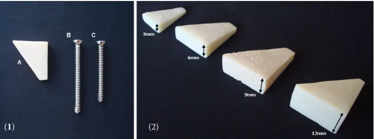
The modification of the conventional TTA em-ployed in the current study consisted of replacement of the titanium cage conventionally used as a spacer in the original TTA technique (Modenato et al. 2005) with a wedge-shaped spacer manufactured using Ricinus communis polyurethane resin, which was fabricated at the Chemistry and Physics Molecular Department, Sao Carlos Chemistry Institute, Sao Paulo University (USP). It was subsequently sent to the University of Sao Paulo State, where the sur-geries were performed. However, the resin polymer was larger than the original titanium cage. Thus, two cortical screws were used for the fixation of the wedge-shaped polyurethane polymer (Figure 1).
RCCL was diagnosed clinically using the cranial drawer and tibial compression tests. All patients were submitted to gait and radiographic assess-ment, besides basic preoperative haematological laboratory tests. The patients were prescribed meloxicam (1 mg/kg, s.i.d., orally) and rest in the preoperative period, from the time of diagnosis to the day of surgery.
Following early preoperative fasting and basic care, the patients were given an association of butor-phanol (Torbugesic Fort Dodge, Sao Paulo, Brazil) (0.4 mg/kg) and levomepromazine(Neozine 5 mg/ml – Sanofi Aventis, Sao Paulo, Brazil) (0.5 mg/kg) in-tramuscularly. Anaesthesia was induced using a
sin-Figure 1. Image of the implants used in the modified technique of tibial tuberosity advancement (TTAm): (1) Ricinus communis polyurethane wedge-shaped spacer (A), two stainless steel cortical screws (B and C); (2) different sizes of the polyurethane polymers (Ricinus communis), sized 3, 6, 9 and 12 mm
[image:2.595.116.498.572.714.2]gle bolus of propofol (PropofolTM Cristalia, Sao Paulo,
Brazil) (5 mg/kg, i.v.) followed by administration of epidural injection of an association of bupivacaine (Neocaina Cristalia, Sao Paulo, Brazil) (1 mg/kg), lidocaine (Xylestesin Cristalia, Sao Paulo, Brazil) (2 mg/kg) and tramadol hydrochloride (Tramadon Cristalia, Sao Paulo, Brazil) (1 mg/kg). No comple-mentary anaesthesia maintenance protocols were used. The affected limb was clipped and aseptically prepared using chlorhexidine gluconate 2% and al-cohol 70% solution.
The animals were placed in dorsal recumbency and the affected limb was slightly lateralised for surgery. The mechanism of action and the deter-mination of the extension of the tibial tuberosity advancement required to keep the patellar ligament perpendicular to the tibial plateau was based on the same principles of the original TTA technique (Modenato et al. 2005).
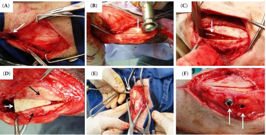
Tibial tuberosity was approached medially by a skin incision started on the tibial plateau area, extending to 1–2 cm beyond the end of the tibial tuberosity (Figure 2A). The periosteum was incised and elevated (Figures 2B and 2C). The medial col-lateral ligament and patellar ligament insertion were preserved. Before starting the tibial tuberos-ity osteotomy, a drill hole (1.0–2.0 mm) was made at the end of the tibial tuberosity using an electric driller (Figure 2D). To protect the patellar ligament and joint capsule enclosing the line of osteotomy, a straight Kelly clamp positioned between the patel-lar ligament and the tibial plateau was used. A line
[image:3.595.64.363.531.757.2]was drawn using a metal ruler and the scalpel blade, extending from the edge of the tibial tuberosity (Figure 3A) to the drill hole. A longitudinal oste-otomy of the tibial tuberosity was carried out using an oscillating saw (Figure 3B). The tibial tuberosity was distracted away creating a space (Figure 3C) using a metal bar; tweezers and/or spatula were used as a distractor. The polymer wedge implant (Figure 3C) was inserted into the gap created be-tween the tibial tuberosity and the body of the tibia. The choice of the thickness of the implant was set before surgery, according to the results of the me-diolateral radiography assessment, with the knee in 135º flexion, as recommended for the original TTA technique (Modenato et al. 2005). The width and length could be adjusted to the gap during surgery if necessary, by scraping the implant using metal rasps. Two stainless steel cortical screws were used for the fixation of the osteotomy tuberosity and im-plant to the tibial body. The screws were placed on the craniocaudal direction through the tuberosity, implant and body of the tibia: (1) one on the site of insertion of the patellar ligament; (2) and the other one 4–12 mm distally and parallel to the first screw, depending on the size of the patient (Figures 3E and 3F). Following stabilisation, the periosteum was reconstituted using synthetic absorbable suture in a simple continuous pattern, covering the implant. Afterwards, subcutaneous tissue was approximated with the same suture material, followed by skin su-ture using non-absorbable synthetic monofilament suture in a simple interrupted pattern.
Figure 2. Images of the medial surface of the tibia of dogs during the intraop-erative period of the TTAm: (A) skin incision (arrow) encompassing the tibial tuberosity area; (B) periosteal incision (arrow) in the tibial tuberosity; (C) edges of the periosteum (arrow) exposing the proximal area of the tibia; (D) drilling using 2 mm drill at the distal part of the tibial tuberosity (arrow)
(A) (B))
Postoperative care included rest, restriction of ex-ercise, and 10-minutes short walks 2–3 times a day. Prescription included oral administration of raniti-dine (Cloridrto de Ranitidina Medley, Campinas, Brazil) (2.2 mg/kg, b.i.d., for 10 days), cefalexin (Cefalexina, Medley, Campinas, Brazil) (30 mg/kg, b.i.d., for 10 days), tramadol hydrochloride (Tramadon Cristalia, Sao Paulo, Brazil) (2–4 mg/kg, b.i.d., for seven days) and dipirone (Dipirona Medley, Campinas, Brazil) (30 mg/kg, b.i.d., for seven days). Wound care included cleansing using sterile gauze and normal saline, followed by topical administration of rifampicin, coverage with sterile gauze and adhesive band (b.i.d., for 10 days) and the use of a Elizabethan collar.
Radiographic control assessment were carried out in the early preoperative period, in the early postoperative period, and 30, 60, 90 and 120 days postoperatively. Radiographs were classified into one out of four scores according to bone forma-tion (presence of bridge) in proximal and distal tibial crest, as carried out in a previous study (Berte et al. 2014): organised callus (score 4); disorganised callus (score 3); mildly disorganised
callus (score 2); and areas of bone growth/reac-tion (score 1). Moreover, descriptive assessment of the areas of resorption in the tibial tuberosity and next to the screws, as well as the integrity of osteoarticular implants and structures, including degenerative joint disease signs, were carried out at all time points.
Gait analyses were performed by observing the patients’ motion in the preoperative, early postop-erative phase and on Days 30, 60, 90 and 120 post-operatively. The gait assessment was always done by the same observer (RMM). The limb weight bearing degree was classified into four scores (Henry 2010; Dal-Bo et al. 2014): excellent (score 4), with full weight bearing without lameness; good (score 3), when mild lameness was present; satisfying (score 2), with moderate lameness; unsatisfactory (score 1), with permanent lameness, keeping the hind limb lifted, without weight bearing.
[image:4.595.72.528.94.325.2]Data from gait analysis and comparison of scores of bone healing were accomplished using the non-parametric Friedman’s test and Dunn’s post-test for multiple comparisons, if P < 0.05, using the statistics software Graph Prism 4.0.
Figure 3. Images of the medial aspect of the proximal tibia of dogs during TTAm: (A) straight Kelly haemostat (arrow) positioned between the patellar ligament and joint capsule, delimiting the proximal limit of the tibial tuberosity (arrow head) in order to guide the osteotomy; (B) longitudinal osteotomy of the tibial tuberosity (arrow) using an oscillating saw; (C) distraction of the tibial tuberosity away from the body of the tibia (arrow); (D) positioning of the polyurethane polymer implant (white arrow) between the tibial tuberosity (arrowhead) and the body of the tibia (black arrow); (E) craniocaudal perforation (arrow), with the aid of a driller for positioning the screws; (F) final aspect following setting the screws (arrows) craniocaudally through the tibial tuberosity, implant and tibia body
(A) (B) (C))
RESULTS
All animals recovered uneventfully, presenting improved outcome. Most patients were able to bear weight on the operated limb already in the early postoperative period. There were no cases of non-healing or bone resorption up to 120 days postoperatively. There were no cases of patellar dislocation following TTAm surgery. Mean overall operative time was 45 ± 5.7 min.
There were differences (P < 0.0001) between the different times of assessment concerning ra-diographic bone healing scores, in both areas, just proximal and distal to the implant. In multiple com-parison among measuring times (Table 1), there was complete healing (score 4) in five (13.1%) and seven (18.4%) knees out of 42 that were operated on at day 90 postoperatively, respectively, in the areas proximal and distal to the implant site. After 120 days, 12 (31.6%) and 21 (55.2%) knees showed complete bone healing in proximal and distal ar-eas, respectively. However, there was no difference between Days 90 and 120 postoperatively.
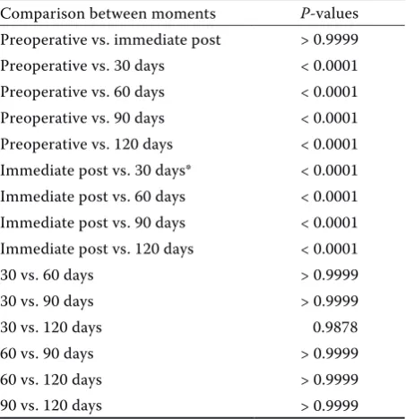
There were differences between time points re-garding gait scores (P < 0.0001). In the multiple com-parison assessment among time points (Table 2), there were differences between the control and
pre-operative, and preoperative and Days 30, 60, 90 and 120 postoperatively. There was a significant progres-sive improvement (P > 0.05) between postoperative time points following 30 days of evaluation.
In preoperative gait evaluation, 28.21% were clas-sified as good, 41.03% as satisfactory and 30.77% unsatisfactory. In early post-operative evaluation, 6.45% out of knees were classified as excellent, 41.94% good, 35.48% satisfactory and 16.13% as unsatisfactory. After 30 days 56.41% were excellent, and no animal was classified as unsatisfactory. The score 1 increased to 85% after 90 days.
Complications included suture dehiscence and local infection in one animal (2.85%); implant fail-ures such as breakage of the proximal screw in two animals (5.71%); and loosening of screws and displacement of the tibial tuberosity in an obese patient (2.85%). All complications were successfully managed and all patients presented improvements both clinically and radiographically.
DISCUSSION
[image:5.595.304.531.140.374.2]The Ricinus communis polyurethane polymer used as a spacer for the TTA was advantageous not Table 1. Results of post-Dunn test for multiple
compari-sons of scores for radiographic bone healing, according to the postoperative time, the proximal and distal sites in relation to the implant, in the knees of dogs operated by TTA technique modified
Comparison between moments
P-values proximal
to the implant to the implantdistal Control* vs. 30 days 0.0124 0.0294 Control vs. 60 days < 0.0001 < 0.0001 Control vs. 90 days < 0.0001 < 0.0001 Control vs. 120 days < 0.0001 < 0.0001
30 vs. 60 days 0.2692 0.1111
30 vs. 90 days < 0.0001 < 0.0001 30 vs. 120 days < 0.0001 < 0.0001
60 vs. 90 days 0.1364 0.2692
60 vs. 120 days 0.0004 0.0001
90 vs. 120 days > 0.9999 0.2952
*control radiograph performed in the immediate post-oper-ative period; significance level: 5%
Table 2. Results of post-test for multiple comparisons Dunn gait scores, according to the postoperative time in the knees of dogs operated by TTA modified technique
Comparison between moments P-values Preoperative vs. immediate post > 0.9999 Preoperative vs. 30 days < 0.0001 Preoperative vs. 60 days < 0.0001 Preoperative vs. 90 days < 0.0001 Preoperative vs. 120 days < 0.0001 Immediate post vs. 30 days* < 0.0001 Immediate post vs. 60 days < 0.0001 Immediate post vs. 90 days < 0.0001 Immediate post vs. 120 days < 0.0001
30 vs. 60 days > 0.9999
30 vs. 90 days > 0.9999
30 vs. 120 days 0.9878
60 vs. 90 days > 0.9999
60 vs. 120 days > 0.9999
90 vs. 120 days > 0.9999
only due to biocompatibility and osseointegration, but also due to easy handling and versatility of the implant, which can be moulded intraoperatively. Thus, it allows perfect anatomical adjustment of the osteotomy site (Dias 2008; Regonato et al. 2009; Teixeira 2012). In contrast, the conventional TTA metal implants cannot be moulded and require precise measuring (Modenato et al. 2005; Kim et al. 2008). Moreover, similarly to the conventional implants, the Ricinus communis polyurethane poly-mer can be autoclaved prior to use (Tepic 2006).
We suggest that the length of the incisions for TTAm should be slightly shorter than those re-quired for the TTA, which encompasses about 2–3 cm of the length of the tibial tuberosity of the patient (Tepic 2006). Conventional TTA requires fixation of a plate distally at the beginning of the middle third of the body of the tibia, while TTAm does not require plate fixation and the stabilisation of the tibial tuberosity and the implant is performed in the immediate cranial area of the tibia, thus re-quiring shorter incision and tissue divulsion.
The application of two screws for stabilisation of the tibial tuberosity and implant, in the cranio-caudal direction, was effective (Dias 2008; Lins et al. 2009) as cortical screws of stainless steel have acceptable biocompatibility and corrosion resist-ance (Durall and Diaz 1996). The use of the pol-yurethane wedge polymer as a spacer made the technique much simpler as a reduced amount of implants and material is required in comparison to the conventional TTA technique.
All patients exhibited radiographic evidence of bone healing. In the cranial cruciate ligament replacement techniques using tendon-bone seg-ments, physiotherapy was necessary for better re-covery of patients in this time period (Berte et al. 2014). Most dogs submitted to TTA (84%) show bone healing at 12 weeks, and 97% of dogs exhib-ited no or only minimal lameness (Lafaver et al. 2007; Henry 2010). In the current study, 95% of the patients had no or minimal lameness during the assessment of gait on Day 90 postoperatively. As in TTA, all patients subjected to TTAm recovered to the same condition as before the injury, accord-ing to the owners’ perceptions (Lafaver et al. 2007; Stein and Schmoekel 2008).
The rates of complications following TTAm were lower than those observed in other studies con-cerning the original TTA technique. Major compli-cations of conventional TTA include injury to the
meniscus, tibial fracture, lick granuloma, implant failure, septic arthritis, and medial patellar luxation; the minor complications include tibial tuberosity fracture fragment without displacement, implant failure without displacement, clicking sounds dur-ing ambulation, inadequate calcification of the os-teotomy with infections of the surgical injury and partial dehiscence of sutures (Lafaver et al. 2007). These authors found a rate of 12.3% for major com-plications and 19.3% for minor comcom-plications using the TTA technique, whereas in patients subjected to TTAm in this work, we found a major complica-tion rate of 9.52% of and a minor complicacomplica-tion rate of 4.76% (Lafaver et al. 2007; Stein and Schmoekel 2008). All complications were successfully managed in the current study and patients exhibited signif-icant improvements in clinical and radiographic evaluations, similarly as reported in another study involving conventional TTA (Modenato et al. 2005).
In conclusion, the modified technique of tibial tuberosity advancement, using the Ricinus com-munis polyurethane wedge spacer stabilised with cortical screws, can be considered for use in the treatment of rupture of the cruciate cranial liga-ment in dogs. It is a versatile alternative to the conventional TTA technique and is less expensive and results in lower rates of complications. No dif-ferences regarding healing and gait score between this technique and conventional TTA were seen. By 90 days post-TTAm most animals (85%) presented with an excellent gait score. All knees exhibited good healing, and no cases of bone resorption were observed. Complete bone healing was observed in more than 50% of knees after 120 days.
REFERENCES
Apelt D, Kowaleski MP, Boudrieau RJ (2007): Effect of tibial tuberosity advancement on cranial tibial subluxation in canine cranial cruciate-deficient stifle joints: An in vitro experimental study. Veterinary Surgery 36, 170–177. Berte L, Salbego FZ, Baumhardt R, Polidoro D, Da Silva
GM, Weiller MA, Dos Santos RP, Vargas CB, Mazzanti A (2014): Physiotherapy after replacement of cranial cru-ciate ligament in tendon segment homologous bone pre-served in 98% glycerin in dogs (In Portuguese). Acta Scientiae Veterinariae 42, 1–8.
ligament rupture and medial meniscus injury in dogs (In Portuguese). Ciencia Rural 44, 1426–1430.
Dias FAC (2008): Cranial cruciate ligament rupture in dogs: tibia tuberosity advencement plateau angle study in dogs (In Portuguese). [112 f. Dissertation. Veterinary Medicine Integrated Masters.] Veterinary Medicine College, Lisbon Technique University.
Durall I, Diaz MC (1996): Early experience with the use of an interlocking nail for the repair of canine femoral shaft fractures. Veterinary Surgery 25, 397–406.
Henry GA (2010): Fracture healing and complications. In: Trahll DE (eds.): Diagnostic on Veterinary Radiology (In Portuguese). 5th ed. Elsevier, Brazil. 284 pp.
Kim SE, Pozzi A, Kowaleski MP, Lewis DD (2008): Tibial osteotomies for cranial cruciate ligament insufficiency in dogs. Veterinary Surgery 37, 111–125.
Kudnig ST (2000): Intra-articular replacement. Australian Veterinary Journal 78, 384–385.
Lafaver S, Miller NA, Stubbs WP, Taylor RA, Boudrieau RJ (2007): Tibial tuberosity advancement for stabilization of the canine cranial cruciate ligament-deficient stifle joint: surgical technique, early results, and complications in 101 dogs. Veterinary Surgery 36, 573–586.
Lins BT, Rahal SC, Louzada MJ, Dalmas JC, Selmi AL (2009): Modified stabilization method for the tibial tuberosity advancement technique: a biomechanical study. Ciencia Rural 39, 473–478.
Miller JM, Shires PK, Lanz OI, Martin RA, Grant JW (2007): Effect of 9 mm tibial tuberosity advancement on cranial tibial translation in the canine cranial cruciate ligament-deficient stifle. Veterinary Surgery 36, 335–340.
Modenato M, Borghetti L, Ballatori C, Romeo T (2005): Tibial tuberosity advancement (TTA) as a possible solu-tion to the cranial cruciate ligament rupture in the dog. Annali Facolta di Medicina Veterinaria 58, 253–262. Muzzi LAL, Rezende CMF, Muzzi RAL (2009):
Physiother-apy after cranial cruciate ligament arthroscopic repair in dogs. I – Clinical, radiographic and ultrasonographic
Corresponding Author:
Maria Eduarda Bastos Andrade Moutinho da Conceicao, University of Sao Paulo State (UNESP) Julio de Mesquita Filho, Jaboticabal Campus (FCAV/UNESP), Jaboticabal, Sao Paulo, Brazil E-mail: [email protected]
avaliation (In Portuguese). Arquivos Brasileiros de Me-dicina Veterinaria e Zootecnia 61, 805–814.
Pereira Jr. OCM, Rahal SC, Iamaguti P, Felisbino SL, Pavan PT, Vulcano LC (2007): Comparison between polyure-thanes containing castor oil (soft segment) and cancellous bone autograft in the treatment of segmental bone defect induced in rabbits. Journal of Biomaterials Applications 21, 283–284.
Regonato E, Canola JC, Chierice GO, Padilha Filho JG (2009): Radiographic evaluation of acetabular coverage on femoral head after triple osteotomy and application triple pelvic osteotomy in dog cadavers (In Portuguese). Pesquisa Veterinaria Brasileira 29, 625–631.
Rodaski S, Barreiros LJ, Torres MBA, Machado VMV, Kleiner JA, Guerios SD, Perroni MA (1999): Studies on the biocompatibility of experimental implant polyure-thane polymer of castor (Ricinus communis) in the tibial diaphysis dog (In Portuguese). Archives of Veterinary Science 4.
Stein S, Schmoekel H (2008): Short-term and eight to 12 months results of a tibial tuberosity advancement as treat-ment of canine cranial cruciate ligatreat-ment damage. Journal of Small Animal Practice 49, 398–404.
Sturion DJ, Buck EL, Tanaka NM, Germani MH, Sturion MAT (1999): Use of polymers on veterinary medicine (In Portuguese). UNOPAR Cientifica, Ciencias Biologicas e da Saude 1, 103–115.
Teixeira VSS (2012): Study of stem rods polymer behavior doped with bioactive glass and hydroxyapatite in the medullary canal on mice femur (In Portuguese). [Dis-sertation. Master in Health and Development.] Mato Grosso do Sul Federal University.
Tepic T (2006): Cranial tibial tuberosity advancement for the cruciate deficient stifle. In: Proceedings World Vet-erinary Orthopedic Congress, 2nd Annual Veterinary
Orthopedic Society Meeting. 44–45.