Original Article
Clinical value of exhaled breath condensate
let-7 in non-small cell lung cancer
Jin-Liang Chen*, Hui-Na Han*, Xue-Dong Lv, Hang Ma, Jin-Nan Wu, Jian-Rong Chen
Department of Respiraology, Second Affiliated Hospital of Nantong University, Nantong, China. *Equal contribu-tors.
Received December 8, 2019; Accepted January 23, 2020; Epub February 1, 2020; Published February 15, 2020
Abstract: Non-small cell lung cancer (NSCLC) is one of the most common causes of tumor-associated mortality worldwide. Early diagnosis is the key focus for improving prognosis. In the present study, the association between exhaled breath condensate (EBC) let-7 and NSCLC diagnosis and clinicopathologic characteristics was investigated in order to explore non-invasive simple technological therapeutic methods. The expression levels of let-7 from 180 samples were analyzed using the reverse transcription-quantitative polymerase chain reaction (RT-qPCR), consist-ing of 30 patients with NSCLC (lung cancer and para-carcinoma tissues, serum and EBC) and 30 healthy volunteers
(serum and EBC). The results revealed that the let-7 levels in tumor tissues, serum, and EBC in NSCLC were signifi -cantly decreased compared with the control group (all, P<0.001). The let-7 expression in lung cancer tissue, serum, and EBC in NSCLC decreased alongside the progression of disease (tumor-node-metastasis stage and lymph node metastasis; all P<0.05). No significant association between let-7 expression and other clinicopathologic character
-istics (age, sex, smoking status and histopathologic classification) was identified. A receiver operating characteristic
curve (ROC) was used to present data and the area under the curve (AUC) of lung cancer tissue let-7 was 0.894,
and the specificity and sensitivity were 90% and 93.3%, respectively. The AUC of serum let-7 in NSCLC diagnosis was 0.771, and the specificity and sensitivity were 86.7% and 60%, respectively. The AUC of let-7 in EBC was 0.750, and the specificity and sensitivity were 76.7% and 66.7%, respectively. In addition, the let-7 expression in EBC
was positively correlated with that in lung cancer tissue (r=0.6048, P<0.001) and positively correlated with that in serum (r=0.6454, P<0.001). Taken together, the results of the present study indicated that detection of let-7 was feasible in EBC and with the advantages associated with EBC, and let-7 in EBC may be a promising biomarker for the diagnosis and evaluation of NSCLC.
Keywords: Non-small cell lung cancer, exhaled breath condensate, let-7
Introduction
Lung cancer ranks first in incidence and second
in cause of mortality of all types of cancer worldwide [1, 2]. Currently, lung cancer
com-prises ~85% non-small cell lung cancer (NSCLC) and 15% SCLC [3]. Methods of diagnosis and
treatment have improved with continued devel-opment of medical technology; however, the majority of patients progress to late stage
due to non-specific clinical features [4].
Consequently, the 5-year survival rate of lung
carcinoma is <18%. Compared with advanced
lung cancer, patients with early-stage lung can-cer possess a satisfactory prognosis through complete surgical resection, with a 5-year
survival rate of between 60 and 83.7% [5].
Investigation into novel methods is required
Gene theory, particularly microRNAs (miRNAs), serves a critical role in the initiation and pro-gression of lung cancer. miRNAs are a class of small endogenous conserved RNAs of between 20 and 25 nucleotide base sequences in
length. The first miRNA, lin-4, was identified in
Caenorhabditis elegans by Lee in 1993. As oncogenes or tumor suppressor genes, miRNAs regulate the post-transcriptional expression of ~1/3 of genes in humans [6], and are also involved in the regulation of the biologic behav-ior of tumor cells including cell proliferation, dif-ferentiation, apoptosis, and invasion. As the
first identified miRNA in humans, the tumor
Exhaled breath condensate let-7 in non-small cell lung cancer
uting to early detection of NSCLC [8]. Additionally, miRNAs in exhaled breath
conden-sate (EBC) have been identified in distinct
respiratory system diseases [9]. The present study investigated the association between let-7 and NSCLC.
Materials and methods
Study subjects
Data from 30 healthy volunteers and 30 patients with NSCLC were gathered from the Department of Thoracic Surgery at the Second
Affiliated Hospital of Nantong University from
May 2016 to August 2018. The present study was approved by the Ethics Committee of The
Second Affiliated Hospital of Nantong University
with consent acquired prior to undertaking the study. Data are presented as the mean ± stan-dard deviation. The group of patients with NSCLC comprised 19 males (age, 65.37±7.10 years) and 11 females (age, 61.73±9.34 years), who had not undergone chemotherapy, radio-therapy, target gene therapy or immunological therapy, or experienced other serious organ disease prior to surgery. Lung cancer was
iden-tified using histopathology following surgical
resection. The tumor-node-metastasis (TNM) stage is based on the International Union against Cancer for Lung Cancer staging of 2009. The healthy control group consisted of 17 males (age, 64.35±7.78 years) and 11 females (age, 62.85±8.91 years). Addi-
tionally, no significant differences were found in
the incidence of potential interference factors (age, smoking status, sex; all P>0.05) between patients with NSCLC and healthy cases. Cancer and para-carcinoma tissue collection
All fresh tumor tissue specimens and
para-car-cinoma (≥3 cm) tissue specimens were collect -ed within 30 min of surgery. Samples were snap-frozen in liquid nitrogen and stored at -70°C until use.
Serum collection
Blood samples were collected from 30 hospi-talized patients with NSCLC prior to surgery and 30 healthy volunteers. The supernatants (serum) were transferred into RNase-free tubes following centrifugation at 3,500 rpm for 5 min.
The sera were immediately stored at -70°C until use. The same process was followed for serum collection for the healthy volunteer group. EBC collection
EBC samples were collected using an EcoScreen condenser (Erich Jaeger GmbH, Hoechberg, Germany) prior to surgery. EBC samples were transferred into RNase-free tubes within 15 and 30 min of normal frequent breathing, and immediately stored at -70°C until use. During the EBC collection procedure, the saliva and sputum were not allowed to mix with the EBC. The same process was followed for EBC collection in the healthy volunteer group.
Total RNA extraction and cDNA synthesis
Total RNA was extracted from tissues, sera, and EBCs, respectively, using a miRcute miRNA isolation kit (Tiangen Biotech Co., Ltd., Beijing, China) according to the manufacturer’s
proto-col. For serum and EBC samples, 2.0 μl External
Control (Tiangen Biotech Co., Ltd.) was added for miRNAs to the mixed liquor of samples (serum or EBC) and Buffer MZ prior to the pres-ent study. The purity (ratio of absorbance at 260 and 280 nm) and concentration of miRNAs were analyzed using a OneDrop™ OD-1000 spectrophotometer system. The cDNA was syn-thesized with oligo (dT) primers using the two-step miRcute miRNA First-Strand cDNA Synthesis kit (Tiangen Biotech Co., Ltd.). The miRNA reverse transcription (RT) mixture (20 µl total) consisted of 2 µl decorated miRNAs with poly(A), 2 µl RT Primer (10×), 2 µl RT Buffer (10×), 1 µl Super Pure dNTPs, 1 µl RNasin (40 U/µl), 0.5 µl Quant RTase and 11.5 µl RNase-free double-distilled water. The reaction was performed at 37.0°C for 60 min.
Quantification of miRNAs using quantitative PCR (qPCR)
Forward primer sequences of let-7, cel-miR-NA39 and U6 were synthesized and purchased from Tiangen Biotech Co., Ltd. U6 was selected as an internal control for tissue samples and cel-miRNA39 was used as an external refer-ence for serum and EBC investigations. The qPCR was performed in 8 cap tubes using Step One Plus system (Applied Biosystems; Thermo
Ct value of let-7, cel-miRNA39 and U6, respec-tively, was determined using the miRcute miRNA qPCR Detection kit (SYBR Green) in accordance with the manufacturer’s protocol (Tiangen Biotech Co., Ltd.). The qPCR (total 20 µl) included 10 µl miRcute miRNA Premix (2×; including SYBR and ROX), 0.4 µl forward primer
(10 μM), 0.4 µl reverse primer (10 μM), 2 µl
cDNA, 1.6 µl ROX reference dye (50×) and 5.6 µl double-distilled water. The PCR procedure included one step at 94°C for 2 min, followed by 40 cycles at 94°C for 20 sec and 64°C for 34 sec. The relative expression levels of let-7 were calculated using 2-ΔΔCt. The Ct value of the target miRNA (let-7) was normalized to U6 for tissue and miR39 for serum and EBC. ΔCt(tissue)
= Ctlet-7 - CtU6. ΔCt(EBC, serum) = Ctlet-7 - Ctcel-miRNA39.
ΔΔCt(tissue) = ΔCt(cancer tissue) - ΔCt(para-carcinoma tissue).
ΔΔCt(EBC, serum) = ΔCt(NSCLC) - ΔCt(healthy)mean.
Statistical analysis
All data were analyzed using SPSS (version 21.0; IBM Corp, Armonk, NY, USA) and
GraphPad Prism (version 5.0; GraphPad Software, Inc., La Jolla, CA, USA). Normally dis-tributed data were analyzed using a Kolmogorov-Smirnov Z test. For non-normally distributed data, associations between groups were calcu-lated using a Mann-Whitney U test or Kruskal-Wallis H (K) test. Normally distributed data were
analyzed using analysis of variance and a χ2
test. Receiver operator characteristic (ROC) curves were created to analyze the diagnostic value of let-7 in patients with NSCLC. P<0.05
was considered a significant difference.
Results
Internal and external references
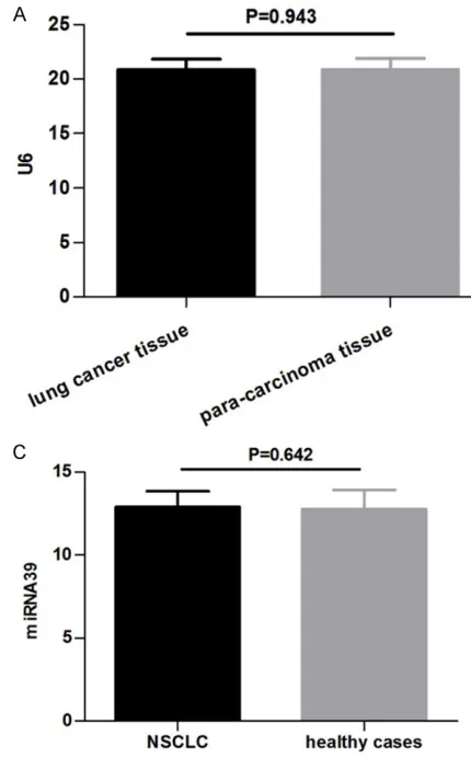
In the present study, U6 was selected to be the
internal control; no significant differences
between the NSCLC group (20.76±0.90) and the healthy controls (20.88±0.94) were
identi-fied (P=0.609; Figure 1A). In the serum, the mean Ct value of cel-miRNA39 in the NS- CLC group was 15.48±0.86 compared with
15.44±0.64 in the healthy group with no signifi
[image:3.612.87.302.67.413.2]Exhaled breath condensate let-7 in non-small cell lung cancer
Similarly, no significant differences in the
expression levels of cel-miRNA39 in EBC
were identified between the NSCLC group
(12.90±0.95) and the healthy controls (12.78±1.16; P=0.642; Figure 1C). These results suggest that U6 levels were consistent between tissue samples and that cel-miRNA39 was not affected by the tumor; U6 and cel-miR-NA39 were able to be used as the control refer-ences for the target gene let-7.
Tissue
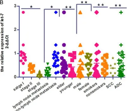
The expression levels of let-7 in tissues from the NSCLC group (0.32±0.21) were markedly decreased compared with adjacent non-can-cerous tissues (P<0.001; Figure 2A). Ad- ditionally, the expression levels of let-7 at stage I and II were increased compared with stage III (P=0.007). The expression of let-7 in patients with lymph node metastasis was decreased
compared with the group without lymph node metastasis (P=0.008). No significant differ -ence between the expression of let-7 in the
elder group (≥65 years) and the younger group (<65 years) was identified (P=0.726). Similarly,
there were also no significant differences
between other clinicopathologic characteri-
stics identified [sex (P=0.296), smoking status (P=0.311) and tumor histopathology (P=0.376); Figure 2B]. The AUC of let-7 in
lung cancer tissue was 0.894 [95%
confi-dence interval (CI), 0.817-0.971; P<0.001].
At a threshold of 0.53, the specificity and sensi
-tivity were 90% and 93.3%, respectively (Figure 2C).
Serum
[image:4.612.307.519.72.258.2]The expression levels of serum let-7 in the NSCLC group [0.40 (0.22, 0.59)] were markedly decreased compared with the healthy control Figure 2. A. Comparison of relative expression of tissue let-7 between lung cancer tissue and para-carcinoma normal tissue in patients with NSCLC. B. Association between tissue let-7 and clinico-pathologic characteristics of patients with NSCLC. *P<0.01; **P>0.05. Elder, ≥65 years; younger, <65 years; stage, tumor-node-metastasis stage; SCC, squamous cell carcinoma; ADC, adenocarcinoma. C.
Diagnostic efficiency of lung cancer tissue let-7 for
group (P<0.001; Figure 3A). With regard to TNM staging, the expression of let-7 in advanced-stage NSCLC was decreased com-pared with the early-stage group (P=0.014). The level of let-7 expression in the group with-out lymph node metastasis (0.83±0.68) was increased compared to the group with lymph node metastasis (0.24±0.12; P=0.003). No
sig-nificant differences between other clinicopath
-ologic features were identified [sex (P=0.747), age (P=0.233), smoking status (P=0.834) or histological type (P=0.864); Figure 3B]. The
AUC of serum let-7 was 0.771 (95% CI,
0.652-0.890; P<0.001). At a threshold of 0.55, the
specificity and sensitivity were 86.7% and 60%,
respectively (Figure 3C). EBC
The expression levels of let-7 in EBC from the NSCLC group (0.46±0.48) were decreased compared with that in the healthy volunteer group (P<0.001; Figure 4A). The expression
levels of EBC let-7 were revealed to be signifi -cantly associated with TNM stage and lymph node metastasis condition, respectively (P<0.001). No significant differences between
EBC let-7 expression and the other
clinicopath-ologic features were identified [sex (P=0.571), age (P=0.325), smoking status (P=0.453) or histological type (P=0.797); Figure 4B]. The
AUC of EBC let-7 was 0.750 (95% CI,
0.625-0.875; P=0.001). At a threshold of 0.61, the
specificity and sensitivity were 76.7% and 66.7%, respectively (Figure 3C).
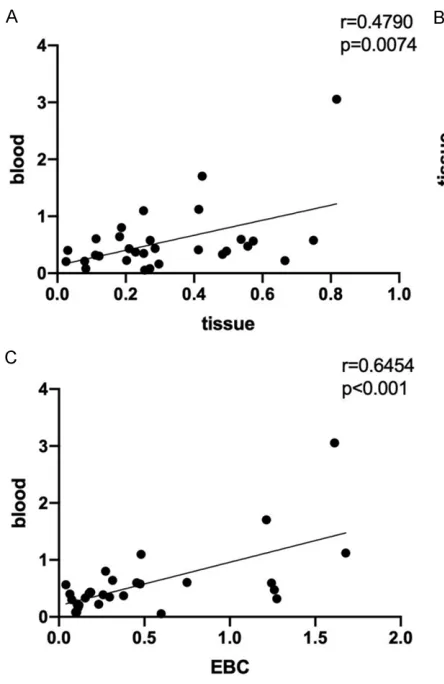
Correlation analysis was done among the expressions of let-7 in EBC, lung cancer tis-sues, and blood. Using Pearson correlation analysis, we obtained a positive correlation between let-7 expression in lung cancer tissues and in blood (r=0.4790, P=0.0074; Figure 5A). Similarly, the let-7 expression in EBC was posi-tively correlated with that in lung cancer tissues (r=0.6048, P<0.001; Figure 5B). A positive cor-relation between let-7 expression in EBC and in blood was also found, and with a higher
correla-tion coefficient value (r=0.6454, P<0.001;
Figure 5C).
Discussion
With the assistance of various enzymes, miR-NAs are synthesized by long primary transcripts Figure 3. A. Comparison of relative expression of se-rum let-7 between patients with NSCLC and healthy volunteers. B. Association between serum let-7 and clinicopathologic characteristics of patients with NSCLC. *P<0.01; **P>0.05. Elder, ≥65 years; younger, <65 years; stage, tumor-node-metastasis stage; SCC, squamous cell carcinoma; ADC, adenocarcinoma. C.
Exhaled breath condensate let-7 in non-small cell lung cancer
Figure 4. A. Comparison of relative expression of EBC let-7 between patients with NSCLC and healthy volunteers. B. Association between EBC let-7 and the clinicopathologic characteristics of patients with non-small cell lung cancer. *P<0.01; **P>0.05. Elder, ≥65 years; younger, <65 years; stage, tumor-node-metastasis stage; SCC, squamous cell carcinoma; ADC, adenocarcinoma.
of the miRNA genes in the nucleus and
cyto-plasm. Mature miRNAs combine specifically
with the 3’-untranslated region (UTR) of target mRNA and serve an important role in the
regu-lation of post-transcriptional gene expression through degradation or translational
[image:6.612.90.312.322.664.2]to maintain pairing to miRNAs [11]. Although the let-7 family consists of numerous members which are located on distinct chromosomes, all share common characteristics in markedly con-served sequence and function between various species [12]. Let-7 is involved in the biological behavior including proliferation, differentiation, apoptosis and invasion of tumor cells through regulation of the binding of numerous mRNAs
to target specific genes [13-15], which include
the oncogenes such as high-mobility group 2
[16, 17], integrin β3 [18], c-Myc, mitogen-acti -vated protein kinase kinase kinase kinase 3, Ras [19, 20], B-cell lymphoma extra-large [21] and homeobox A1 [22], and distinct signaling pathways cyclin D1, cell division cycle 25 homo-log A, cyclin-dependent kinase 2/homeobox
A1/let-7c [22], nuclear factor-κB-Ras-let7 [23],
phosphoinositide 3-kinase/protein kinase B [24] and mitogen-activated protein kinase kinase/extracellular-signal-regulated kinase. Previous studies have demonstrated that the expression level of let-7 is higher in non-carci-noma cases compared with patients with lung cancer and that let-7 may contribute to the diagnosis of lung cancer. Additionally, de- creased expression of let-7 is associated with advanced-stage lung cancer, lymph node me- tastasis, and progression-free and overall sur-vival rates [13, 25]. Let-7 is also associated with response to chemotherapy [26] and target gene therapy [25].
As with blood, there are numerous substances
in the EBC which reflect the pathology of respi -ratory disease. Extracted from the lower
respi-ratory airway lining fluid through refrigerated
devices [27], the collection of EBC is easy, non-invasive, safe and repeatable compared with induced sputum, bronchoscopy and
thoracen-tesis. There are numerous markers identified
from the EBC in patients with NSCLC, including methylated p16 [28], EGFR [29], and VEGF [30]. These abnormal genes are associated with NSCLC.
The results of the present study demonstrated that the level of let-7 in the serum and lung can-cer tissue were decreased compared with non-cancer cases, and that the level of let-7 decreased with the deterioration of lung can-cer, which included lymph node metastasis and
clinical stage. No significant association
between serum let-7 and other
clinicopatho-logic characteristics were identified (sex, age,
smoking status and histopathological type). Similarly, the expression of let-7 in EBC of patients with NSCLC was decreased compared with that of healthy controls. The levels of let-7 in EBC decreased with the severity of NSCLC (TNM stage and lymph node metastasis). No
significant association between EBC let-7 and
other clinicopathologic characteristics was
identified (sex, age, smoking status and histo -pathological type). In addition, the level of let-7 in EBC in NSCLC patients was positively corre-lated with that in lung cancer tissues and serum, suggesting that let-7 analysis in EBC may be effective in estimating NSCLC.
Taken together, the results of the present study indicated that detection of let-7 was feasible in EBC and with the advantages associated with EBC, and let-7 in EBC may be a promising bio-marker for the diagnosis and evaluation of NSCLC. Limitations of the present study include
the following: first, as EBC consists of >90% water and <1% aerosol particles, the level of
let-7 is low and there are no standardized col-lection devices available so various devices are used; secondly, the study size is small, and the data are not extensive; the patients with NSCLC receiving chemotherapy or target gene therapy should be followed-up for life.
Acknowledgements
This work was supported by the project of Natural Science of Jiangsu Province (BK20- 191207).
Disclosure of conflict of interest
None.
Address correspondence to: Jian-Rong Chen, De-
partment of Respiraology, Second Affiliated
Hospi-tal of Nantong University, 6 North Road Haier- xiang, Nantong 226001, Jiangsu, China. Tel: +86-13706291312; E-mail: drchenjr@163.com
References
[1] Akamine T, Toyokawa G, Tagawa T, Yamazaki K, Seto T, Takeo S and Mori M. Lorlatinib for the treatment of patientswith non-small cell lung cancer. Drugs Today (Barc) 2019; 55: 107-116.
Exhaled breath condensate let-7 in non-small cell lung cancer
J. Tumor autophagy is associated with survival outcomes in patients with resected non-small cell lung cancer. Lung Cancer 2019; 129: 85-91.
[3] Wu Z, Tian Y, Yu Q, Li H, Tian Z, Jiang H, Tian D and Yang X. The expression and correlation be-tween chemokine CCL7 and ABCE1 in non-small cell lung cancer. Exp Ther Med 2018; 16: 3004-3010.
[4] Krawcyzk A, Nowak D, Nowak PJ, Padula G and Kwiatkowska S. Elevated exhalation of hydro-gen peroxide in patients with non-small cell lung cancer is not affected by chemotherapy. Redox Rep 2017; 22: 308-314.
[5] Palma JF, Das P and Liesenfeld O. Lung cancer screening: utility of molecular applications in conjunction with low-dose computed tomogra-phy guidelines. Expert Rev Mol Diagn 2016; 16: 435-447.
[6] Nobili V, Alisi A, Mosca A, Della Corte C, Veraldi S, De Vito R, De Stefanis C, D’Oria V, Jahnel J, Zohrer E, Scorletti E and Byrne CD. Hepatic farnesoid X receptor protein level and
circulat-ing fibroblast growth factor 19 concentration in
children with NAFLD. Liver Int 2018; 38: 342-349.
[7] Zhao J, Wang K, Liao Z, Li Y, Yang H, Chen C, Zhou YA, Tao Y, Guo M, Ren T and Xu L. Pro-moter mutation of tumor suppressor microR-NA-7 is associated with poor prognosis of lung cancer. Mol Clin Oncol 2015; 3: 1329-1336. [8] Zhang XY, Wang Q and Zhang SJ. MicroRNAs in
sputum specimen as noninvasive biomarkers for the diagnosis of non-small cell lung cancer: an updated meta-analysis. Medicine 2019; 98: 6.
[9] Korba K, Dizdas TN, Baysal E, Uzun UC, Kaya OO, Ozyilmaz B, Kutbay YB, Ozdemir TR, Kir-biyik O, Erdogan KM, Guvenc MS, Kocal GC and Basbinar Y. cfDNA in exhaled breath con-densate (EBC) and contamination by ambient air: toward volatile biopsies. J Breath Res 2019; 13: 036006.
[10] Lamichhane SR, Thachil T, De leso P, Gee H, Moss SA and Milic N. Prognostic Role of mi-croRNAs in human non-small-cell lung cancer: a systematic review and meta-analysis. Dis Markers 2018; 2018: 8309015.
[11] Lin S and Gregory RI. MicroRNA biogenesis pathways in cancer. Nat Rev Cancer 2015; 15: 321-333.
[12] Inamura K. Diagnostic and therapeutic poten-tial of microRNAs in lung cancer. Cancers 2017; 9: 49.
[13] Xia Y, Zhu Y, Zhou X and Chen Y. Low expres-sion of let-7 predicts poor prognosis in patients with multiple cancers: a meta-analysis. Tumor Biology 2014; 35: 5143-5148.
[14] Zhou Y, Liang H, Liao Z, Wang Y, Hu X, Chen X, Xu L and Hu Z. MiR-203 enhances let-7 biogen-esis by targeting LIN28B to suppress tumor growth in lung cancer. Sci Rep 2017; 7: 42680. [15] Xie P, Li XL, Tan XF, Sun X, Wang C and Yu J.
Sequential serum let-7 is a novel biomarker to predict accelerated reproliferation during frac-tional radiotherapy in lung cancer. Clin Lung Cancer 2016; 17: e95-e101.
[16] Di Fazio P, Maass M, Roth S, Meyer C, Grups J, Rexin P, Bartsch DK and Kirschbaum A. Ex-pression of hsa-let-7b-5p, hsa-let-7f-5p, and hsa-miR-222-3p and their putative targets HMGA2 and CDKN1B in typical and atypical carcinoid tumors of the lung. Tumour Biol 2017; 39: 1010428317728417.
[17] Chen Z, Wang D, Gu C, Liu X, Pei W, Li J, Cao Y, Jiao Y, Tong J and Nie J. Down-regulation of let-7 microRNA increased K-ras expression in lung
damage induced by radon. Environ Toxicol
Pharmacol 2015; 40: 541-548.
[18] Zhao B, Han H, Chen J, Zhang Z, Li S, Fang F, Zheng Q, Ma Y, Zhang J, Wu N and Yang Y. Mi-croRNA let-7c inhibits migration and invasion of human non-small cell lung cancer by
target-ing ITGB3 and MAP4K3. Cancer Lett 2014;
342: 43-51.
[19] Huang J, Lin H, Zhong M, Huang J, Sun S, Lin L and Chen Y. Role of Lin28A/let-7a/c-Myc path-way in growth and malignant behavior of papil-lary thyroid carcinoma. Med Sci Monit 2018; 9: 8899-8909.
[20] Yang G, Zhang W, Yu C, Ren J and An Z. Mi-croRNA let-7: regulation, single nucleotide polymorphism, and therapy in lung cancer. J Cancer Res Ther 2015; 11 Suppl 1: C1-6. [21] Tian N, Han Z, Li Z, Zhou M and Fan C. Lin28/
let-7/Bcl-xL pathway: the underlying mecha-nism of drug resistance in Hep3B cells. Oncol Rep 2014; 32: 1050-1056.
[22] Zhan M, Qu Q, Wang G, Liu YZ, Tan SL, Lou XY, Yu J and Zhou HH. Let-7c inhibits NSCLC cell proliferation by targeting HOXA1. Asian Pac J Cancer Prev 2013; 14: 387-92.
[23] Jiang R, Li Y, Zhang A, Wang B, Xu Y, Xu W, Zhao Y, Luo F and Liu Q. The acquisition of cancer stem cell-like properties and neoplastic trans-formation of human keratinocytes induced by arsenite involves epigenetic silencing of let-7c
via Ras/NF-κB. Toxicol Lett 2014; 227: 91-98. [24] Shen H, Liu J, Wang R, Qian X, Xu R, Xu T, Li Q,
Wang L, Shi Z, Zheng J, Chen Q and Shu Y.
Ful-vestrant increases gefitinib sensitivity in
non-small cell lung cancer cells by upregulating
let-7c expression. Biomed Pharmacother 2014;
68: 307-213.
development of lung cancer: a systematic re-view and meta-analysis. Eur Rev Med Pharma-col Sci 2018; 22: 8353-8366.
[26] Stahihut C and Slack FJ. Combinatorial action of microRNAs let-7 and miR-34 effectively syn-ergizes with erlotinib to suppress non-small cell lung cancer cell proliferation. Cell Cycle 2015; 14: 2171-2180.
[27] Chen JL, Lv XD, Ma H, Chen JR and Huang JA. Detection of cancer embryo antigen and endo-thelin-1 in exhaled breath condensate: a novel approach to investigate non-small cell lung cancer. Mol Clin Oncol 2016; 5: 124-128. [28] Xiao P, Chen JR, Zhou F, Lu CX, Yang Q, Tao GH,
Tao YJ and Chen JL. Methylation of P16 in ex-haled breath condensate for diagnosis of non-small cell lung cancer. Lung Cancer 2014; 83: 56-60.
[29] Smyth RJ, Toomey SM, Sartori A, O’Hanrahan E, Cuffe SD, Breathnach OS, Morgan RK and Hennessy BT. Brief report on the detection of the EGFRT790M mutation in exhaled breath condensate from lung cancer patients. J Tho-rac Oncol 2018; 13: 1213-1216.