Original Article
Rab5a promotes the migration and invasion
of hepatocellular carcinoma by up-regulating Cdc42
Xiao Yang1*, Zhengshu Liu3*, Yongguo Li2, Ke Chen1, Hong Peng1, Liying Zhu1, Huihao Zhou1, Ailong Huang1, Hua Tang1
1Key Laboratory of Molecular Biology for Infectious Diseases (Ministry of Education), 2Department of forensic Medicine, Chongqing Medical University, Chongqing, China; 3Department of physical Examination, The First Affili-ated Hospital of Chongqing Medical University, Chongqing, China. *Equal contributors.
Received October 11, 2017; Accepted November 17, 2017; Epub January 1, 2018; Published January 15, 2018
Abstract: There are many factors participating in the process of human hepatocellular carcinoma(HCC) occurrence and development. In this study, we found that Rab5a expression was higher in the HCC tissues and 3 cell lines than normal liver tissues. Overexpression of Rab5a promoted cell invasion and migration in vitro and in vivo. In contrary, inhibition of Rab5a suppressed cell invasion and migration. Mechanistic studies revealed that Rab5a was positively regulated cell division cycle 42 (Cdc42) expression by enhancing its promoter activity. These data suggested that Rab5a is highly expressed in HCC, and promote the invasion and migration by targeting Cdc42 expression.
Keywords: HCC, Rab5a, invasion, migration, Cdc42
Introduction
HCC is the most common tumor and the third primary cause of cancer-related deaths world-wide [1]. It is highly prevalent in Eastern and South-Eastern Asia, with incidence rates of 31.9/100,000 and 22.2/100,000 respectively [2]. Although there are many established thera-peutic strategies including surgery, chemother-apy and radiotherchemother-apy, the 5-year overall surviv-al has reached 30%-50%, the 5-year recurrent rate is still up to 70-85% due to recurrence and distant metastasis of HCC [3, 4]. To date, the precise mechanisms lead to the development and progression of HCC still unclear [5]. Rab5 belongs to Rab GTPases family, which contains three subtypes including Rab5a, Rab5b and Rab5c [6]. Rab5a is localized at human chromosome 3p24.3 and its molecular weight is 23.66 kDa [7]. Rab5a is abnormally expressed in a variety of tumors (such as breast cancer, lung cancer and ovarian cancer), which plays a vital role in the pathogenesis and dis-tant metastasis [8-10]. However, there is rare report concerning the role Rab5a in HCC. In this study, we examined the expression of Rab5a in HCC and determined the role of
Rab5a in invasion and migration. The results showed that Rab5a may act as a tumor stimula-tor in HCC.
Materials and methods
Cell lines
All the cell lines (LO2, SMMC7721, HepG2, SK-Hep1) for this study were preserved in our lab, which were cultured in Dulbecco’s modified Eagle’s medium (DMEM) (Hyclone, China), sup-plemented with 10% fetal bovine serum (FBS, Cell-box), 100 units/mL penicillin and 100 μg/ ml streptomycin. A humidified incubator with 5% CO2 at 37°C was used for maintaining the cells.
RNA extraction and qpCR
samples were executed by using UltraSYBR mixture (Cwbio, China) and conducted using the CFX Connet TM real-time PCR system (Bio-Rad). The quantification analysis was cal-culated by the 2-ΔΔCT. All experiments were performed in triplicate and repeated at least 3 times.
Immunohistochemistry
The tissue sections of hepatocellu-lar carcinoma patients were obta- ined from pathology Department of Chongqing Medical University. The human subject protocol was ap- proved by the Clinical Research Ethics Committee of Chongqing Me- dical University. Written consent was obtained from each patient. The tis-sue of transplanted tumors and human liver cancer tissue were paraformaldehyde fixed and paraffin embedded. Then the slides were deparaffinized with dimethylben-zene, followed by gradient alcohol dehydration and analyzed for Rab- 5a (Bioword, 1:100 dilution) and Cdc42 (Bioword, 1:100 dilution) expression. Visualized with 3,3-dia- minobenzidine substrate (ZSGB-Bio, DAB) and counterstained with hema-toxylin of the slides. The PBS ing was used as the negative stain-ing control. Mounted and visualized under an inverted microscope.
Western blot analysis
[image:2.612.94.350.99.709.2]Cells were lysed with RIPA Lysis Buffer (Beyotime, China) including 1 mmol/L PMSF, and protein concen-tration was measured using the BCA Assay Kit (Beyotime). Protein sam-ples were separated by 12% SDS-PAGE and transferred to a PVDF membrane. Blots in PVDF membr- anes were incubated with primary antibody, followed by anti-rabbit HRP secondary antibody (Proteinte- ch, 1:4000) or anti-mouse HRP sec-ondary antibody (Bioword, 1:3,000) for 2:30 h at room temperature, and the signals were detected with an ECL Detection Reagent (Millipore, Billerica, MA).
Table 1. Primer sequences used for PCR or constructions of various plasmids
Amplifier primers Primer sequence
Rab5a-F TGCTCTAGACGCCACCATGGCTAGTCGAGGCGCAAC Rab5a-R TGCTCTAGATTAGTTACTACAACACTGATTC
plasmids, small interfering RNA (siRNA) and transfection
The CDS section of Rab5a was amplified by PCR with primers (Table 1). The PCR product was purified and linked into the XhaI sites of pcDNA3.1 (-) expression vector. Fragment of si-Rab5a and negative control (si-NC) were pur-chased from TsingKe (Table 1). Transfections were performed with a Lipofectamine2000 kit (Invitrogen, Carlsbad, CA) according to the man-ufacturer’s instructions.
Wound healing assays
Transfected cells were plated in 6-well plates and incubated 24 h. When cells grew in full monolayer on cover slips in six well plates and then created the wound with using a 200 μl
TK. Cells were collected in 48 h after transfec-tion and analyzed using the Dual-Luciferase Reporter Assay System (Promega).
Recombinant lentiviral overexpression vectors
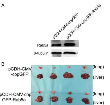
The Rab5a gene fragment was ligated into pCDH-CMV-MCS-EF1-copGFP Vector. Sequenc- ing was performed in 293T cells (Human embry-onic kidney stem cells) after packaging, virus solution was collected and virus titer detected. Then the virus was infected with the target SK-Hep1 cells. Recombinant lentiviral overex-pression vectors in cells can be selected for GFP positive cells by FACS.
Tumor migration assay
[image:3.612.91.375.69.412.2]Male nude mice (8 weeks old) were purchas- ed from the Laboratory Animal Services Cen- Figure 1. The expressions of Rab5a in HCC tissues and HCC cell lines. A,
B. The expressions of Rab5a in HCC tissues by IHC and qRT-PCR, paracar-cinoma tissue and N were used as control; C, D. Rab5a mRNA and pro-tein expressions in LO2 and HCC cell lines by qRT-PCR and Western blot
(SMMC7721, SK-Hep1 and HepG2). β-tubulin was used as the endogenous control. *P < 0.05, **P < 0.01.
pipette tip. PBS washed the cells gently and cells were replaced incubated in DMEM with 2% FBS. Then take pho-tos at 0 h, 24 h and 48 h after wounding.
Transwell assays
After 24 h transfection, 1 × 10⁵ cells suspending in 200 μl of serum-free DMEM were seeded to the 8 μm transwell migration chambers (Costar). 600 μl of DMEM media con-taining 10% FBS was added to the bottom chamber. After cul-tured 24 h at 37°C incubator, the membrane was cleaning out with PBS. Migrated cells were fixed with 4% parafor-maldehyde for 30 min and stained with crystal violet for 10 min. Then washed with dis-tilled water and took photo-graph, counting the cells with using the inverted microsc- ope.
Luciferase reporter assay
pRL-ter of CQMU. Animal handling and experimen- tal procedures were approved by the Animal Experimental Ethics Committee of Chongqing Medical University. pCDH-CMV-copGFP and pCDH-CMV-copGFP-Rab5a with a total of 0.8 × 10⁷ cells, respectively, were injected into the liver of nude mice, and sacrificed them at 30 days.
Statistical analysis
All results were expressed as the means ± sd of at least three independent experiments. Statistical analysis was performed by X2 analy-sis and Student’s t test. p < 0.05 was consid-ered statistically significant.
Rab5a). Over-expression and siRNA efficiencies were measured by qRT-PCR and Western blot in SK-Hep1 and SMMC7721 cells. The results sh- owed that pCMV-Sport6-Rab5a increased ap- proximately 15 times in SK-Hep1 cells (Figure 2A) and 20 times in SMMC7721 cells (Figure 2C). In contrast, si-Rab5a resulted in an obvi-ous inhibition of Rab5a in SK-Hep1 cells (Fig- ure 2B) and SMMC7721 cells (Figure 2D), spe-cially si-Rab5a (661). Then, we chose si-Rab5a (661) to undergo the following experiments.
Up-regulation of Rab5a promoted the invasion and migration in HCC cells
[image:4.612.90.372.72.446.2]To clarify whether Rab5a could affect the inva-sion and migration in HCC cells, transwell assay Figure 2. The efficiencies of over-expression and siRNA of Rab5a. A, B. The
efficiencies of PCMV-Sport6-Rab5a and siRNA by qRT-PCR and WB in SK-Hep1 cells; C, D. The efficiencies of PCMV-Sport6-Rab5a and siRNA by qRT-PCR and WB in SMMC7721 cells. *P < 0.05, **P < 0.01.
Results
Up-regulation of Rab5a in HCC tissue and HCC cell lines
Firstly, we examined Rab5a expression level by both IHC and qRT-PCR in human HCC tissues and precancerous tis-sues. Compared with the para- carcinoma tissues, the expres-sion of Rab5a was significant-ly up-expressed in human HCC tissues (Figure 1A, 1B). Equ- ally, qRT-PCR and western bl- otting were done in HCC cell lines to detect the expression of Rab5a. The results showed that Rab5a mRNA and protein levels were markedly up-regu-lated in 3 HCC cell lines (SM- MC7721, SK-Hep1 and Hep- G2) compared with the hepat-ic immortal cell line L02 (Fig- ure 1C, 1D).
Over-expression and siRNA efficiency in SK-Hep1 and SMMC7721 cells
(si-and wound healing assay were performed. pCMV-Sport6-Rab5a (pCMV-Sport6 as control) and si-Rab5a (Si-NC as control) were transfect-ed in HepG2 and SK-Hep1 cells respectively. We found that Rab5a overexpression promoted the invasion activity while Rab5a silencing repressed the invasion in HepG2 and SK-Hep1 cells (Figure 3A). On the other hand, wound healing assay showed that pCMV-Sport6-Rab5a boosted the migration while pCMV-Sport6-Rab5a silencing depressed the migration of SK-Hep1 cells (Figure 3B). These findings indicated that Rab5a promoted the invasion and migration of HCC cells.
Rab5a up-regulated Cdc42 expression by enhancing its promoter activity
We measured a large number of genes related with invasion and migration biologic functions by qRT-PCR in SK-Hep1 cells, such as FOXC2, TP53, VEGF, MMp2, Cdc42, etc. As a result, the expressions of TP53, Cdc42, RhoA, Twist1 and KLF4 were all increased in pCMV-Sport6-Rab5a group (Figure 4A) and decreased in si-Rab5a
migration in vivo
To further definite the effects of Rab5a on tumorigenesis in vivo, nude mice were subcuta-neously injected with SK-Hep1 cells stably transfected with the lentiviral vector express- ing Rab5a (pCDH-CMV-copGFP-Rab5a) (Figure 5A). 30 days after infection, nude mice were sacrificed and the migration potential were analyzed (Figure 5B). Tumor cell migration in liver was detected by IHC staining assay. As a result, Cdc42-staining increased in the pCDH-CMV-copGFP-Rab5a group compared with con-trol (Figure 5C). Therefore, Rab5a promoted development in HCC in vivo by regulating Cdc42 expression.
Discussion
The occurrence and development of HCC is a complex process based on multiple genetic changes. The invasion and metastasis of HCC is a more complicated procedure, which in- volved the mutation and activation of onco-genes, deletion of anti-oncoonco-genes, and disor-Figure 3. Rab5a promoted invasion and migration in HCC cells. A. Cell
migra-tion was detected by transwell assay in HepG2 and SK-Hep1 cells; B. Wound healing assay in SK-Hep1 cells transfected over-expressing Rab5a, si-Rab5a and their controls.
(Figure 4B). Then, Western blotting was used to further confirm. However, only three genes (TP53, Cdc42 and Rh- oA) protein level correspond-ed with the mRNA level (Figure 4C). We finally chose Cdc42 to undergo the following experi- ments.
To investigate the potential mechanism of Cdc42 up-regu-lated by Rab5a, we construct-ed pGL3-Basic-Cdc42 vector which contains the Cdc42 pro-moter (-1002 bp~+20 bp) for luciferase reporter assay. As a result, Cdc42 promoter activi-ty was higher in the pcDNA3.1-Rab5a and pGL3-Basic-Cdc42 co-transfected group compa- red to pcDNA3.1 and pGL3-Basic-Cdc42 co-transfected group (Figure 4D). These data suggested that Rab5a could up-regulate Cdc42 expression by increasing its promoter activity.
der of apoptosis regulation mechanism. With the development of molecular biology, more and more genes closely related to HCC metas-tasis were found to provide a new method for the treatment of individuals.
As a member of Rab family, Rab5a regulates the formation and transport in early vesicles, adjusts the fusion of endocytosis and early endosomes, and participates in the process of receptor internalization, intracellular transport of substances, signal transduction and cyto-skeletal remodeling. In a word, Rab5a is a sig-nal regulator in protein transmembrance path-ways [7]. In recent years, it is reported that aberrant expression of Rab5a contributes to
[image:6.612.93.519.73.467.2]tumor progression and distant metastasis [8]. Yang et al [9] have found that Rab5a is closely related to breast cancer axillary lymph node metastasis. Zhao [10] have showed that Rab5a can transfer tumor cells from G1 phase to S phase via regulating APPL1-related epidermal growth factor (EGF) signaling pathway in ovari-an covari-ancer. Yu [11] revealed that increased expression of Rab5a was significantly correlat-ed with poor prognosis in colorectal cancer patients. Lu [12] explained vacuolin-1 activated Rab5a to block autophagosome-lysosome fusion in cancer cells. Fukui et al [13] found that Rab5a enhance the EGF signaling and Rab5a upregulation predict poor prognosis in HCC patients.
In our study, we examined the expression of Rab5a in HCC tissues and cell lines.
The results showed that the expression of Rab5a mRNA and protein were upregulated in HCC tissues as well as that of HCC cell lines, which can be used as a molecular marker in HCC. In addition, it is well known that metasta-sis is severely responsible for HCC reoccur-rence. We found that overexpression of Rab5a promoted cell invasion and migration in vitro
and in vivo by accelerating Cdc42 expression via enhancing its promoter activity. Cell division cycle 42 (Cdc42) is a member of the Rho sub-family, which is known to regulate the dynamic organization of the cytoskeleton and mem-brane trafficking for physiologic processes such as tumor cell proliferation, motility, polari-ty, cytokinesis and growth [14]. A large number of studies found that Cdc42 was overexpressed in tumors (breast cancer, lung cancer, testicular cancer, bladder cancer and so on), and it plays an important role in cell proliferation, transfor-mation, invasion and metastasis [15-18]. More interesting, Cdc42 is a member of Rho subfam-ily belongs to Ras super famsubfam-ily GTPase as well as Rab5a, which may plays a similar role in the occurrence and development of HCC. The next step is to elucidate the interaction mechanism of Rab5a and Cdc42, to promote the develop-ment of clinical treatdevelop-ment in HCC.
Based on all the above results, Rab5a presents an oncogenic function and plays a significant role in HCC development.
Acknowledgements
This work were supported by the Scientist Culture Plan of Chongqing Medical University (162014) and the Young Scientist Culture Plan of Chongqing (cstc2014kjrc-qnrc10007). Disclosure of conflict of interest
None.
Address correspondence to: Hua Tang, Department of Infectious Diseases, Institute for Viral Hepatitis, Key Laboratory of Molecular Biology for Infectious Diseases (Ministry of Education), The Second Affiliated Hospital, Chongqing Medical University, 1 Yixueyuan Road, Yuzhong Area, Chongqing 400016, China. Tel: +86 23 68486780; Fax: +86 23 68486780; E-mail: tanghua86162003@cqmu. edu.cn
References
[1] Torre LA, Bray F, Siegel RL, Ferlay J, Lortet-Tieu-lent J, Jemal A. Global cancer statistics, 2012. CA Cancer J Clin 2015; 65: 87-108.
[image:7.612.95.306.71.272.2][2] Farrell GC, Chan HL, Yuen MF, Amarapurkar DN, Chutaputti A, Fan JG, Hou JL, Han KH, Kao
JH, Lim SG, Mohamed R, Sollano J, Ueno Y. Prevention of hepatocellular carcinoma in the Asia-Pacific region: consensus statements. J Gastroenterol Hepatol 2010; 25: 657-663. [3] Llovet JM, Ducreux M, Lencioni R, Di Bisceglie
AM, Galle PR, Dufour JF, Greten TF, Raymond E, Roskams T, De Baere T, Ducreux M, Mazzafer-ro V, Bernardi M, Bruix J, Colombo M, Zhu A. EASL-EORTC clinical practice guidelines: man-agement of hepatocellular carcinoma. Eur J Cancer 2012; 48: 599-641.
[4] Shan SY, Huang LF, Xia Q. Salvage liver trans-plantation leads to poorer outcome in hepato-cellular carcinoma compared with primary liver transplantation. Sci Rep 2017; 7: 44652. [5] Li S, Li J, Fei BY, Shao D, Pan Y, Mo ZH, Sun BZ,
Zhang D, Zheng X, Zhang M, Zhang XW, Chen L. MiR-27a promotes hepatocellular carcino-ma cell proliferation through suppression of its target gene peroxisome proliferator-activated receptor γ. Chin Med J 2015; 128: 941-947. [6] Pfeffer S, Aivazian D. Targeting Rab GTPases
to distinct membrane compartments. Nat Rev Mol cell Biol 2004; 5: 886-896.
[7] Ali BR, Wasmeier C, Lamoreux L, Strom M, Seabra MC. Multiple regions contribute to membrane targeting of Rab GTPses. J Cell Sci 2004; 117: 6401-6412.
[8] Yu L, Hui-chen F, Chen Y, Zou R, Yan S, Chun-xiang L, Wu-ru W, Li P. Differential expression of RAB5A in human lung adenocarcinoma cells with different metastasis potential. Clin Exp Metastasis 1999; 17: 213-219.
[9] Yang PS, Yin PH, Tseng LM, Yang CH, Hsu CY, Lee MY, Horng CF, Chi CW. Rab5A is associated with axillary lymph node metastasis in breast cancer patients. Cancer Sci 2011; 102: 2172-2178.
[10] Zhao Z, Liu XF, Wu HC, Zou SB, Wang JY, Ni PH, Chen XH, Fan QS. Rab5a overexpression pro-moting ovarian cancer cell proliferation may be associated with APPL1-related epidermal growth factor signaling pathway. Cancer Sci 2010; 101: 1454-1462.
[11] Minhao Yu, Yang L, Shaolan Q, Ming Z. In-creased expression of Rab5A predicts metas-tasis and poor prognosis in colorectal cancer patients. Int J Clin Exp Pathol 2015; 8: 6974-6980.
[12] Lu Y, Dong S, Hao B, Li C, Zhu K, Guo W, Wang Q, Cheung KH, Wong CW, Wu WT, Markus H, Yue J. Vacuolin-1 potently and reversibly inhib-its autophagosome-lysosome fusion by activat-ing RAB5A. Autophagy 2014; 10: 1895-1905. [13] Fukui K, Tamura S, Wada A, Kamada Y, Igura T,
Kiso S, Hayashi N. Expression of Rab5A in he-patocellular carcinoma: possible involvement in epidermal growth factor signaling. Hepatol Res 2007; 37: 957-965.
[14] Soniya S, Wannian Y. Cellular signaling for acti-vation of Rho GTPase Cdc42. Cell Signal 2008; 20: 1927-1934.
[15] Fritz G, Brachetti C, Bahlmann F, Schmidt M, Kaina B. Rho GTPases in human breast tu-mors: expression and mutation analyses and correlation with clinical parameters. Br J Can-cer 2002; 87: 635-644.
[16] Cuiyan Z, Jie H, Fang Z, Kezhi Z, Junting W, Su-sheng S, Xiaoli F, Ning L, Xinhua M, Zhaoli C, Kang S, Bin Q, Baozhong L, Sheng C, Meihua X, Jie H. Overexpression of RhoE in non-small cell lung cancer (NSCLC) is associated with smok-ing and correlates with DNA copy number changes. Cancer Biol Ther 2007; 6: 335-342. [17] Kamai T, Yamanishi T, Shirataki H, Takagi K,
Asami H, Ito Y, Yoshida K. Overexpression of RhoA, Rac1, and Cdc42 GTPases is associated with progression in testicular cancer. Clin Can-cer Res 2004; 10: 4799-4805.