Original Article
YM155 down-regulates survivin and
induces PUMA-dependent apoptosis
in oral squamous cell carcinoma cells
Xinghua Yang1, Lin Peng2, Yanli Shi3
1School of Dentistry and Oral Health, Shandong University, Shandong, China; 2Clinical Labarotory, Laiwu Hospital Affiliated to Taishan Medical College, Shandong, China; 3Stomatology, Shandong Provincial Hospital Affiliated to Shandong University, Shandong, China
Received July 1, 2016; Accepted July 15, 2016; Epub October 1, 2016; Published October 15, 2016
Abstract: Objective: YM155, which inhibits the anti-apoptotic protein survivin, is known to exert anti-tumor effects in various cancers. However, there were few reports describing the inhibitory effect of YM155 on human oral squa-mous cell carcinoma cells that highly express survivin. In this study, we investigated the anti-tumor effects of YM155 on oral squamous cell carcinoma cells and explored its molecular mechanisms. Methods: Oral squamous cell car-cinoma SCC9 cells was treated with series of concentration of YM155 (0.01, 0.1, 1 and 10 ng/ml) for 6, 12 and 24 h. The effect of YM155 on growth of SCC9 cells was detected by MTT and colony formation assay. Cell apoptosis
was detected by flow cytometric analysis and the terminal deoxynucleotidyl transferase-mediated dUTP-biotin nick end labeling (TUNEL) assays. Western blot was used to detect the expression of survivin, p53 and PUMA protein. Caspase-3 activity was measured by cleavage of the caspase-3 substrate. To test the role of PUMA and caspase-3 on YM155-induced apoptosis and growth inhibition, the SCC9 cells was transfected with PUMA siRNA or caspase-3
siRNA or control siRNA for 16 hs before YM155 (1 and 10 ng/ml) treatment for 24 h. In addition, we also investi-gated the effect of YM155 in an in vivo xenograft model. Results: Treatment of YM155 efficiently reduced survivin
expression in the SCC9 cells. The downregulation of survivin induced remarkable apoptosis and growth inhibition
of SCC9 cells and resulted in significant upregulation of PUMA expression and caspase-3 activation. However, the induction of cell apoptosis of growth inhibition was reversed by PUMA siRNA or caspase-3 transfection. In addition, YM155 efficiently retarded tumor growth in established tumors of human SCC9 cell xenografted mice. Conclusion:
YM155 is a potent inhibitor of progression of SCC9 cells, which could be due to attenuation of survivin, and
activa-tion of PUMA/caspase-3 cellular signaling processes. This study suggests that YM155 may be a potential molecular
target with therapeutic relevance for the treatment of oral squamous cell carcinoma.
Keywords: Oral squamous cell carcinoma, survivin, YM155, p53, PUMA, apoptosis
Introduction
Oral cancer is one of the 10 most common cancers in the world. Its high mortality rate and
the disfigurement that survivors may suffer
gives rise to a considerable global public health burden [1]. About 90% of malignant oral neo-plasms are oral squamous cell carcinomas (OSCC), followed by adenocarcinoma and, rare-ly, other types of tumor [2]. Despite advances in treatment for OSCC, the 5-year survival rate remains poor [3]. As an important hallmark of
OSCC, apoptosis resistance restricts the effi -cacy of traditional therapies [4]. Survivin (BI- RC5), a member of the inhibitor of apoptosis
protein (IAP) gene family, has been shown to inhibit apoptosis, enhance proliferation and promote angiogenesis [5-7]. Survivin is selec-tively expressed in fetal and proliferating tis-sues and in various solid tumors, such as OSCC [8], and elevated survivin expression in OSCC has been associated with poor prognosis, high recurrence rate and chemotherapy and radia-tion resistance [8]. Therefore, targeting survivin
is promisingly beneficial for OSCC therapies. Sepantronium bromide (YM155) is the
have demonstrated that YM155 alone or in combination decreases tumor growth, induces apoptosis, sensitizes resistant cells to apopto-sis, and prolongs survival of tumor-bearing mice [11-13]. However, its molecular mecha-nism of action is still unclear.
In vitro studies revealed that YM155 trigger- ed apoptosis of head neck squamous cell carcinoma (HNSCC) cells in mitochondria and death receptor-dependent manner. In addition, YM155 not only downregulated the express- ion of survivin but also remarkably suppress- ed the activation of the mTOR signaling path-way in vitro and in vivo [14]. In human oral cancer cell lines, YM155 inhibited the growth and caused caspase-dependent apoptosis in the MC3 and HN22 cells, the mechanism that YM155 causes apoptosis of human oral can- cer cell lines was through downregulation of Sp1 and Mcl-1 [15]. Tang et al. has showed YM155 exhibited its anti-tumor activities in oral cancer cell lines by downregulation of Mcl-1 [16]. In adenoid cystic carcinoma (ACC)
cells, YM155 caused significant
autophagy-de-pendent cell death. In addition, YM155-indu- ced autophagy and cell death in vivo was cor-related with the suppression of Erk1/2 and S6 activation as well as increased TFEB nuclear translocation [17].
PUMA (p53 upregulated modulator of apopto -sis) is a pro-apoptotic member of the BH3-only subgroup of the Bcl-2 family. It is a key media-tor of p53-dependent and p53-independent
apoptosis [18, 19]. PUMA transduces death
signals primarily to the mitochondria, where it acts indirectly on the Bcl-2 family members Bax and/or Bak by relieving the inhibition im- posed by antiapoptotic members. It directly binds and antagonizes all known antiapoptotic Bcl-2 family members to induce mitochondrial dysfunction and caspase activation [20]. It has been shown that survivin inhibits Fas (CD95)-mediated apoptosis by supporting cas-pase3/p21 formation as a result of interaction with cdk4 [21]. In addition, survivin was shown to suppress the cell death induced by Bax [22]. A recent study has reported that targeting sur-vivin resulted in increased transcription of p53 targets, such as BAX, PUMA, NOXA and p21, and increased p53-dependent breast cancer
cells apoptosis [23], suggesting PUMA signals
may be regulated by survivin.
In this study, we evaluated the anticancer effects of YM155 in oral squamous cell carcino-mas cell line in vitro and xenografts in vivo. In addition, we investigated the effect of YM155
on PUMA signals expression to determine whether YM155 affects the PUMA pathways.
Materials and methods
Cell culture and reagents
The study was conducted in accordance with the guidelines in the Declaration of Helsinki. The human oral squamous cell carcinomas (OSCC) SCC9 cell was purchased from the In- situte of Biochemistry and Cell Biology (Sh- anghai, China). It was maintained in DMEM-F12 (Gibco), and supplemented with 10% fetal bo- vine serum and maintained at 37°C in a 5% CO2 humidified incubator. Antibodies against Survivin, PUMA, activated-caspase-3 and
β-actin was from Santa Cruz (Shanghai, China). YM155 were purchased from Selleck Chemi-
cals (Shanghai, China). PUMA siRNA, caspase-3
siRNA or control siRNA were purchased from Santa (Shanghai, China).
siRNA transfection
PUMA siRNA or caspase-3 siRNA or
misma-tched siRNA (control siRNA) were transiently transfected into SCC9 cells using Lipofecta- mine 2000 reagent (Invitrogen, Inc., Carlsbad,
CA, USA) as the manufacture’s instruction. Briefly, SCC9 cells (2 × 10³) were plated in
each well of a 96-well plate. Experimental conditions were set in quadruplicate. After cells were attached, the culture medium was
replaced with serum-free medium plus 3 μl of siRNA (20 μM) and mixed with 1 μl transfec-tion reagent and 100 μl Lipofectamine
me-dium supplied by the kit. Then, the siRNA-trans-fection reagent complex was incubated with
500 μl of diluted cells (5 × 10⁴ cells/well) for 24
hours at 37°C and 5% CO2. The cells without siRNA transfection were used as the control.
The knockdown effect was verified by
Wes-tern blot analysis. Stable SCC9 cell line trans-fected with siRNA were screened by adminis- tration of G418 (Invitrogen, Carlsbad, CA). Western blot analysis
SCC9 cells were treated with 0.01, 0.1 ,1 and 10 ng/ml YM155 for 6, 12 and 24 hs,
respec-tively, or transfected with PUMA/caspase-3
cells were lysed and protein was analyzed by SDS-polyacrylamide gel electrophoresis (SDS-PAGE). The following antibodies were appli- ed: monoclonal human anti-survivin, anti-p53
and anti-PUMA, anti-activated caspase-3 and anti-β-actin. Secondary antibodies (dilution 1:
20,000) were horseradish peroxidase-conju-gated (Hangzhou, China).
Caspase-3 activity assay
Caspase-3 activity was measured by cleavage
of the caspase-3 substrate. Briefly, SCC9 cells (2 × 10⁴) were treated as the methods above.
Reactions were spiked with DMSO or the
Caspase-3 specific inhibitor Ac-DMQD-CHO at a final concentration of 2 mM. Measurements
were done in triplicate. The mean of three bio-logical replicates is shown.
Cell yiability assay
Cell viability was assessed with the 3-(4,5- dimethylthiazol-2-yl)-2,5-diphenyltetrazolium bromide assay (MTT) according to the
manu-facture’s instruction. Briefly, SCC9 cells (1 ×
104) were plated in each well of a 96-well plate.
The following day, the cells were incubated with increasing concentrations of YM155 (ranging from 0.01 to 10 ng/ml) and incubated at
37°C for 6-24 hs. To assess the role of PUMA/
caspase-3 on YM155-induced growth of SCC9
cells, the SCC9 cells (1 × 10⁴) were transfect-
ed PUMA siRNA orcaspase-3 siRNA or control
siRNA for 16 h before the incubation with the concentrations of YM155 (1 and 10 ng/ml)
for 24 hs. Then 50 μL of 0.15%
3-(4,5-dimeth-ylthiazol-2-yl)-2,5-diphenyltetrazolium bromide (Sigma-Aldrich) was added to each well (after exposure to YM155 for 72 hs) and incubated for 4 hours at 37°C. The medium containing 3-(4,5-dimethylthiazol-2-yl)-2,5-diphenyltetra-zolium bromide was then removed. Then 200 uL of DMSO was added to each well after the medium was removed. The optical density (OD) values were measured at 570 nm on a scan-ning multi-well spectrophotometer (BioRad
Model 550, USA). Each assay was performed in
triplicate. Absorbance values were normalized to the values for the vehicle-treated cells to determine the percent of survival.
Colony-formation assay
SCC9 cells, stable PUMA siRNA or caspase-3
siRNA transfected SCC9 cells were seeded at 500 cells/well in 6-well plates and allowed to
adhere for 24 h. Cells were subsequently treat-ed with YM155 (1 and 10 ng/ml) for a period of 24 h after which, media was aspirated, cells were washed and incubated in drug-free media for approximately 2 weeks to allow colony
for-mation. Colonies were fixed with methanol and
stained with 0.1% crystal violet in 20% metha-nol, and then were photographed and counted.
All visible colonies were quantified.
In vitro apoptosis assay
The measurement of phosphatidylserine redis-tribution in a plasma membrane was conduct-ed according to the protocol outlinconduct-ed by the manufacturer of the Annexin V-FITC/PI apopto-sis detection kit (Abcam, Hangzhou, Zhejiang,
China). Briefly, SCC9 cells were treated with
0.01, 0.1, 1 and 10 ng/ml YM155 for 6, 12 and
24 hs, respectively, or transfected with PUMA/
caspase-3 siRNA or control siRNA for 16 h before YM155 (1 and 10 ng/ml) treatment for
24 h. Then the cells (1 × 10⁵) were suspended
in 500 ml of Annexin V binding buffer. 5 ml of Annexin V-FITC and 5 mL of PI were added and incubated with for 15 min in dark. 400 mL bind-ing buffer was added to each sample. The
stained cells were analyzed directly by flow cytometry using the Cell Quest program (Be-cton Dickinson, San Jose, CA, USA).
TUNEL staining
For in vitro assay, SCC9 cells, PUMA siRNA or
caspase-3 siRNA transfected SCC9 cells were treated with YM155 (1 and 10 ng/ml) for 24 h. Cell apoptosis was detected using Terminal
deoxynucleotidyl transferase dUTP nick end labeling (TUNEL) in situ cell death detection kit according to the manufacturer’s instructions.
The apoptotic index was determined by dividing the number of apoptotic cells by the total num-ber of cells in the cells of at least 20 randomly
selected fields (× 200).
For in vivo assay, five-micron-thick frozen sec -tions were cut on a cryostat, placed on Super-
frost Plus slides (Fisher Scientific, Pittsburgh, Pennsylvania, USA), and stored at -70°C.
TU-NEL was carried out according to
manufactur-er’s recommendations. TUNEL+ immunoreac-
tivity was detected and counted under light microscopy.
In vivo experiments
Animal experiments were approved by the
and were carried out according to the China guidelines for animal care and protection in order to minimize distress for the animals. 4 to
6 week-old severe combined immunodeficient
(SCID) female mice were utilized for all
experi-ments. SCC9 (1 × 10⁷) cells were injected into
the flank. Tumor growth was monitored by mea
-suring tumor volume (length × width2/2/mm3).
When subcutaneous tumors reached a size of
50 mm3 (day 0), xenografted animals were
ran-domly allocated into vehicle (saline) and YM155 (50 mg/kg) groups. YM155 was subcutane-ously administered as a 3-day continuous infu-sion per week for 2 weeks using the Alzet Osmotic Pump® (Model 1003D). At experiment
termination, mice were dissected and tumor tissue was processed for
immunohistochemis-try (IHC) and TUNEL assay. For statistical
ana-lysis of tumor growth, two-way ANOVA and Bonferroni post correction were applied.
Immunohistochemistry
Immunohistochemistry (IHC) was performed
using standard procedures. Briefly, 4 μm sec -tions from xenograft tumor blocks were
depar-affinized and rehydrated, heated for 10 min in
10 mM citrate buffer (pH 6.0) in a pressure cooker for epitope retrieval, and then incubated for 60 min at room temperature with
anti-sur-vivin and anti-PUMA antibodies. Antibody bind
-ing was detected by means of the UltraVision
LP detection system according to the
manufac-turer’s recommendations.
Statistical analysis
The significance of the results was determined by the Student’s t test). Values are expressed
as mean ± SD from at least three separate experiments and A P value less than 0.05 was
considered to be statistically significant.
Results
YM155 inhibits growth and colony formation, and induces apoptosis of SCC9 cells
SCC9 cells were first treated with 0.01, 0.1, 1
and 10 ng/ml YM155 for 24 hs. YM155 treat-ment resulted in 18-86% decrease in cell via- bility (P<0.05 and P<0.01, Figure 1A), suggest-ing that YM155 treatment reduced viability of SCC9 cells in a dose-dependent manner. To
confirm cell growth inhibition, we have also con
-ducted the cell colony formation assay. We
found similar results as MTT assay using this method (P<0.05 and P<0.01, Figure 1B). Next, we examined whether the inhibition of cell growth was also accompanied by the induc-Figure 1. Effect of YM155 on growth and colony formation, and induces apoptosis of SCC9 cells. SCC9 cells were treated with 0.01, 0.1, 1 and 10 ng/ml YM155 for 24 hs. A: MTT assay; B: Colony formation assay; C: FACS analysis;
D: TUNEL assay. The survival rate and apoptotic rates are the means ± SD of 3 independent experiments. vs control,
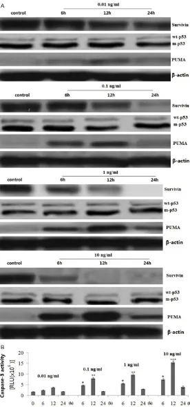
[image:4.612.92.518.76.302.2]Figure 2. Effect of YM155 on survivin, p53 and PUMA expression and
caspase-3 activity. SCC9 cells were treated with 0.01, 0.1, 1 and 10 ng/
ml YM155 for 6, 12 and 24 h. A: Survivin, p53 and PUMA protein expres -sion was detected by western blot assay. B: Caspase-3 activity was detected by cleavage of the caspase-3 substrate. vs control, *P<0.05, **P<0.01 and ***P<0.001.
tion of apoptosis induced by
YM155. SCC9 cells were first
treated with 0.01, 0.1, 1 and 10 ng/ml YM155 for 24 hs. After treatment, the degree of apoptosis was measured. The induction of apoptosis was found to be
dose-depen-dent by flow cytometry assay
(P<0.05 and P<0.01, Figure 1C) and TUNEL assay (P< 0.05 and P<0.01, Figure 1D). These results provided con-vincing data showing that YM155 could induce apopto-sis in SCC-9 cells.
We also treated the SCC9
cells with 0.01, 0.1, 1 and 10 ng/ml YM155 for 6, 12 and 24 hs. The results show- ed that YM155 resulted in cell growth inhibition and in- creased apoptosis in a time-dependent manner (data not shown).
YM155 inhibits survivin and induces PUMA/caspase-3 upregulation
SCC9 cells were treated with 0.01, 0.1, 1 and 10 ng/ml YM155 for 6, 12 and 24 h.
Survivin and PUMA expres -sion was detected by western blot assay. Caspase-3 activity was measured by cleavage of the caspase-3 substrate. The results showed that survivin was overexpressed in the SCC9 cells, and YM155 treat-ment caused survivin
inhibi-tion and PUMA upregulainhibi-tion
in dose- and time-dependent manner (Figure 2A). Activa- tion of caspase-3 was also in dose- and time-dependent manner (Figure 2B),
indicat-ing the specificity of the apop -totic effect of YM155 by cas-pase activation in SCC9 cells.
PUMA was regulated through
p53-dependent and
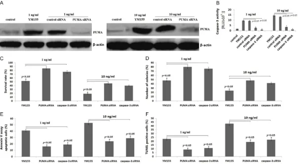
[image:5.612.94.370.72.662.2]Figure 3. Effect of PUMA/caspase-3 on YM155-induced apoptosis and growth inhibition of SCC9 cells. SCC9 cells were transfected with PUMA siRNA or caspase-3 siRNA for 16 h, then treated the transfected cells with YM155 (1 and 10 ng/ml) for 24 h. A: PUMA protein expression was detected by western blot assay; B: Cas -pase-3 activity was detected by cleavage of the cas-pase-3 substrate. C: Cell survival rate was detected by MTT assay; D: Colony formation assay; E: Apoptosis was
next investigated whether p53 was regulated by YM155/Survivin. As shown in Figure 2A, treatment with 0.01, 0.1, 1 and 10 ng/ml YM155 for 6, 12 and 24 h in the SCC9 cells did not affect wt-p53 expression, suggesting
that PUMA was p53-independent regulation
by YM155 treatment.
We also found that treatment with YM155 (10
and 1 ng/ml) for 24 h resulted in completely survivin inhibition. In addition, treatment with
YM155 rapaidly increased the PUMA expres -sion in the SCC9 cells at 6 h, and reached the
highest level at 12 h, and significantly
de-creased at 24 h (Figure 2A). In addition, cas-pase-3 activity was also in the highest level at
12 h, significantly decreased at 24 h (Figure 2B). The reason of why PUMA/caspase-3
ex-pression was downregulated after 24 h is not clear.
YM155 induces apoptosis and inhibits growth of SCC9 cells through PUMA/caspase-3 activa-tion
As shown in Figure 2A, treatment with 1 and 10 ng/ml of YM155 for 24 h could completely inhibit survivin expression. So we selected 1 and 10 ng/ml of YM155 for further study. To
confirm the mechanism responsible for
YM155-mediated apoptosis and growth inhibition, we
transfected PUMA siRNA or caspase-3 siRNA
or control siRNA into SCC9 cells, then treat- ed the transfected cells with YM155 (1 and 10 ng/ml) for 24 h. As shown in Figure 3A, tar-
geting PUMA by PUMA siRNA transfection in-hibited YM155-induced PUMA expression and
caspase-3 activation (Figure 3B). In addition, targeting caspase-3 by caspase-3 siRNA trans-fection also inhibited YM155-induced cas-pase-3 activation (Figure 3B). Transfection of
SCC9 cells with PUMA siRNA or caspase-3 siRNA significantly reduced the ability of
YM-155 to induce cell apoptosis and inhibit grow- th in SCC9 cells (Figure 3C-F). The control
siRNA did not cause any significant change
in cell viability and cell apoptosis (data not
shown). Although PUMA siRNA or caspase-3 siRNA significantly reduced the ability of YM155
to induce cell apoptosis and growth inhibition,
PUMA siRNA or caspase-3 siRNA could not
completely revered the effect of YM155, sug-gesting that the apoptosis-inducing effect by YM155 is partly mediated through the
activa-tion of PUMA/caspase-3 pathway.
YM155 treatment inhibited growth of SCC9 xenograft tumor in nude mice
To confirm the anti-tumor effects of YM155 in vivo, we used a SCID mouse xenograft model. Animals bearing SCC9 tumors were adminis-tered subcutaneously as a 3-day per week con-tinuous infusion for 2 weeks using the Alzet Osmotic Pump® (Model 1003D). Animals
treat-ed with YM155 showtreat-ed more than 60% tumor growth inhibition compared to the controls (Figure 4A). TUNEL assay showed the more
apoptotic cells was found in YM155 treated tumors (P<0.01, Figure 4B). In addition, Survivin
expression was inhibited and PUMA
express-ion was increased with YM155 treatment by immunohistochemistrical assay (Figure 4C,
4D).
Discussion
Survivin is one of the most frequently over-expressed genes in all types of cancer. In- creased survivin expression in cancer patients is an unfavorable prognostic marker correlat- ing with decreased overall survival in several malignancies, including pancreatic cancer [24], breast carcinomas [25], non-small cell lung [26], colorectal, hepatocellular carcinoma [27] and neuroblastoma [28]. Increased survivin expression was also associated with increased risk of recurrence, lymph node invasion and metastasis. Survivin has been shown to inhibit cell apoptosis and promote cell proliferation and angiogenesis, all of which make survivin a potentially attractive target for therapy [29]. In OSCC, survivin was found to be strik-ingly increased in malignant cells, and incre- ased survivin expression was also associated with bad prognosis [30]. Therefore, targeting
survivin is promisingly beneficial for OSCC
therapies.
Several strategies have been used to suppress survivin expression [31]. One approach is to use antisense oligonucleotides or siRNA to knock down survivin expression, resulting in cell growth arrest and increased apoptosis in a broad range of tumor cell lines [32]. YM155
is a survivin suppressant identified in a
YM155 suppressed in vivo SCC9 cell growth by inducing apoptosis without apparent body weight loss (data not shown). Our results strongly suggest that survivin expression con-tributes to human SCC9 tumor progression, and that survivin inhibition by YM155, a novel survivin suppressant, provides an antitumor effect on human SCC9 cells via induction of apoptosis.
To further explore the mechanism of YM155-induced cell apoptosis and growth inhibition,
possible proteins involved were detected in
vitro and in vivo by Western blot. Previous data
suggest that survivin depletion triggers p53 activation and sensitizes cancer cells to of PARP inhibition [33]. In our study, YM155 treat-ment did not affect wt-p53 expression in the SCC9 cells, suggesting that there was no rela-tion between p53 and YM155/survivin-induced apoptosis.
PUMA as a BH3-only Bcl-2 family protein that
[image:8.612.94.520.78.517.2]plays an essential role in p53-dependent and Figure 4. YM155 inhibits xenograft growth of SCC9 cells. A: Tumor xenografts were established by s.c. injection of
SCC9 cells into the flanks of the mice. Animals bearing SCC9 tumors were administered subcutaneously as a 3-day
per week continuous infusion for 2 weeks. Tumor size was measured every two days. The tumor growth curve was shown. *P<0.05 compared with the control group. B: Apoptotic cells was detected by TUNEL assay, P<0.01. C:
Sur-vivin was detected by immunohistochemistrical staining assay. D: PUMA was detected by immunohistochemistrical
-independent apoptosis [18, 19]. In the present
study, the expression of PUMA was
up-regu-lated in SCC9 cells when treated with YM155
for 6-24 h. Therefore, PUMA upregulation was
p53-independent. To assess whether YM155-induced apoptosis and growth inhibition was
PUMA-dependent, SCC9 cells was transfected with PUMA siRNA to knockdown of PUMA expression, only to find YM155-induced apop -tosis and growth inhibition was partly rever-
sed with PUMA inhibition. We propose that
the mechanism of YM155-induced apoptosis may be attributable to the down-regulation of
survivin as well as the upregulation of PUMA.
Because survivin inhibits apoptosis through both direct and indirect inhibitions of
cas-pase-3, a downstream gene of PUMA. We
next investigated whether YM155/survivin inhi-bition could upregulate caspase-3 activity. In the present study, increases of caspase-3 activity by YM155 treatment were observed,
which was consistent with the results of PUMA,
demonstrating YM155-induced apoptosis and growth inhibition may be partly due to inhibi-
tion of survivin/PUMA/caspase-3 pathway.
Although apoptosis provoked by
YM155/sur-vivin depended on the activation of PUMA/
caspase-3 signal, but how survivin regulated
PUMA/caspase-3 has remained unknown.
Studies have reported that targeting the Ras/
Raf/MEK/ERK pathway induces
PUMA-depen-dent apoptosis in cancer cells irrespective of
p53 status [34, 35], suggesting that PUMA was negatively regulated by ERK signals. Wang
et al. has reported that YM155-induced auto- phagy and cell death in vivo was correlated with the suppression of survivin/Erk1/2 and
S6 activation [36]. We supposed in the
pre-sent study that YM155/survivin might down-regulate ERK1/2 signal, resulting in
upregula-tion of PUMA/caspase-3 expression and
acti-vation. The hypothesis need further
investiga-tion in the further. We also demonstrated in
our study although YM155 induced apoptosis
and growth inhibition, targeting PUMA or cas -pase-3 could partly revevse the function of YM155, suggesting that some other proapop-totic signals may take part in YM155/survivin-induced apoptosis and growth inhibition of SCC9 cells.
It is interesting that in our study PUMA/cas -pase-3 expression reached the peak at 12 h
with YM155 treatment, and was dramatically
decreased at 24 h. The cause of why PUMA/
caspase-3 expression was downregulated at 24 h was not clear. A previous study has re- ported that activation of ERK/Slug signal by
Cytarabine contributed to the PUMA downre-gulation in HL-60 cells after 48 h [37]. Whether
some anti-apoptotic signals which inhibited
PUMA upregulation were induced by YM155
or YM155/survivin in our study need further investigation.
Conclusions
In summary, we presented experimental evi-dence, which strongly supports the antitumor effects of YM155 in SCC9 cells in vitro and in vivo. Thus, we believe that YM155 could po- tentially be an effective therapeutic agent for the inactivation of survivin and activation of
PUMA/caspase-3 signal, resulting in the
in-duction of cell apoptosis and inhibition of cell growth. Our study suggests that YM155 re- present a promising novel agent that should be developed for the treatment of OSCC.
Acknowledgements
This study was granted from the National Na- ture Scientific Research Fund (No: 82350- 713). This research was supported by the National Natural Science Foundation of China (NSFC, No: 82350713).
Disclosure of conflict of interest
None.
Address correspondence to: Xinghua Yang, School
of Dentistry and Oral Health, Shandong University,
Shandong, China. E-mail: yangxinghuakq@gmail. com
References
[1] Kademani D. Oral cancer. Mayo Clin Proc 2007; 82: 878-887.
[2] Petersen PE. Oral cancer prevention and
con-trol-the approach of the World Health
Orga-nization. Oral Oncol 2009; 45: 454-460. [3] Chen GS, Chen CH. A study on survival rates of
oral squamous cell carcinoma. Kaohsiung J Med Sci 2006; 12: 317-325.
[5] Deveraux QL, Reed JC. IAP family proteins--sup -pressors of apoptosis. Genes Dev 1999; 13: 239-252.
[6] Altieri DC. Validating survivin as a cancer ther-apeutic target. Nat Rev Cancer 2003; 3: 46-54.
[7] Lo Muzio L, Pannone G, Staibano S, Mignogna MD, Rubini C, Mariggiò MA, Procaccini M, Ferrari F, De Rosa G, Altieri DC. Survivin ex-pression in oral squamous cell carcinoma. Br J Cancer 2003 ; 89: 2244-2248.
[8] Dohi T, Okada K, Xia F, Wilford CE, Samuel T, Welsh K, Marusawa H, Zou H, Armstrong R,
Matsuzawa S, Salvesen GS, Reed JC, Altieri DC. An IAP-IAP complex inhibits apoptosis. J Biol Chem 2004; 279: 34087-34090.
[9] Nakahara T, Kita A, Yamanaka K, Mori M, Amino N, Takeuchi M, Tominaga F, Kinoyama I, Matsuhisa A, Kudou M, Sasamata M. Broad spectrum and potent antitumor activities of YM155, a novel small-molecule survivin sup-pressant, in a wide variety of human cancer cell lines and xenograft models. Cancer Sci 2011; 102: 614-621.
[10] Minematsu T, Iwai M, Sugimoto K, Shirai N,
Nakahara T, Usui T, Kamimura H.
Carrier-mediated uptake of YM155 monobromide, a novel small-molecule survivin suppressant, into human solid tumor and lymphoma cells. Drug Metab Dispos 2009; 37: 619-628. [11] Chen J, Pise-Masison CA, Shih JH, Morris JC,
Janik JE, Conlon KC, Keating A, Waldmann TA.
Markedly additive antitumor activity with the combination of a selective survivin suppres-sant YM155 and alemtuzumab in adult T-cell leukemia. Blood 2013; 121: 2029-2037. [12] Mir R, Stanzani E, Martinez-Soler F, Villanueva
A, Vidal A, Condom E, Ponce J, Gil J, Tortosa A, Giménez-Bonafé P. YM155 sensitizes ovarian cancer cells to cisplatin inducing apoptosis and tumor regression. Gynecol Oncol 2014; 132: 211-220.
[13] Koike H, Nitta T, Sekine Y, Arai S, Furuya Y, Nomura M, Matsui H, Shibata Y, Ito K, Oyama T, Suzuki K. YM155 reverses rapamycin resis-tance in renal cancer by decreasing survivin. J Cancer Res Clin Oncol 2014; 140: 1705-1713. [14] Zhang L, Zhang W, Wang YF, Liu B, Zhang WF,
Zhao YF, Kulkarni AB, Sun ZJ. Dual induction of apoptotic and autophagic cell death by target-ing survivin in head neck squamous cell carci-noma. Cell Death Dis 2015; 6: e1771.
[15] Sachita K, Yu HJ, Yun JW, Lee JS, Cho SD. YM155 induces apoptosis through
downregu-lation of specificity protein 1 and myeloid cell
leukemia-1 in human oral cancer cell lines. J Oral Pathol Med 2015; 44: 785-791.
[16] Tang H, Shao H, Yu C, Hou J. Mcl-1 downregula-tion by YM155 contributes to its synergistic
anti-tumor activities with ABT-263. Biochem Pharmacol 2011; 82: 1066-1072.
[17] Wang YF, Zhang W, He KF, Liu B, Zhang L,
Zhang WF, Kulkarni AB, Zhao YF, Sun ZJ. Induction of autophagy-dependent cell death by the survivin suppressant YM155 in salivary adenoid cystic carcinoma. Apoptosis 2014; 19: 748-758.
[18] Jeffers JR, Parganas E, Lee Y, Yang C, Wang J,
Brennan J, MacLean KH, Han J, Chittenden T, Ihle JN, McKinnon PJ, Cleveland JL, Zambetti GP. Puma is an essential mediator of p53-de-pendent and -indep53-de-pendent apoptotic path-ways. Cancer Cell 2003; 4: 321-328.
[19] Villunger A, Michalak EM, Coultas L, Mullauer F, Bock G, Ausserlechner MJ, Adams JM, Strasser A. p53- and Drug-Induced Apoptotic Responses Mediated by BH3-Only Proteins Puma and Noxa. Science 2003; 302: 1036-1038.
[20] Yu J, Zhang L. PUMA, a potent killer with or without p53. Oncogene 2008; 27: S71-83. [21] Suzuki A, Ito T, Kawano H, Hayashida M,
Hayasaki Y, Tsutomi Y, Akahane K, Nakano T, Miura M, Shiraki K. Survivin initiates procas-pase 3/p21 complex formation as a result of interaction with Cdk4 to resist Fas-mediated cell death. Oncogene 2000; 19: 1346-1353. [22] Tamm I, Wang Y, Sausville E, Scudiero DA,
Vigna N, Oltersdorf T, Reed JC. IAP-family pro-tein survivin inhibits caspase activity and apoptosis induced by Fas (CD95), Bax, caspas-es, and anticancer drugs. Cancer Res 1998; 58: 5315-5320.
[23] Véquaud E, Desplanques G, Jézéquel P, Juin P, Barillé-Nion S. Survivin contributes to DNA re-pair by homologous recombination in breast cancer cells. Breast Cancer Res Treat 2016; 155: 53-63.
[24] Lee MA, Park GS, Lee HJ, Jung JH, Kang JH, Hong YS, Lee KS, Kim DG, Kim SN. Survivin
ex-pression and its clinical significance in pancre -atic cancer. BMC Cancer 2005; 5: 127. [25] Nassar A, Sexton D, Cotsonis G, Cohen C.
Survivin expression in breast carcinoma: cor-relation with apoptosis and prognosis. Appl Immunohistochem Mol Morphol 2008; 16: 221-226.
[26] Dai CH, Li J, Shi SB, Yu LC, Ge LP, Chen P. Survivin and Smac gene expressions but not livin are predictors of prognosis in non-small cell lung cancer patients treated with adjuvant chemotherapy following surgery. Jpn J Clin Oncol 2014; 5: 327-335.
[27] Augello C, Caruso L, Maggioni M, Donadon M, Montorsi M, Santambrogio R, Torzilli G, Vaira V, Pellegrini C, Roncalli M, Coggi G, Bosari S. Inhibitors of apoptosis proteins (IAPs)
-tocellular carcinoma. BMC Cancer 2009; 9: 125.
[28] Islam A, Kageyama H, Takada N, Kawamoto T, Takayasu H, Isogai E, Ohira M, Hashizume K, Kobayashi H, Kaneko Y, Nakagawara A. High expression of Survivin, mapped to 17q25, is
significantly associated with poor prognostic
factors and promotes cell survival in human neuroblastoma. Oncogene 2000; 19: 617-623.
[29] Pennati M, Folini M, Zaffaroni N. Targeting survivin in cancer therapy. Expert Opin Ther Targets 2008; 12: 463-476.
[30] Xie S, Xu H, Shan X, Liu B, Wang K, Cai Z. Clinicopathological and prognostic significance
of survivin expression in patients with oral-squamous cell carcinoma: evidence from a meta-analysis. PLoS One 2015; 10: e0116517. [31] Ryan BM, O’Donovan N, Duffy MJ. Survivin: a
new target for anti-cancer therapy. Cancer Treat Rev 2009; 35: 553-562.
[32] Lares MR, Rossi JJ, Ouellet DL. RNAi and small interfering RNAs in human disease therapeutic applications. Trends Biotechnol 2010; 28: 570-579.
[33] Véquaud E, Desplanques G, Jézéquel P, Juin P, Barillé-Nion S. Survivin contributes to DNA re-pair by homologous recombination in breast cancer cells. Breast Cancer Res Treat 2016; 155: 53-63.
[34] Chen D, Wei L, Yu J, Zhang L. Regorafenib in-hibits colorectal tumor growth through PUMA -mediated apoptosis. Clin Cancer Res 2014; 20: 3472-3484.
[35] Dudgeon C, Peng R, Wang P, Sebastiani A, Yu J, Zhang L. Inhibiting oncogenic signaling by sorafenib activates PUMAvia GSK3β and NF-κB to suppress tumor cell growth. Oncogene 2012; 31: 4848-4858.
[36] Wang YF, Zhang W, He KF, Liu B, Zhang L,
Zhang WF, Kulkarni AB, Zhao YF, Sun ZJ. Induction of autophagy-dependent cell death by the survivin suppressant YM155 in salivary adenoid cystic carcinoma. Apoptosis 2014; 19: 748-758.
[37] Liu GJ, Pan GJ, Wang J, Wang LN, Xu Y, Tang Y.