Original Article
Effects of high-fat diet on the morphological
characteristics of cerebral microvasculature
without hyperlipidemia in Wistar rat
Yin He1*, Dong Han2*, Maojing Shi3, Zhifu Sun4, Jiayi Xie2, Jihong Zhu1, Yuansheng Liu1
1Department of Emergency, Peking University People’s Hospital, 11 South Xi Zhi Men Street, Xicheng District,
Beijing 100044, The People’s Republic of China; 2National Center for Nanoscience and Technology, Beijing, The
People’s Republic of China; 3Department of Emergency, Peking University International Hospital, Beijing 100044,
The People’s Republic of China; 4Otorhinolaryngology Head and Neck Surgery, Beijing Anzhen Hospital, Capital
Medical University, Beijing, The People’s Republic of China. *Equal contributors.
Received May 19, 2016; Accepted August 1, 2016; Epub November 1, 2016; Published November 15, 2016
Abstract: A high-fat diet (HFD) is associated with cerebrovascular disorders, however; its effect on the microvascu-lature in the brain is unknown. The aim of this study was to investigate the cerebral microvessel pathology by scan-ning electron microscope. Ten male Wistar rats were randomly divided into two groups: standard diet (n=5) and HFD (n=5). All of the rats were fed either standard rodent chow diet or HFD for 10 weeks and the brains were examined for microvascular pathology at the end of experiment. Coronal sections of a region of the cortex were observed by employing scanning electron microscope. Cerebral microvessels in the standard diet group exhibited the luminal en-dothelial surface with microvilli, indentations, and occasional vacuoles. The microvessels in the HFD group showed varying degrees of endothelial cell pathology including altered nuclear swelling as well as degenerative changes in the luminal wall. There was also thickening of the vascular basal laminae and expanded perivascular spaces often filled with amorphous debris. On a HFD, the luminal endothelial surface had more microvilli, indentations, and vacu-oles. A HFD can affect the morphology of cerebral microvasculature and arteries in the Wistar rat in the absence of hyperlipidemia and lead to a series of pathological changes and microvascular remodeling, which can increase the likelihood of blood flow retardation and platelet aggregation, and ultimately thrombosis. The effects of HFD on microvasculature is more serious than that on large and medium-sized artery. Scanning electron microscope is a tool for studying microvasculature and artery pathology.
Keywords: High-fat diet, microvasculature, cerebrovascular disorder, hyperlipidemia, scanning electron micro-scope
Introduction
Hyperlipidemia, an established risk factor for cardiovascular diseases, is also implicated in cerebrovascular disorders [1]. It is well estab-lished that hyperlipidemia can cause athero-sclerosis in medium to large sized arteries; however, its role on cerebral microvasculature is less studied [2]. There is a growing body of evidence that hypercholesterolemia also leads to microvascular dysfunction long before the appearance of atherosclerotic lesions in large vessels and that the altered microvascular function during hypercholesterolemia produces exaggerated tissue injury responses to isch-emia and reperfusion [3]. RL Reddick et al
and vacuoles, and so on. Through bisecting the blood vessels, the vascular muscle layer can be observed to determine whether there is vascu-lar muscle layer thickening, muscuvascu-lar fibrosis with or without muscle cell hypertrophy, athero-matous plaque, or calcification plaque.
The microvasculature, a network of blood ves-sels less than 100 μm in diameter, is a part of circulation and its main function is to transport oxygen and nutrients, hormones, and metabol-ic products through the exchange of circulating blood and parenchyma cells [6]. According to anatomical features and the direction of the blood flow, the microvasculature is subdivided into arterioles, capillaries, and venules. Adequ- ate blood flow in capillaries can ensure the nor-mal perfusion and organ function. Capillaries consist of a single layer of epithelium and a basement membrane, which allow the exchange of molecules between blood and tissues. En- dothelial dysfunction (ED) is a disorder, which is defined as reduction of bioavailability of vasodi-lators, mainly nitrous oxide, and is character-ized by a state of endothelial activation where a pro-inflammatory, proliferative and pro-coagu-lant milieu is predominant [7]. It is an early stage of the occurrence and development of atherosclerosis [8].
However, at present there are few reports on the effects of hyperlipidemia for cerebral micro-vasculature, and even fewer studies on the effect of a high-fat diet on the cerebral micro-vasculature. Based on the previous studies, in our research, the hypothesis is that without hyperlipidemia, high-fat diet can damage the cerebral microcirculation. The aim of this study was to investigate the effects of high-fat diet on morphological characteristics of the cerebral microvasculature and vasculature in the Wistar rat in the absence of hyperlipidemia through scanning electron microscopy.
Materials and methods
Dietary and environmental manipulations
Male Wistar rats were purchased from Vital River Laboratories. All procedures were in accordance with the Guide for Care and Use of Animal Experimentation of the Peking University People’s Hospital. All rats were randomly assigned to one of two groups: standard diet (n=5) or high-fat diet (n=5). The standard diet
rats were fed a chow (Research Diets, D12451) that contained 11.2% fat, 55.5% carbohydrate, and 33.3% protein (kcal); whereas the high-fat diet rats received a chow which contained 45.2% fat, 28.6% carbohydrate, and 26.2% protein (kcal). The high-fat diet was high calorie food (high-fat diet =4.5 kcal/g versus standard diet =3.3 kcal/g). All rats were housed in indi-vidual cages in a climate-controlled room with purified air (23 ± 3°C; 60 ± 5% relative humidi-ty) with a 12 h light/dark cycle (lights on at 6:00 am). The rats in each group were fed the indi-cated diet with sufficient water and food ad libi-tum for 10 consecutive weeks. Food tion was calculated daily; while water consump-tion was evaluated once a week.
Measurement the levels of serum index analy-sis
After the experimental period of 10 weeks, the rats were sacrificed and blood samples were collected and centrifuged at 3,000 rpm at 4°C for 15 min and the supernatants were stored at -70°C for biochemical analysis. The levels of tri-glyceride (TG), total cholesterol (TC), low-densi-ty lipoprotein cholesterol (LDL-C) and high-den-sity lipoprotein cholesterol (HDL-C) in the serum were measured by the enzymatic colorimetric method using commercial reagents assay kits (Beijing Biosino Bio-technology & Science Inc, China) according to the manufacturer’s instru- ctions.
Scanning electron microscope
were cut using a region of the cortex. The speci-mens were then observed through scanning electron microscope Philips XL-30 FEG at 10 kV.
Statistical analysis
The serum triglyceride (TG), total cholesterol (TC), low-density lipoprotein cholesterol (LDL-C), and high-density lipoprotein cholesterol (HDL-C) in the standard diet groups and the HFD group were collected, and the levels (means ± SD for three independent experi-ments) were analyzed. Statistical significance of observed differences among these groups was calculated using a two-tailed unpaired Student’s t test. A p value of less than 0.05 was considered to be statistically significant. The statistical calculations were performed with SPSS 13.0 for Windows (SPSS, Inc, Chicago, IL, USA).
Results
The levels of serum index
We analyzed the values of triglyceride (TG), total cholesterol (TC), low-density lipoprotein cholesterol (LDL-C), and high-density lipopro-tein cholesterol (HDL-C) in the serum and observed no statistically significant differences between standard and high-fat diet (HFD) chows (P>0.05) (data not shown).
HFD changes cerebral microvessel morpho-logical characteristics
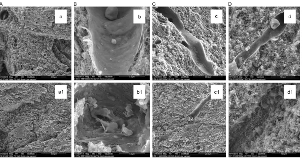
Microvessels of a defined lumen diameter were included in the analysis (rodent: d<7 μm, human d<12 μm). In the HFD group, we observed the reduction of functional capillary density and microvascular rarefaction (Figure 1 a versus
a1) with a significant increase in macromolecu-lar permeability and cerebral microvessels exhibiting finger-like endothelial processes pro-truding the vessel lumen (microvilli). Further- more, the luminal endothelial surface appeared to have more microvilli, indentations, and vacu-oles compared with the standard diet group (Figure 1 b versus b1, d versus d1). Additional changes observed in the HFD group include microthrombus caused by red blood cell and platelet aggregation formed in the microvascu-lature, thinner vascular walls, more pronounced gaps among vascular endothelial cells, and
vis-ible binding of collagen fibers in the capillary lumen that results in luminal stenosis (Figure 1C).
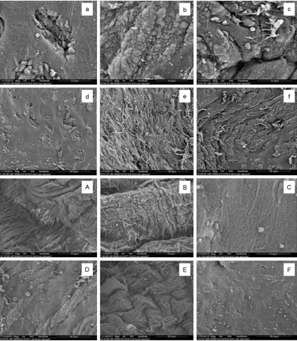
At the ultrastructural level, microvessels in the HFD group showed varying degrees of endothe-lial cell pathology including altered nuclear swelling as well as degenerative changes in the luminal wall. We also observed increased thick-ness of the vascular basal laminae and expand-ed perivascular spaces often fillexpand-ed with amor-phous debris. The luminal endothelial surface appeared to contain more microvilli, indenta-tions, and vacuoles in the HFD group (Figure 2).
Cerebral vascular
In contrast with the standard diet, a high fat diet can lead to cerebral vascular pathology, including arterial wall thickening, stiffness, and loss of elasticity. Furthermore, HFD leads to stenosis, a rougher cerebral vascular endothe-lial surface, which would result in hemodynam-ic change and increase the risk of mhemodynam-icrocircula- microcircula-tion blood clots, with more microvilli, indenta-tions, occasional vacuoles, and more frequent and wider gaps between endothelial cells. In addition, more folds were present on chamber surface with the HFD than the standard diet. At the ultrastructural level, the HFD cerebral vas-culature had more apparent nuclear swelling and a rougher, disordered surface as well as increased collagen fibers, scattered lipid drops and thrombocytes; however, the cerebral vas-culature of the standard diet group appeared smoother and neater (Figure 2).
Discussion
including the thickening of the vascular basal
[image:5.612.90.522.71.567.2]ty between blood and vessel wall, secretion of effector molecules, as well as regulation of blood vessel tone and blood flow, but also par-ticipates in the processes of inflammation, immunity, thrombogenesis, and angiogenesis [9-12]. All endothelial pathology can increase the likelihood of blood flow retardation and platelet aggregation, and ultimately thrombo-sis. Furthermore, the HFD group has reduced functional capillary density and microvascular rarefaction compared with the standard diet group. Before hyperlipidemia, microvascular remodeling and rarefaction occurs in brain tis-sue (Figure 2). Oxidative stress, endothelial dysfunction, and apoptosis are possible mech-anisms implicated on microvascular rarefac-tion [13].
Previous research also showed that a high fat diet can lead to microcirculation dysfunction, including endothelial dysfunction characterized by blunted vasodilatatory responses and increased shear stress by raising blood flow, reduction of functional capillary density and microvascular rarefaction, a significant aug-mentation of macromolecular permeability as well as prostanoid-mediated vasoconstriction in conductance vessels of skeletal muscles [14]. Increased cholesterol levels induced by a high-fat diet activate a number of cerebral vas-cular events, including oxidative stress, endo-thelial dysfunction, blood-brain barrier distur-bances and vascular inflammation, which as a result worsens stroke mortality [15]. Actually, an obvious induction of CYP2E1 was caused by hyperglycemia in the HFD rats combined with cerebral ischemia-reperfusion injury [16]. Con- sequently, the induction of CYP2E1 oxidizes mitochondrial DNA, proteins, and lipids and triggers pro-inflammatory cytokines, such as TNF-α, IL-1, IL-6, TGF-β, which further increases mitochondrial ROS (reactive oxygen species) [16, 17]. On the other hand, oxidative low-den-sity lipoprotein (ox-LDL) could enhance the expression of multiple adhesive factors and cytokines in cells through activating the vascu-lar endothelial cells. Furthermore, ox-LDL could significantly elevate the level of TNF-α, IL-6, and sICAM-1 in the supernatant after the interven-tion with ox-LDL [18]. Which will lead to endo-thelial injury and increase the risk of athero-sclerosis. Besides, oxidative stress can lead to severe and immediate damage to cellular pro-teins, DNA, and lipids by causing overproduc-tion of free radicals that overwhelms the
detoxi-fication capacity of cellular antioxidant enzymes [19]. Other research showed that hyperlipid-emia could exacerbate cerebral ischhyperlipid-emia- ischemia-reperfusion (I/R)-induced injury by the synergis-tic effect on CYP2E1 induction, as a result, reactive oxygen species formation, oxidative stress, inflammation, and neuronal apoptosis by coexistence of hyperlipidemia and cerebral I/R have been induced [20]. At present, there has not been related research on effects of a high-fat diet on morphological characteristics of cerebral microvasculature in the absence of hyperlipidemia. The purpose of this experiment was to approach the subject. A HFD increases O-GlcNAc-modified proteins which contribute to intensifying contractile responses and cerebro-vascular dysfunction [21]. Hyperlipidemia is best known for its role in atherosclerotic vascu-lar disease, a process that is the most appar-ent in larger arteries such as the aorta or the coronary arteries [4]. However, the processes involved in atherosclerosis (arteriosclerosis affecting arteries) may be somewhat different to those in arteriolosclerosis (arteriosclerosis affecting smaller arteriolar branches) and the relationship between dyslipidaemia and arteri-olosclerosis is less clear. There is some evi-dence that hypercholesterolemia may also cause both functional and structural changes in the peripheral microvasculature [22]. Chole- sterol is generally not thought to cause pathol-ogy in the microvasculature. In particular, how a high-cholesterol diet induces microvascular pathology in brain is not known since arterial lesions develop in apoE-deficient mice fed regu-lar mouse chow.
much as that of the white matter. The endothe-lial cells are surrounded by a 30- to 40-nm-thick basement membrane, which is often a target of investigation due to frequently obse- rved malformations under pathophysiological conditions [25]. While we observed increased microvessel diameter, microvascular density and length were not consistently affected. However, degenerative changes and thickened vascular basement membranes were present ultrastructurally [4]. In the cerebral cortex of wild-type mice, the vascular walls of microves-sels are composed of flat endothelium and smooth muscle cells rich in fine myofilaments [26]. Oh Young Bang et al showed that cerebral micro- and macroangiopathy often coexist and share common risk factors, including athero-sclerosis [27]. Obesity and high fat intake could cause functional and structural changes in the vasculature [28-32]. High intake of fat in the presence or absence of obesity can also lead to vascular dysfunction [33, 34].
Taken together, a high-fat diet can change the morphology of cerebral microvasculature and arteries in Wistar rat in the absence of hyperlip-idemia. Additionally, it can lead to a series of pathological changes and microvascular remo- deling which can increase the likelihood of blood flow retardation and platelet aggregation, and ultimately thrombosis. This causes increas- ed incidence of related cerebrovascular dis-ease. Furthermore, the effects of a high-fat diet are more serious on the microvasculature than that on large and medium-sized artery.
Acknowledgements
We thank the National Natural Science Fo- undation of China for valuable support [NO. 11372012].
Disclosure of conflict of interest
None.
Address correspondence to: Yuansheng Liu, De- partment of Emergency, Peking University People’s Hospital, 11 South Xi Zhi Men Street, Xicheng District, Beijing 100044, The People’s Republic of China. E-mail: [email protected]
References
[1] Constantinescu E, Safciuc F and Sima AV. A hy-perlipidemic diet induces structural changes in
cerebral blood vessels. Curr Neurovasc Res 2011; 8: 131-144.
[2] Franciosi S, Gama Sosa MA, English DF, Oler E, Oung T, Janssen WG, De Gasperi R, Schmeidler J, Dickstein DL, Schmitz C, Gandy S, Hof PR, Buxbaum JD and Elder GA. Novel cerebrovas-cular pathology in mice fed a high cholesterol diet. Mol Neurodegener 2009; 4: 42.
[3] Ishikawa M, Stokes KY, Zhang JH, Nanda A and Granger DN. Cerebral microvascular respons-es to hypercholrespons-esterolemia: rolrespons-es of NADPH oxidase and P-selectin. Circ Res 2004; 94: 239-244.
[4] Reddick RL, Zhang SH and Maeda N. Athero-sclerosis in mice lacking apo E. Evaluation of lesional development and progression. Arterio-scler Thromb 1994; 14: 141-147.
[5] Rodriguez-Baeza A, Reina-de la Torre F, Poca A, Marti M and Garnacho A. Morphological fea-tures in human cortical brain microvessels af-ter head injury: a three-dimensional and im-munocytochemical study. Anat Rec A Discov Mol Cell Evol Biol 2003; 273: 583-593. [6] Lymphatic system and microcirculation.
Pro-ceedings of the Third International Symposium on Daflon 500 held during the First Mediterra-nean Congress of Angiology. Corfu, Greece, May 31, 1988. Int Angiol 1989; 8: 1-75. [7] Anderson TJ. Assessment and treatment of
en-dothelial dysfunction in humans. J Am Coll Car-diol 1999; 34: 631-638.
[8] Hirase T and Node K. Endothelial dysfunction as a cellular mechanism for vascular failure. Am J Physiol Heart Circ Physiol 2012; 302: H499-505.
[9] Ross R. Atherosclerosis--an inflammatory dis-ease. N Engl J Med 1999; 340: 115-126. [10] Lusis AJ. Atherosclerosis. Nature 2000; 407:
233-241.
[11] Falk E. Morphologic features of unstable ath-erothrombotic plaques underlying acute coro-nary syndromes. Am J Cardiol 1989; 63: 114E-120E.
[12] Sumpio BE, Riley JT and Dardik A. Cells in fo-cus: endothelial cell. Int J Biochem Cell Biol 2002; 34: 1508-1512.
[13] DeLano FA, Parks DA, Ruedi JM, Babior BM and Schmid-Schonbein GW. Microvascular dis-play of xanthine oxidase and NADPH oxidase in the spontaneously hypertensive rat. Microcir-culation 2006; 13: 551-566.
[14] Costa RR, Villela NR, Souza M, Boa BC, Cyrino FZ, Silva SV, Lisboa PC, Moura EG, Barja-Fidal-go TC and Bouskela E. High fat diet induces central obesity, insulin resistance and micro-vascular dysfunction in hamsters. Microvasc Res 2011; 82: 416-422.
[16] Tiku ML, Liesch JB and Robertson FM. Produc-tion of hydrogen peroxide by rabbit articular chondrocytes. Enhancement by cytokines. J Immunol 1990; 145: 690-696.
[17] Becker KJ. Targeting the central nervous sys-tem inflammatory response in ischemic stroke. Curr Opin Neurol 2001; 14: 349-353.
[18] Jiang YR, Miao Y, Yang L, Xue M, Guo CY, Ma XJ, Yin HJ, Shi DZ and Chen KJ. Effect of chinese herbal drug-containing serum for activating-blood and dispelling-toxin on ox-LDL-induced inflammatory factors’ expression in endotheli-al cells. Chin J Integr Med 2012; 18: 30-33. [19] Chan PH. Reactive oxygen radicals in signaling
and damage in the ischemic brain. J Cereb Blood Flow Metab 2001; 21: 2-14.
[20] Cao XL, Du J, Zhang Y, Yan JT and Hu XM. Hy-perlipidemia exacerbates cerebral injury thro- ugh oxidative stress, inflammation and neuro-nal apoptosis in MCAO/reperfusion rats. Exp Brain Res 2015; 233: 2753-2765.
[21] Lima VV, Giachini FR, Matsumoto T, Li W, Bres-san AF, Chawla D, Webb RC, Ergul A and Tostes RC. High-fat diet increases O-GlcNAc levels in cerebral arteries: a link to vascular dysfunction associated with hyperlipidaemia/obesity? Clin Sci (Lond) 2016; 130: 871-880.
[22] Leung H, Wang JJ, Rochtchina E, Wong TY, Klein R and Mitchell P. Dyslipidaemia and mi-crovascular disease in the retina. Eye (Lond) 2005; 19: 861-868.
[23] Song SH and Roach MR. Surface features of human aortic atherosclerosis as seen with scanning electron microscopy. Yonsei Med J 1998; 39: 430-438.
[24] Reina-De La Torre F, Rodriguez-Baeza A and Sahuquillo-Barris J. Morphological characteris-tics and distribution pattern of the arterial ves-sels in human cerebral cortex: a scanning elec-tron microscope study. Anat Rec 1998; 251: 87-96.
[25] Farkas E and Luiten PG. Cerebral microvascu-lar pathology in aging and Alzheimer’s disease. Prog Neurobiol 2001; 64: 575-611.
[26] Mato M, Ookawara S, Mashiko T, Sakamoto A, Mato TK, Maeda N and Kodama T. Regional difference of lipid distribution in brain of apoli-poprotein E deficient mice. Anat Rec 1999; 256: 165-176.
[27] Bang OY, Chung JW, Ryoo S, Moon GJ, Kim GM, Chung CS and Lee KH. Brain microangiopathy and macroangiopathy share common risk fac-tors and biomarkers. Atherosclerosis 2016; 246: 71-77.
[28] Wilde DW, Massey KD, Walker GK, Vollmer A and Grekin RJ. High-fat diet elevates blood pressure and cerebrovascular muscle Ca(2+) current. Hypertension 2000; 35: 832-837. [29] Li W, Prakash R, Chawla D, Du W, Didion SP,
Filosa JA, Zhang Q, Brann DW, Lima VV, Tostes RC and Ergul A. Early effects of high-fat diet on neurovascular function and focal ischemic brain injury. Am J Physiol Regul Integr Comp Physiol 2013; 304: R1001-1008.
[30] d’Uscio LV, Smith LA and Katusic ZS. Hypercho-lesterolemia impairs endothelium-dependent relaxations in common carotid arteries of apo-lipoprotein e-deficient mice. Stroke 2001; 32: 2658-2664.
[31] Osmond JM, Mintz JD, Dalton B and Stepp DW. Obesity increases blood pressure, cerebral vascular remodeling, and severity of stroke in the Zucker rat. Hypertension 2009; 53: 381-386.
[32] Kitayama J, Faraci FM, Lentz SR and Heistad DD. Cerebral vascular dysfunction during hy-percholesterolemia. Stroke 2007; 38: 2136-2141.
[33] Naderali EK, Brown MJ, Pickavance LC, Wilding JP, Doyle PJ and Williams G. Dietary obesity in the rat induces endothelial dysfunction with-out causing insulin resistance: a possible role for triacylglycerols. Clin Sci (Lond) 2001; 101: 499-506.