Int J Clin Exp Pathol 2016;9(12):12390-12398 www.ijcep.com /ISSN:1936-2625/IJCEP0040180
Original Article
Helicobacter pylori infection associated miR-451
suppressed the proliferation of gastric cancer
by targeting CASC4
Daping Xiao1*, Zhihua Yun2*, Ting Wu1, Jin Zhang1, Jie Shao1
1Department of Clinical Laboratory, 359 Hospital of PLA, Zhenjiang, Jiangsu Province, China; 2Department of
Clinical Laboratory, Wujin Hospital Affiliated to Jiangsu University, Changzhou, Jiangsu Province, China. *Equal
contributors.
Received September 18, 2016; Accepted September 27, 2016;Epub December 1, 2016; Published December 15, 2016
Abstract: Aberrant microRNA (miRNA) expression has been observed in gastric cancer (GC) but fragmentary infor-mation is available on the miRNA dysregulation occurring with each phenotypic change involved in gastric carci-nogenesis with Helicobacter pylori (H.pylori). In this study we mainly aimed to investigate the potential function of miRNA in GC patients with H.pylori. Based on the microarray screening, we found miR-451 was suppressed in the tumor tissues GC patients with H.pylori and was further confirmed in a larger sample size and GC cell lines. Ectopic expression of miR-451 dramatically suppressed cell proliferation in vitro. Bioinformatical analysis revealed that CASC4 might be the potential target gene of miR-451. We also found that miR-451 strongly reduced the expression of CASC4 oncogene in GC cells. In clinical specimens, CASC4 was over-expressed in tumors and H.pylori positive tissues. Univariate analysis indicated that low miR-451 expression in GC patients with H.pylori were significantly negative prognostic predictors for overall survival in patients. Taken together, our results indicated that miR-451 functions as a growth-suppressive miRNA in H.pylori related GC mediated mainly by repressing CASK expression and might act as a poor fingerprint for the prognosis.
Keywords: miRNA, CASC4, proliferation, prognosis, H.pylori
Introduction
Gastric cancer is the fourth most common can-cer and second leading cause of cancan-cer-related death worldwide, and about 90% of non-cardia GCs are the ultimate consequence of long-standing Helicobacter pylori (H.pylori) infection [1, 2]. Chronic H.pylori infection of the gastric epithelium is strongly associated with the development of gastritis, peptic ulcers, muco-sa-associated lymphoid tissue lymphoma and gastric cancer. Despite recent extensive inves-tigations on the molecular landscape of GC [3, 4], the molecular grounds for the various steps in H.pylori-related carcinogenesis have yet to be fully elucidated, and no consistent and reli-able biomarkers have become availreli-able for use in GC secondary prevention strategies [5-7]. MicroRNAs (miRNAs) are short, non-coding RNA molecules that regulate gene expression
bioin-miR-451 in
H.pylori
associated GC
formatic approaches and studying gastric epi-thelial cell lines, stomach tissue based on the high throughput screening. In this comprehen-sive study, we demonstrate that miR-451 is a critical miRNA, which regulates gastric epitheli-al cell proliferation by targeting potentiepitheli-al onco-genes. Furthermore, our work provides sub-stantial evidence for the causal involvement of miR-451 in predicting the prognosis of patients with gastric malignancies induced by chronic Hp infection.
Materials and methods
Study subjects
A total of 130 GC cases from The 359th Hospital of PLA and Wujin Hospital Affiliated to Jiangsu University were enrolled in this study. Patients were consecutively recruited between July
2012 and August 2015 at The 359th Hospital of PLA and Wujin Hospital Affiliated to Jiangsu University. All cases are incident ones during enrollment of the current case-control study. The diagnosis of all patients was histological confirmed. All participants have provided their written informed consents to participate in this study. This study was approved by the institu-tional Review Board of the 359th Hospital of PLA and Wujin Hospital Affiliated to Jiangsu University.
Cell culture and reagents
[image:2.612.93.514.68.411.2]Gastric cancer cell lines including HGC-27, MKN45, AGS, 7901, SNU-1 and MGC80-3 were purchased from the Chinese Academy of Sciences Cell Bank (Shanghai, China). All cells were cultured in Dulbecco modified Eagle me- dium (DMEM) purchased from Gibco (CA, USA)
miR-451 in
H.pylori
associated GC
supplemented with 10% fetal bovine serum (Invitrogen, Carlsbad, USA) and grown in humid-ified 5% CO2 at 37°C. The miR mimics, inhibitor and normal control were obtained from Genepharma (Shanghai, China). The transfec-tion was conducted by using Lipofectamine 2000 (Invitrogen Corp, CA, USA).
TaqMan low density array (TLDA)
In the screening stage, TLDA Chips (Life Technologies) was used to screen differentially expressed miRNAs from the four grouped sam-ples (five samsam-ples in each group). Total RNA was extracted from the tissues samples using the TRIzol Reagent. Megaplex RT reactions and pre-amplification reactions were run according to the manufacture’s protocol, in which 75-ul 0.16 * TE was added to PreAmp product, and 9-ul diluted PreAmp product was used to run the Real time Polymerase Chain Reaction (RT-PCR) reactions by dispensing 100 μl of the PCR reaction mix into each port of the TaqMan MicroRNA Array. The default PCR procedure was used, and the analysis was performed by using RQ manager software (Life Technologies).
Quantitative RT-PCR and Western blot analysis
Total RNA was extracted using Trizol (TAKARA, Japan). Levels of mature miR-451 were mea-sured using TaqMan MicroRNA Assay (Applied Biosystems, CA, USA) by normalizing to the lev-els of U6. SYBR Green PCR Kit (TAKARA, Japan) was used to quantify the mRNA levels of CASC4 by normalizing to GAPDH. The PCR reactions were performed and analyzed using ABI Step-one system. The relative expression ratio of miR-451 in paired tissues and cells was calcu-lated by the 2-ΔΔCT method. Western blots were performed as described previously. Briefly, total protein was separated on a precast 10% poly-acrylamide gel and blotted with antibodies for CASC4 (diluted 1:1000, cell signaling technolo-gy) and β-action (diluted 1:1000, cell signaling technology). Densitometric analysis of protein bands was performed via Image J software. Cell proliferation assays
[image:3.612.96.513.70.334.2]The ability of cells proliferation was assayed using CCK8 (Beyotime, Nantong, China) and EDU (Millipore, Massachusetts, America)
Figure 2.H.pylori infection associated miR-451 suppressed cell proliferation. A: Relative expression of miR-451 in different GC cell lines. B and C: miR-451 expression in MKN45 and 7901 cells infected with different MOIs of
miR-451 in
H.pylori
associated GC
according to the manufacturer’s instructions. The mock and infected cells were seeded at a density of 1 × 10⁴ cells/well in 96-well flat-bot-tom and respectively cultured for EDU assays and CCK8 assay.
Construction of luciferase-based reporter plasmids
The total fragment of the CASC4 3’UTR was amplified. The PCR production was cloned into the pGL3-promoter luciferase-based plasmid (Promega, CA, USA). The amplified fragment was verified by DNA sequencing.
Dual-luciferase reporter assay
For luciferase activity analysis, HGC-27 and SGC-7901 cells were co-transfected with 100
ng of luciferase reporter constructs 5 ng con-trol plasmid and 10 pmol of miRNAs with 1 µl lipofectamine 2000 according to the manufac-turer’s instructions (Invitrogen, NY, USA). After incubation for 48 h, we carried out the lucifer-ase assay using the luciferlucifer-ase reporter assay system (Promega, Madison, WI) according to the manufacturer’s protocol. Measurements of luminescence and absorbance of control plas-mid were performed on a luminometer (Glomax 20/20; Promega). Three independent experi-ments were performed in triplicate.
Statistical analysis
[image:4.612.92.512.71.442.2]The Mann-Whitney U-test or Fisher’s exact test was used for between-group comparisons where appropriate, and correlation between
miR-451 in
H.pylori
associated GC
the results obtained with the two different anal-yses determined with Spearman’s test. Follow-up studies of patients were performed until the time of writing or patient death. Cancer-specific survival outcomes were evaluated by applying the Kaplan-Meier method for all patients, except those who died from surgical
[image:5.612.90.512.72.534.2]complica-tions. The log-rank test was used to compare the prognostic significance of individual vari-ables on survival. Cox’s proportional hazards model was applied in multivariate analysis to identify independent predictors of survival. P-values<0.05 were considered statistically significant. Statistical analysis was performed
miR-451 in
H.pylori
associated GC
with SPSS 13.0 and SAS software (version 9.1.3; SAS Institute, Cary, NC, USA). The graphs were generated by Graphpad Prism 5.0 (Gra- phpad Software, Inc.).
Results
miR-451 was down-regulated in tumor tissues of H.pylori-positive gastric cancer patients.
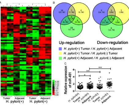
The TLDA microarray was applied to screen the dysregulated miRNAs in GC patients with H.pylori-positive or H.pylori-negative. As pre-sented in Figure 1A, each group contained five GC tumor tissues or corresponding adjacent tumor tissues. Clustering analysis revealed an aberrant different expression miRNA profile in these four groups. We further analyzed the dif-ferent expressed miRNAs in four groups by using Venny diagram by pairwise comparison. We found that only six miRNAs were upregulat-ed and four miRNAs were downregulatupregulat-ed with the following characteristics: 1) upregulated/ downregulated in H.pylori-positive GC tumor tis-sues comparing with H.pylori-negative GC tumor tissues; 2) upregulated/downregulated in H.pylori-positive GC adjacent tumor tissues comparing with H.pylori-negative GC adjacent tumor tissues; 3) upregulated/downregulated in H.pylori-positive GC tumor tissues comparing with H.pylori-positive adjacent GC tumor tis-sues; 4) with the cutoff 4/0.25 (Figure 1B). Among the ten candidate miRNAs, miR-451 presented the most significant, we next detect-ed the expression of miR-451 in a larger sam-ple size with 65 samsam-ples in each group, as
pre-451. Consequently, we chose MKN45 and 7901 cells for further functional studies. We then infected 7901 and MKN45 cells with dif-ferent MOIs of H.pylori (0, 1:1, 1:50, 1:100) and we found that miR-451 expression gradually decreased with increased MOIs (Figure 2B and
2C, P<0.05) indicating that miR-451 was down-regulated in H.pylori infected state and its down-regulated was significantly associated with GC progression.
Ectopic miR-451 inhibits growth GC cells in vitro
To explore the effect of miR-451 on cell growth, MKN45 and 7901 cells were transiently trans-fected with miR-451 mimic or inhibitor, respec-tively. The expression of miR-451 validated after mimic or inhibitor transfection was con-firmed. The results of CCK8 assay displayed that miR-451 significantly inhibited cell growth in 7901 cells and in MKN45 cells (P<0.05), whereas miR-451 inhibitor promoted cell growth in these two cells (P<0.05). By contrast, negative control (NC) or inhibitor NC had no effect on cell growth, indicating that the effect caused by miR-451 was specific (Figure 2D and
2E).
[image:6.612.90.374.74.223.2]We next used lentiviral vectors to stably restore the expression of miR-451 in MKN45 and 7901 cells and examined cell growth rate. We showed that the expression levels of miR-451 were increased in 7901 and MKN45 cells respec-tively in a dose-dependent manner and reached a very high level at MOI 100. Therefore, the
Figure 5.H.pylori infection induced miR-451 expression might be an ideal prognostic indicator of GC. The Kaplan-Meier curve for the overall survival of patients was presented by using different indicators as miR-451 expression combined with H.pylori infection.
sented in Figure 1C, and we found that miR-451 was aber-rant decreased in H.pylori-positive GC tumor tissues comparing with either H.py- lori-negative GC tumor tis-sues or H.pylori-positive GC adjacent tumor tissues. MiR-451 was decreased in H.pylori positive cells
In order to investigate the detailed function of miR-451, we first analyzed the expres-sion of miR-451 in multiple GC cell lines. As presented in
miR-miR-451 in
H.pylori
associated GC
same condition (MOI=100) was applied for fur-ther experiments. The growth inhibition induced by miR-451 overexpression was similar to that induced by miR-451 mimic transfection (Figure 3A). As demonstrated in EDU assay (Figure 2E), miR-451 overexpressed 7901 and MKN45 cells displayed much fewer and smaller stain compared with control groups (Figure 3B). CASC4 was the direct target of miR-451 in GC cells
To explore the mechanism of growth inhibition induced by miR-451, we predicted the potential target genes by using bioinformatical analysis. The following tools were employed including TargetScan, PicTar, miRTarget and miRBase. After screened by ranking index in different databases, we found that CASC4 might be the target gene of miR-451. We first detected the expression of CASC4 in GC patients, we found that CASC4 expression were significant inverse-ly with miR-451 with the highest expression in tumor tissues of H.pylori-positive GC patients (Figure 4A). We further performed luciferase reporter assay to determine whether miR-451 could directly target the 3’UTR of CASC4 in GC cells. The target sequence of CASC4 3’UTR (wt 3’UTR) or the mutant sequence (mut 3’UTR) was cloned into a luciferase reporter vector (Figure 4B). 7901 cells were then transfected with wt or mut 3’UTR vector and miR-451 mimic. Ectopic expression of miR-451 led to a dose-dependent decrease in CASC4 protein levels. Moreover, inhibition of endogenous miR-451 resulted in up-regulated expression of CASC4 in 7901 cells (Figure 4C). The luciferase reporter assay showed a significant decrease of luciferase activity when compared with miRNA control (Figure 4D). The activity of mut 3’UTR vector was unaffected by a simultane-ous transfection with miR-451. Moreover, co-transfection with miR-451 inhibitor and wt 3’ UTR vector in 7901 cells led to a 2-fold increase of luciferase activity. Taken together, all these results strongly suggested that CASC4 was a direct target of miR-451 in GC cells.
miR-451 predicted poor prognosis of GC patients
Since miRNA was reported to be indicator of various diseases recently, we also accessed miR-451 in GC patients with different H.pylori infection status. Assessment of the 95%
confi-dence interval (CI) in the healthy control group was the threshold from discriminating normal from suppressed systemic levels. The GC patients were divided into four groups accord-ingly: H.pylori-positive with miR-451 low (39 patients); H.pylori-positive with miR-451 high (32 patients); H.pylori-negative with miR-451 low (29 patients); H.pylori-negative with miR-451 high (30 patients). We assessed the 5-year survival rate in the four groups. The 5-year sur-vival rate in the H.pylori-positive with miR-451 low group was significantly lower than in the other three groups. The 5-year survival rate in the H.pylori-negative with miR-451 high was significantly higher than in the other three groups (Figure 5) indicating that miR-451 might be a poor predictor for the prognosis of GC patients together with H.pylori infection.
Discussion
GC is defined as cancer that forms in the tis-sues lining the stomach. Globally, GC is the fifth leading cause of cancer and the third leading cause of cancer mortality, comprising 7% of cases and 9% of deaths. In 2012 GC occurred in 950,000 people and caused 723,000 deaths [15, 16]. The most common cause is infection by H.pylori, which accounts for>60% of cases [17]. Most clinical evidence suggested that H.pylori infection is related to GC, but the underlying molecular mechanism remained largely unknown.
miR-451 in
H.pylori
associated GC
However, miRNAs may function according to a combinatorial circuit model, in which a single miRNA may target multiple mRNAs, and several co-expressed miRNAs may target a single mRNA. Recent studies have suggested that the biological concept of “one hit-multiple targets” could be used in clinical therapeutics.
In addition to the oncogenic effects of CASC4 in GC cells, CASC4 (cancer susceptibility candi-date 4) encodes a transmembrane protein pre-dicted to localize to the Golgi apparatus. Researchers have found that overexpressing CASC4 could increase acinar size and prolifera-tion, and decreased apoptosis, partially reca-pitulating SRSF1’s oncogenic effects in breast cancer [24, 25]. Since no evidence has been identified in human gastric cancer, additional work needs to be conducted in the future focus-ing on the detailed mechanism of CASC4 in human gastric cancer.
In conclusion, we mainly focused on the screened miR-451 in GC patients with different H.pylori infection status. We identified that sup-pressed miR-451 was highly associated with H.pylori infection. Overexpressed miR-451 could cause an inhibition of cell proliferation via targeting CASC4 in vitro which was also associated with the H.pylori lower expression of miR-451 in H.pylori positive GC patients indi-cated the worst prognosis.
Acknowledgements
This work was partly supported by Medical sci-ence and technology innovation grants Nanjing military region to JS.
Disclosure of conflict of interest
None.
Address correspondence to: Jie Shao, Department of Clinical Laboratory, 359 Hospital of PLA, Zhenjiang, Jiangsu Province, China. Tel: +86 511 83335901; Fax: +86 511 83335901; E-mail: medicine1970@163.com
References
[1] Caldeira J, Figueiredo J, Bras-Pereira C, neiro P, Moreira AM, Pinto MT, Relvas JB, Car-neiro F, Barbosa M, Casares F, Janody F and Seruca R. E-cadherin-defective Gastric Cancer Cells Depend on Laminin to Survive and In-vade. Hum Mol Genet 2015; 24: 5891-900.
[2] Snyder EL, Watanabe H, Magendantz M, Ho-ersch S, Chen TA, Wang DG, Crowley D, Whit-taker CA, Meyerson M, Kimura S and Jacks T. Nkx2-1 represses a latent gastric differentia-tion program in lung adenocarcinoma. Mol Cell 2013; 50: 185-199.
[3] Hartgrink HH, Jansen EP, van Grieken NC and van de Velde CJ. Gastric cancer. Lancet 2009; 374: 477-490.
[4] Elimova E and Ajani JA. Time-to-Treatment Fail-ure As the Primary End Point of a First-Line Ad-vanced Gastric Cancer Randomized Trial: How Confused Would You Want Us to Be? J Clin On-col 2015; 33: 2410.
[5] Zhao CM, Hayakawa Y, Kodama Y, Muthupalani S, Westphalen CB, Andersen GT, Flatberg A, Jo-hannessen H, Friedman RA, Renz BW, Sandvik AK, Beisvag V, Tomita H, Hara A, Quante M, Li Z, Gershon MD, Kaneko K, Fox JG, Wang TC and Chen D. Denervation suppresses gastric tumorigenesis. Sci Transl Med 2014; 6: 250ra115.
[6] Graham DY. Helicobacter pylori update: gastric cancer, reliable therapy, and possible benefits. Gastroenterology 2015; 148: 719-731 e713. [7] Sung H, Yang HH, Hu N, Su H, Taylor PR and
Hyland PL. Functional annotation of high-qual-ity SNP biomarkers of gastric cancer suscepti-bility: the Yin Yang of PSCA rs2294008. Gut 2015; 65: 361-4.
[8] Gibbings D, Mostowy S, Jay F, Schwab Y, Cos-sart P and Voinnet O. Corrigendum: Selective autophagy degrades DICER and AGO2 and regulates miRNA activity. Nat Cell Biol 2015; 17: 1088.
[9] Wu SL, Fu X, Huang J, Jia TT, Zong FY, Mu SR, Zhu H, Yan Y, Qiu S, Wu Q, Yan W, Peng Y, Chen J and Hui J. Genome-wide analysis of YB-1-RNA interactions reveals a novel role of YB-1 in miR-NA processing in glioblastoma multiforme. Nu-cleic Acids Res 2015; 43: 8516-28.
[10] Tang W, Tang J, He J, Zhou Z, Qin Y, Qin J, Li B, Xu X, Geng Q, Jiang W, Wu W, Wang X and Xia Y. SLIT2/ROBO1-miR-218-1-RET/PLAG1: a new disease pathway involved in Hirschs- prung’s disease. J Cell Mol Med 2015; 19: 1197-1207.
[11] Deng X, Zhao Y and Wang B. miR-519d-mediat-ed downregulation of STAT3 suppresses breast cancer progression. Oncol Rep 2015; 34: 2188-94.
[12] Imaoka H, Toiyama Y, Okigami M, Yasuda H, Saigusa S, Ohi M, Tanaka K, Inoue Y, Mohri Y and Kusunoki M. Circulating microRNA-203 predicts metastases, early recurrence, and poor prognosis in human gastric cancer. Gas-tric Cancer 2015; 19: 744-53.
Diag-miR-451 in
H.pylori
associated GC
nosis and Recurrence of Non-Small Cell Lung Cancer. PLoS One 2015; 10: e0134220. [14] Liu WJ, Xu Q, Sun LP, Dong QG, He CY and Yuan
Y. Expression of serum let-7c, let-7i, and let-7f microRNA with its target gene, pepsinogen C, in gastric cancer and precancerous disease. Tumour Biol 2015; 36: 3337-3343.
[15] McLean MH and El-Omar EM. Genetics of gas-tric cancer. Nat Rev Gastroenterol Hepatol 2014; 11: 664-674.
[16] Sahin IH, Hassan MM and Garrett CR. Impact of non-steroidal anti-inflammatory drugs on gastrointestinal cancers: current state-of-the science. Cancer Lett 2014; 345: 249-257. [17] Wang F, Meng W, Wang B and Qiao L.
Helico-bacter pylori-induced gastric inflammation and gastric cancer. Cancer Lett 2014; 345: 196-202.
[18] Tsai MM, Huang HW, Wang CS, Lee KF, Tsai CY, Lu PH, Chi HC, Lin YH, Kuo LM and Lin KH. Mi-croRNA-26b inhibits tumor metastasis by tar-geting the KPNA2/c-jun pathway in human gastric cancer. Oncotarget 2016; [Epub ahead of print].
[19] Kiga K, Mimuro H, Suzuki M, Shinozaki-Ushiku A, Kobayashi T, Sanada T, Kim M, Ogawa M, Iwasaki YW, Kayo H, Fukuda-Yuzawa Y, Yashiro M, Fukayama M, Fukao T and Sasakawa C. Epi-genetic silencing of miR-210 increases the proliferation of gastric epithelium during chronic Helicobacter pylori infection. Nat Com-mun 2014; 5: 4497.
[20] Mamoori A, Gopalan V, Lu CT, Chua TC, Morris DL, Smith RA and Lam AK. Expression pattern of miR-451 and its target MIF (macrophage mi-gration inhibitory factor) in colorectal cancer. J Clin Pathol 2016; [Epub ahead of print]. [21] Wang J, Zhao X, Shi J, Pan Y, Chen Q, Leng P
and Wang Y. miR-451 suppresses bladder can-cer cell migration and invasion via directly tar-geting c-Myc. Oncol Rep 2016; 36: 2049-2058.
[22] Ren C, Chen H, Han C, Fu D, Wang D and Shen M. High expression of miR-16 and miR-451 predicating better prognosis in patients with gastric cancer. J Cancer Res Clin Oncol 2016; [Epub ahead of print].
[23] Bandres E, Bitarte N, Arias F, Agorreta J, Fortes P, Agirre X, Zarate R, Diaz-Gonzalez JA, Ramirez N, Sola JJ, Jimenez P, Rodriguez J and Garcia-Foncillas J. microRNA-451 regulates macro-phage migration inhibitory factor production and proliferation of gastrointestinal cancer cells. Clin Cancer Res 2009; 15: 2281-2290. [24] Anczukow O, Akerman M, Clery A, Wu J, Shen
C, Shirole NH, Raimer A, Sun S, Jensen MA, Hua Y, Allain FH and Krainer AR. SRSF1-Regu-lated Alternative Splicing in Breast Cancer. Mol Cell 2015; 60: 105-117.