Original Article
Nephroblastoma overexpressed gene (NOV) enhances
RCC cell motility through upregulation of ICAM-1 and
COX-2 expression via Akt pathway
Shuai Liu1*, Liping Han2*, Xiaoqing Wang3, Zheng Liu1, Sentai Ding1, Jiaju Lu1, Dongbin Bi1, Yikun Mei4,
Zhihong Niu1
1Department of Urology, Shandong Provincial Hospital Affiliated to Shandong University, Jinan 250021, P. R. China; 2Department of neurology, Qianfoshan Hospital Affiliated to Shandong University, Jinan, People’s Republic
of China; 3Shandong University, Jinan 250000, P. R. China; 4West China School of Public Health, Sichuan Univer -sity, Chengdu, Sichuan, P. R. China. *Equal contributors.
Received December 13, 2014; Accepted February 5, 2015; Epub February 1, 2015; Published February 15, 2015
Abstract: Renal cell carcinoma (RCC) carries a high risk of malignancy and metastasis. The inducible isoform of prostaglandin synthase, cyclooxygenase (COX)-2, and ICAM-1 may be involved in tumor metastasis. CCN3, also called nephroblastoma overexpressed gene (NOV), has been found to regulate the proliferation and differentiation of cancer cells. The effects of NOV on RCC cell migration and expression of COX-2 and ICAM-1 have not described yet in detail. But here, NOV was found to promote the migration and expression of COX-2 and ICAM-1 in human RCC cells. Akt inhibitor was found to interfere with this NOV-induced migration and up-regulation of COX-2 and ICAM-1 in RCC cells. NOV stimulation was here found to promote the phosphorylation of Akt. RCC tissue chips were subjected to IHC staining, which showed COX-2 expression in RCC tissues to be a significantly closely correlated with NOV ex -pression, with significance determined using Pearson correlation testing (P < 0.05). The results of the current work indicate that NOV activates COX-2 and ICAM-1 through Akt, promoting the migration of RCC cells.
Keywords: NOV, renal cell carcinoma, cell motility, COX-2, ICAM-1, akt pathway
Introduction
Renal cell carcinoma (RCC) is a common type of kidney cancer. Metastasis often goes undetect-ed until the time of diagnosis or until the patient develops distant metastases after removal of the primary tumor. Metastatic, locally
unresect-able RCC is extremely difficult to treat [1].
Better therapeutic options must be based on a thorough understanding of the basic biology underlying RCC.
Most studies of the molecular foundation of cancer focus on the causes of oncogenic
trans-formation and tumor development [2]. However,
this approach may overlook the complexities of tumor cell invasion. Invading cells render them-selves more motile by inducing changes in the extracellular matrix (ECM), altering their cell-cell adhesion properties, reorganizing their own
cytoskeletons, and suppressing anoikis [3]. The
rate-limiting step in the conversion of arachi-donic acid to prostaglandins (PGs) is controlled by cyclooxygenases (COXs). The catalyst COX-2 is an inducible enzyme activated by
extracellu-lar stimuli including proinflammatory cytokines and growth factors [4]. Overexpression of COX-2
is associated with poor prognosis and short survival times. COX-2 is overexpressed in many types of cancer, including breast, colon,
pancre-atic, lung, renal, head, and neck cancers [5-7].
This indicates that COX-2 may affect tumorigen-esis, and disrupting its expression may prevent metastasis.
Intercellular adhesion molecule 1 (ICAM-1, also called CD54) is a cell surface glycoprotein in the immunoglobulin superfamily and mediates adhesion-dependent cell-to-cell interactions
[8-10]. Previous studies have shown that
and lung cancer [11, 12]. Knockdown of the
ICAM-1 expression decreased the invasive
activity of breast cancer cells [11]. Moreover,
ICAM-1 antibody or antisense ICAM-1 cDNA has also been reported to rescue the invasiveness
of breast cancer cells [13]. Thus, ICAM-1 may
play a critical role in tumorigenesis and tumor cell motility.
NOV is a cysteine-rich matrix cellular protein in the CCN (Cyr61, CTGF, Nov) family. Recent stud-ies have shown that proteins from the CCN fam-ily also play important roles in various process-es associated with tumorigenprocess-esis, including cancer cell adhesion, survival, proliferation,
and invasion [14, 15]. Most CCN are secreted
and associated with the extracellular matrix. They may connect signaling pathways and facil-itate crosstalk between the stroma and
epithe-lium [16]. The NOV molecule is expressed in the
blood vessels, and in the nervous and musculo-skeletal systems, though it may perform
differ-ent functions in each. When first described,
NOV was believed to be antiproliferative and associated with differentiation and arrest of growth in Wilm’s tumor, rhabdomyosarcomas
and chondrosarcomas [17-19]. However, it is
now believed to be associated with increased
proliferative index in 3T3 fibroblasts and in car
-cinomas of the prostate and kidney [20, 21].
CCN1 (Cyr61) and CCN2 (CTGF) modulate cell migration and invasion in human cancer cells
and promotes migration among RCC cells [22, 23], and our published paper also indicated
that CCN3 (NOV) regulates proliferation,
adhe-sion, migration and invasion in RCC cells [24].
However, the underlying mechanisms of this regulation have not described yet in detail. NOV was here found to promote migration and to upregulate COX-2 and ICAM-1 expression in human RCC cells, with the Akt signaling path-way involved.RCC tissue chips were subjected immunohistochemistry (IHC) staining, which
showed COX-2 expression to be significantly
closely correlated with NOV expression in RCC tissues.
Materials and methods
Materials
Rabbit polyclonal antibodies specific to β-actin, COX-2, p-Akt, ICAM-1 and Akt were purchased from Santa Cruz Biotechnology (Santa Cruz, CA, U.S.). Akt inhibitor was purchased from Cal-
biochem (San Diego, CA, U.S.). Recombinant human NOV was purchased from PeproTech (Rocky Hill, NJ, U.S.). All other chemicals were purchased from Sigma-Aldrich (St. Louis, MO, U.S.).
Cell lines
The RCC cell lines (786-O cell and ACHN cell) were purchased from the cell bank of the Chinese Academy of Sciences. The cells were kept in RPMI-1640 medium supplemented with 20 mM HEPES and 10% heat-inactivated FCS, streptomycin (100 mg/ml) and 2 mM-gluta-mine, penicillin (100 U/ml) at 37°C with 5% CO2.
Real-time RT-PCR analysis
Quantitative real-time RT-PCR was carried out using SYBRGreen and a standard protocol (Roche Diagnostics GmbH, Mannheim, Ger- many). Total RNA was isolated and then cDNA
was reverse-transcribed from 1 μg of RNA using
RT-PCR standard methods. Real-time PCR was carried out in triplicate using gene expression assays on an ABI 7500 machine (Applied Biosystems, Foster City, CA, U.S.). Real-time PCR primers for COX-2 were 5’-GCACCCCGA- CATAGAGAGC-3’ (forward) and 5’-TTCCACGTC- AAAGGACTGCAT-3’ (reverse). ICAM-1 was 5’-AT- GCCCAGACATCTGTGTCC-3’ (forward) and 5’- GGGGTCTCTATGCCCAACAA-3’ (reverse). PCR
products were detected by measuring the fluo -rescence emitted at the end of each reaction cycle. The threshold cycle (Ct) here refers with
the number of cycles required to detect a fluo -rescence signal greater than baseline. The 786-O cell line served as a calibrator and the housekeeping gene glyceraldehyde-3-phos-phate dehydrogenase (GAPDH) served as an endogenous control. A negative control was prepared without a cDNA template and run with each assay. Data analysis was performed with SDS version 1.3.0.
Migration assay
Migration assays were performed with Trans- well in 24-well dishes (Costar, NY, U.S.; pore size, 8 mm). The RCC cell lines 786-O and ACHN were pretreated for 12 h with NOV at different concentrations and for 30 min with Akt
cells were seeded in 100 ml of serum-free RPMI-1640 medium in the upper chamber. Approximately 300 ml of the same medium containing 10% FBS was added to the lower chamber. Plates were incubated for 4 h at 37°C in 5% CO2. Cells that remained adherent to the
upper sides of the filters were removed using cotton-tipped swabs. Inserts were fixed in
methanol and stained H&E. The cells that came to attach to the other side of the insert were
counted in 8 random fields under a light micro
-scope at a magnification of 200. Each clone
was plated three times per experiment, and each experiment was performed at least three
times [25].
Western blot analysis
Cellular lysates were prepared as described in
previous works [26]. Proteins were resolved on
SDS-PAGE and then transferred to Immobilon
polyvinyldifluoride (PVDF) membranes.
Concen-trations were determined using a BCA protein assay kit and Bradford protein method (Sigma,
St Louis, MO, U.S.). Approximately 30 μg protein
was loaded onto a pre-cast Bis-Tris polyacryl-amide gel (8%), transferred to a PVDF mem-brane, and blotted with rabbit human anti-bodies against CAM-1, Akt, p-Akt (1:1000), COX-2, and (HRP)-conjugated secondary anti-bodies (Abcam) (1:2000). The immunoblots
were visualized using enhanced chemilumines-cence. Quantitative data were recorded using ImageQuant software Image J and a computing densitometer.
IHC staining on RCC tissue chips
The study cohort included 40 patients who had undergone radical or partial nephrectomy for clear cell renal cell carcinoma (ccRCC) at the Shandong Provincial Hospital between 1993 and 2005. This protocol was approved by the Shandong Provincial Hospital ethical commit-tee. A custom tissue array (TMA) was built using samples collected from these 40 patients. Two benign renal tissue samples served as con-trols. The immunohistochemical procedures were performed using an IHC kit (Santa Cruz, ImmunoCruz™ Rabbit ABC Staining System: sc-2018). Levels of NOV and COX-2 expression were measured using Image-Pro Plus software. The correlation between NOV expression and COX-2 was studied here.Furthermore, ICAM-1 expression were measured either, but our data
did not show so significant difference between
each tissues.
Statistical analysis
Results are here presented as means ± SD. Statistical analyses, including the student’s t-test, were performed using the Statistical Package for the Social Sciences (SPSS for Windows release 10.0; SPSS Inc., Chicago, IL,
U.S.) as indicated in the results section and fig -ure legends. P < 0.05 was set as the threshold
of statistical significance. The associations
among clinicopathological factors NOV, and
COX-2 were evaluated using the χ2 test or
Fisher’s exact test where appropriate. Corre- lations among variables were assessed using the Spearman correlation test.
Results
NOV-directed RCC cell migration
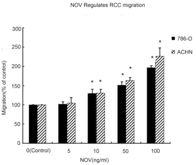
First ACHN and 786-O cells pretreated with NOV (Figure 1), NS-398, and Akt inhibitor were counted in transwell chambers (Figure 2). Cells
pretreated with NOV showed a significant
[image:3.612.91.290.72.240.2]increase in cell motility over parental cells (P < 0.01) (Figure 2). The Akt inhibitor NS-398 reduced the magnitude of this NOV-induced increase in cell migration (P < 0.05) (Figure 2).
Figure 2. Transwell migration assays were performed to determine the motility of 786-O and OS-RC-2 cells. A.The COX-2 specific inhibitors NS-398 and Akt pathway inhibitor were used in RCC cells 786-O and ACHN. The dose of NOV was keeped in 100 ng/ml. Untreated cells were used as control. Values represent the means ± SD of 3 inde-pendent experiments (*P < 0.05 vs. Control, #P < 0.05 vs. NOV treated cells.). B. Migration assays were performed using transwell cham bers.The cells were pretreated with NOV, NOV+NS398 or NOV+Akt inhibitor (Untreated cells were used as control) for 8 h and then trypsinized and plated in the upper wells in serum-free medium at a con-centration of 1×10⁵/well. After 4 h, the numbers of cells migrating to the lower wells were counted following H&E
NOV-directed RCC cell migration and upregula-tion of COX-2 and ICAM-1
Previous works have shown that COX-2 can
mediate the motility of human cancer cells [27, 28]. It was here hypothesized that COX-2 may
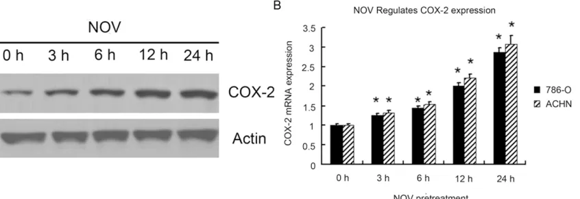
be involved in NOV-mediated RCC migration. When RCC cells were treated with NOV the rate of expression of COX-2 mRNA and protein increased (Figure 3). Over-expression of COX-2 was found to increase the motility of RCC cells (Figure 3). The COX-2 specific inhibitors NS-398 were used to confirm that it was COX-2 that
mediated NOV-induced cell migration. Admi- nistration of NS-398 reduced the magnitude of NOV-induced increases in cell migration (Figure 2). Similar trend has occurred to ICAM-1.We measured at different time and dose, though, here, exposure time haven’t yet to be the criti-cal factor, when treated with NOV, the rate of ICAM-1 protein and mRNA expression increased with the dose of NOV (Figure 4). And the COX-2
[image:5.612.97.521.132.308.2] [image:5.612.96.284.394.609.2]specific inhibitors NS-398 have nothing with Figure 3. COX-2 expression in 786-O and ACHN cells was assessed by Western blotting and Real-time PCR. 786-O and ACHN cells were incubated with same dose of NOV (100 ng/ml) for 24 h. Western blotting and Real-time PCR were used to detect the expression of COX-2 protein and mRNA. 0 h treated cells were used as control. Bars indicate the means ± SD of 3 independent experiments (*P < 0.05 versus control).
Figure 4.ICAM-1 expression in 786-O and ACHN cells was assessed by Western blotting and Real-time PCR. 786-O and ACHN cells were treated with different dose of NOV (0 ng/ml, 5 ng/ml, 10 ng/ml, 50 ng/ml, 100 ng/ml) for 8 h. Western blotting and Real-time PCR were used to detect the expression of ICAM-1 protein and mRNA. 0h treated cells were used as control. Untreated cells were used as control. Values represent the means ± SD of 3 independent experiments. *P < 0.05 vs. Control.
Figure 5. Western blotting were performed to deter-mine the expression of p-Akt, Akt, COX-2 and ICAM-1 in 786-O cells. The COX-2 specific inhibitors NS-398 and Akt pathway inhibitor were used in RCC cells 786-O.The dose of NOV was keeped in 100 mg/nl. Untreated cells were used as control. The intensities of proteins were quantified by densitometry analysis using Image J software. Bars indicated the means ± SD from 3 independent experiments.NS-398 and Akt pathway inhibitor treated cells p-AKT, COX-2 and
ICAM-1 expression. These results suggest that NOV-induced migration of cancer cells may involve upregulation of COX-2 and ICAM-1.
Akt signaling pathway, ICAM-1, NOV-mediated upregulation of COX-2 and migration of RCC
As indicated by the results of the migration assays, exposure to Akt inhibitor interfered with NOV-induced increases in cell migration. This suggested that the Akt signaling pathway
may have been involved. To confirm this, p-Akt
expression was measured in 786-O cells over-expressing NOV, 786-O and in ACHN RCC cell lines pretreated with NOV. Results showed higher levels of p-Akt expression than in paren-tal cells (P < 0.01) (Figure 5). Articularly, the variation has similar trend with ICAM-1. That is
to say, through Akt pathway, NOV also influenc -es ICAM expr-ession to increase cell migration (Figure 5). Data also showed that COX-2 expres-sion was also upregulated in these cells. When the Akt pathway was partially blocked by the inhibitor, there was less NOV-mediated COX-2 upregulation (Figure 5), suggesting that the Akt signaling pathway affects NOV-mediated COX-2 upregulation and migration of RCC.
Immunohistochemical expression, clinicopath-ological variables, and associations among NOV and COX-2 expression (Table 1)
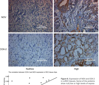
Examples of reactivity to NOV and COX-2 are given in (Figure 6).
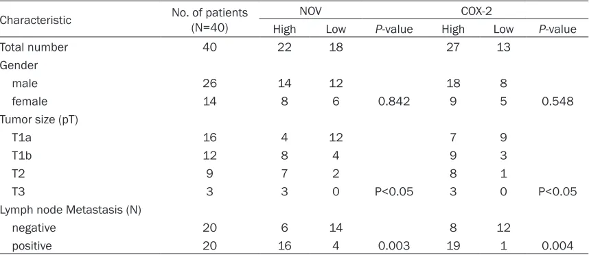
[image:6.612.93.522.96.282.2]Protein expression values and clinicopathologi-cal data for each patient are summarized in Table 1. As shown, levels of NOV and COX-2 expression showed no association with gen- der.
NOV expression was noted in 27 patients (74%), and high levels of NOV expression were
observed in 22 cases (55%). There was signifi -cantly more expression in larger, later-stage tumors (pT) (P < 0.05). NOV was also signifi -cantly higher in patients with lymph node metastasis (P = 0.003).
COX-2 expression was noted in 32 of 40 cases (80%). COX-2 expression was found to be
sig-nificantly closely correlated with both tumor
stage and tumor size (P < 0.05). There was
sig-nificantly more NOV expression in cases with
lymph node metastasis (P = 0.004).
Another strong correlation was found between COX-2 and NOV expression in ccRCC tissues (R = 0.393, P < 0.001).
Discussion
Renal cell carcinoma is a malignant, high-grade renal neoplasm. Existing chemotherapies are
not sufficiently effective, and 20% of all patients
die of metastasis. Effective adjuvant therapies capable of preventing RCC metastasis must be developed. It is here hypothesized that NOV and NOV receptors may direct the migration of RCC cells. The results of the current work Table 1. Immunohistochemical expression, clinicopathological variables, and associations among NOV and COX-2 expression
Characteristic No. of patients (N=40) NOV COX-2
High Low P-value High Low P-value
Total number 40 22 18 27 13
Gender
male 26 14 12 18 8
female 14 8 6 0.842 9 5 0.548
Tumor size (pT)
T1a 16 4 12 7 9
T1b 12 8 4 9 3
T2 9 7 2 8 1
T3 3 3 0 P<0.05 3 0 P<0.05
Lymph node Metastasis (N)
negative 20 6 14 8 12
positive 20 16 4 0.003 19 1 0.004
showed that NOV could promote the migration of human ccRCC cells. Previous results showed that overexpression of NOV could induce an increase in the migration of ccRCC cells by as much as 5-fold. The results of the current work
were verified by pretreating ccRCC cells with
NOV protein. Transcriptional upregulation of COX-2 and ICAM-1 and activation of Akt path-ways was found to be one of the mechanisms underlying the effects of NOV on migration. Treatments involving COX-2 and ICAM-1 selec-tive inhibitors may be suitable for the preven-tion of ccRCC metastasis.
The extracellular domain of ICAM-1 is crucial for leukocytes and has been shown to be correlat-ed with the transendothelial migration of leuko-cytes from the capillary bed to the surrounding
tissue [29]. ICAM-1 may also promote the migration of other cell types [30, 31]. It has
been reported that ICAM-1 plays an important
role in lung cancer cell invasion [32]. In oral
cancer, prostaglandin E2 (PGE2) induced cell
motility through ICAM-1 up-regulation [33]. And
[image:7.612.101.525.74.439.2]our data shows that ICAM-1 expression had a considerable correlation with NOV expression, which is the key director of cell motility. All in all,
these results suggest that ICAM-1 is an impor-tant target for treating cancer metastasis, especially for our renal cancer.
COX-2 is a pleiotropic enzyme. It mediates physiological functions such as augmentation of angiogenesis, inhibition of cell apoptosis, and increases in cell motility. Reports have indi-cated that COX-2 expression is associated with
metastasis in human cancer cells [27, 28].
Here, overexpression of COX-2 was found to induce cell motility in ccRCC. Human ccRCC cells treated with NOV showed more COX-2 expression. However, COX-2 inhibitors were found to antagonize NOV-induced reductions in cell motility. It is here reported that NOV can increase COX-2-related production of PGD2, PGI2, and PGE2 production in osteosarcoma cells. COX-2-induced PGs may be NOV-res- ponsive mediators. They may promote cancer migration and metastasis. This conclusion will
be confirmed in ccRCC in a subsequent study.
The results of the current work demonstrate that pretreatment of ccRCC with Akt inhibitor can antagonize increases in cell migration and COX-2 expression under NOV stimulation
condi-tions. This was confirmed when exposure to Akt
inhibitor was found to reduce the magnitude of the increase in COX-2 expression and cell migration under NOV stimulation conditions. Current results also showed that the cytoplas-mic kinase Akt can be activated by NOV stimu-lation in patients with ccRCC. These effects indicate that Akt activation is part of in NOV-mediated induction of COX-2 expression and cell migration. These results indicate that the Akt pathway might be a key to the ability of NOV to induce COX-2 activation in human ccRCC cells.
The data collected here also demonstrate that increased expression of COX-2 and NOV was correlated with tumor stage and lymph node metastasis. In ccRCC tissues, COX-2 expres-sion showed a considerable correlation with NOV expression, which is consistent with con-clusions drawn in previous works. This indicat-ed that COX-2 and NOV are an important part of ccRCC migration and invasion, that they pro-mote the metastasis of ccRCC cells.
Because of this, the prognosis of patients with distant ccRCC metastasis is very poor. Pre- venting human ccRCC metastasis is highly
important. The current study shows that NOV can increase the expression of COX-2 and ICAM-1 through the Akt pathway, so promoting the migration of human ccRCC cells. The dis-covery of the NOV-mediated pathway may facili-tate understanding of the mechanism of human ccRCC metastasis and may foster development of effective therapies.
Acknowledgements
At the point of finishing this paper, I’d like to
express my sincere thanks to all those who have lent me hands in the course of my writing this paper. Furthermore, thanks for the Shan- dong Provincial Natural Science Foundation (Y2007C067 and ZR2014HP015), Science and Technology Development Plan Project of Shan- dong Province, China (2014GGH218036), and Medical and health technology development projects of Shandong Province (2014WS0341),
the source of our financial support.
Disclosure of conflict of interest None.
Address correspondence to: Dr. Zhihong Niu, De- partment of Urology, Provincial Hospital affiliated to Shandong University, 324 Jingwu Street, Jinan 250021, China. Tel: 86-531-85186310; Fax: 86-531-87068707; E-mail: [email protected]
References
[1] Ek ET and Choong PF. The role of high-dose therapy and autologous stem cell transplanta-tion for pediatric bone and soft tissue sarco-mas. Expert Rev Anticancer Ther 2006; 6: 225-37.
[2] Caswell PT, Vadrevu S and Norman JC. Integ-rins: masters and slaves of endocytic trans-port. Nat Rev Mol Cell Biol 2009; 10: 843-53. [3] Tsuji T, Ibaragi S and Hu GF.
Epithelial-mesen-chymal transition and cell cooperativity in me-tastasis. Cancer Res 2009; 69: 7135-9. [4] Turini ME and DuBois RN. Cyclooxygenase-2: a
therapeutic target. Annu Rev Med 2002; 53: 35-57.
[5] Sano H, Kawahito Y, Wilder RL, Hashiramoto A, Mukai S and Asai K. Expression of cyclooxygen -ase-1 and -2 in human colorectal cancer. Can-cer Res 1995; 55: 3785-9.
[7] Hwang D, Scollard D, Byrne J and Levine E. Ex-pression of cyclooxygenase-1 and cyclooxygen-ase-2 in human breast cancer. J Natl Cancer Inst 1998; 90: 455-60.
[8] Zimmerman T and Blanco FJ. Inhibitors target-ing the LFA-1/ICAM-1 cell-adhesion interac-tion: Design and mechanism of action. Curr Pharm Des 2008; 14: 2128-2139.
[9] Lawson C and Wolf S. ICAM-1 signaling in en-dothelial cells. Pharmacol Rep 2009; 61: 22-32.
[10] Van de Stolpe A and van der Saag PT. Intercel-lular adhesion molecule-1. J Mol Med 1996; 74: 13-33.
[11] Rosette C, Roth RB, Oeth P, Braun A, Kammer -er S, Ekblom J and Denissenko MF. Role of ICAM1 in invasion of human breast cancer cells. Carcinogenesis 2005; 26: 943-950. [12] Grothey A, Heistermann P, Philippou S and
Voigtmann R. Serum levels of soluble intercel-lular adhesion molecule-1 (ICAM-1, CD54) in patients with non-small-cell lung cancer: Cor-relation with histological expression of ICAM-1 and tumour stage. Br J Cancer 1998; 77: 801-807.
[13] Rosette C, Roth RB, Oeth P, Braun A, Kammer -er S, Ekblom J and Denissenko MF. Role of ICAM1 in invasion of human breast cancer cells. Carcinogenesis 2005; 26: 943-950. [14] Kleer CG, Zhang Y, Pan Q and Merajver SD.
(CCN6) is a secreted tumor suppressor protein that modulates IGF signaling in inflammatory breast cancer. Neoplasia 2004; 6: 179-85. [15] Kleer CG, Zhang Y, Pan Q, van Golen KL, Wu ZF
and Livant D. WISP3 is a novel tumor suppres-sor gene of inflammatory breast cancer. Onco -gene 2002; 21: 3172-80.
[16] Holbourn KP, Acharya KR and Perbal B. The CCN family of proteins: structure-function rela-tionships. Trends Biochem Sci 2008; 33: 461-73.
[17] Joliot V, Martinerie C, Dambrine G, Plassiart G, Brisac M and Crochet J. Proviral rearrange-ments and overexpression of a new cellular gene (nov) in myeloblastosis-associated virus type 1-induced nephroblastomas. Mol Cell Biol 1992; 12: 10-21.
[18] Chevalier G, Yeger H, Martinerie C, Laurent M, Alami J and Schofield PN. novH: differential ex -pression in developing kidney and Wilm’s tu-mors. Am J Pathol 1998; 152: 1563-75. [19] Yu C, Le AT, Yeger H, Perbal B and Alman BA.
NOV (CCN3) regulation in the growth plate and CCN family member expression in cartilage neoplasia. J Pathol 2003; 201: 609-15. [20] Liu C, Liu XJ, Crowe PD, Kelner GS, Fan J and
Barry G. Nephroblastoma overexpressed gene (NOV) codes for a growth factor that induces
protein tyrosine phosphorylation. Gene 1999; 238: 471-8.
[21] Maillard M, Cadot B, Ball RY, Sethia K, Edwards DR and Perbal B. Differential expression of the ccn3 (nov) proto-oncogene in human prostate cell lines and tissues. Mol Pathol 2001; 54: 275-80.
[22] Tan TW, Yang WH, Lin YT, Hsu SF, Li TM and Kao ST. Cyr61 increases migration and MMP-13 expression via alphavbeta3 integrin, FAK, ERK and AP-1-dependent pathway in human chondrosarcoma cells. Carcinogenesis 2009; 30: 258-68.
[23] Tan TW, Lai CH, Huang CY, Yang WH, Chen HT and Hsu HC. CTGF enhances migration and MMP-13 up-regulation via alphavbeta3 integ-rin, FAK, ERK, and NF-kappaB-dependent pathway in human chondrosarcoma cells. J Cell Biochem 2009; 107: 345-56.
[24] Liu S, Liu Z, Bi DB, Yuan XD, Liu XW, Ding ST, Lu JJ and Niu ZH. CCN3 (NOV) regulates prolifera-tion, adhesion, migration and invasion in clear cell renal cell carcinoma. Oncol Lett 2012; 35: 1099-1104.
[25] Chuang JY, Yang WH, Chen HT, Huang CY, Tan TW and Lin YT. CCL5/CCR5 axis promotes the motility of human oral cancer cells. J Cell Physi-ol 2009; 220: 418-26.
[26] Chiu YC, Lin CY, Chen CP, Huang KC, Tong KM and Tzeng CY. Peptidoglycan enhances IL-6 production in human synovial fibroblasts via TLR2 receptor,focal adhesion kinase, Akt, and AP-1- dependent pathway. J Immunol 2009; 183: 2785-92.
[27] Yang SF, Chen MK, Hsieh YS, Chung TT, Hsieh YH and Lin CW. PGE2/EP1 signaling pathway enhances ICAM-1 expression and cell motility in oral cancer cells. J Biol Chem 2010; 285: 29808-29816.
[28] Liu JF, Fong YC, Chang CS, Huang CY, Chen HT and Yang WH. Cyclooxygenase-2 enhances al-pha2beta1 integrin expression and cell migra-tion via EP1dependent signaling pathway in human chondrosarcoma cells. Mol Cancer 2010; 9: 43.
[29] Duperray A, Languino LR, Plescia J, McDowall A, Hogg N, Craig AG, Berendt AR and Altieri DC. Molecular identification of a novel fibrinogen binding site on the first domain ofICAM-1 regu -lating leukocyte-endothelium bridging. J Biol Chem 1997; 272: 435-441.
[30] Rauch BH, Muschenborn B, Braun M, Weber AA and Schror K. ICAM-1 and p38 MAPK medi -ate fibrinogen-induced migration of human vascular smooth muscle cells. Eur J Pharmacol 2007; 577: 54-57.
ICAM-1-depen-dent pathway. Thromb Haemost 2003; 89: 1089-1097.
[32] Huang WC, Chan ST, Yang TL, Tzeng CC and Chen CC. Inhibition of ICAM-1gene expression, monocyte adhesion and cancer cell invasion by targeting IKKcomplex: molecular and func -tional study of novel alpha-methylene-gam- mabutyrolactone derivatives. Carcinogenesis 2004; 25: 1925-1934.