Original Article
Expression of microRNA-200c in human pancreatic
ductal adenocarcinoma and its prognostic significance
Jun-Ping Liu1, Jia Shang1, Yi Kang1, Chong-Shan Mao1, Hui-Bin Ning1, Chao Ma2
1Department of Infectious Diseases, Henan Provincial People’s Hospital, Zhengzhou, China; 2Department of He-patopancreatobiliary Surgery, The Affiliated Tumor Hospital of Zhengzhou University, Zhengzhou, China
Received February 19, 2016; Accepted July 6, 2016; Epub September 1, 2016; Published September 15, 2016
Abstract: Introduction: MicroRNAs (miRNAs) have emerged as potential therapeutic candidates due to their ability to regulate multiple targets involved in tumor progression. MiRNA-200c (miR-200c) was previously shown to be cor-related with aggressive clinicopathological features and poor prognosis in several cancers. The aim of this study was to analyze miR-200c expression in pancreatic ductal adenocarcinoma (PDAC) and to determine whether miR-200c has an independent prognostic value in PDAC. Methods: Quantitative real-time PCR (qRT-PCR) assay was performed to detect the expression of miR-200c in human PDAC cells and tissue samples. The association of miR-200c expres-sion with clinicopathologic features was analyzed. Kaplan-Meier analyses were used to assess survival of patients. Univariate and multivariate analyses were performed using the Cox proportional hazards model to analyze the
prognostic significance of miR-200c expression. Results: Our data showed that the relative level of miR-200c in PDAC cells was significantly lower than that in normal human pancreatic duct epithelial cell line. The expression of miR-200c in PDAC tissues was significantly lower than that in adjacent non-tumor tissues. By statistical analyses,
low miR-200c expression was observed to be closely correlated with clinical stage, liver metastasis and lymphnode metastasis. Kaplan-Meier survival analysis revealed that patients with low miR-200c expression had a poor overall survival compared with the high miR-200c group (P < 0.05). Univariate and multivariate analyses showed that low
miR-200c expression was an independent poor prognostic factor for PDAC patients. Conclusion: Our data demon -strated that reduced miR-200c in PDAC tissues was correlated with tumor progression, and miR-200c might be a potential molecular biomarker for predicting the prognosis of patients.
Keywords: microRNA-200c, overall survival, pancreatic cancer, pancreatic ductal adenocarcinoma
Introduction
Pancreatic ductal adenocarcinoma (PDAC) is the fourth leading cause of cancer-related death in Western countries and the sixth in China, is characterized by aggressive invasion, early metastasis, and lack of specific symp -toms [1, 2]. The 5-year survival of PDAC is only approximately 5%, and this figure has remained nearly unchanged over the past two decades, but the incidence of PDAC has been rising worldwide [3, 4]. Pancreatic carcinogenesis is known to be a multi-step process involving mul-tiple genetic and epigenetic alterations [5]. The identification of biomarkers that accurately pre -dict disease recurrence or response to chemo-therapy would be of substantial aid in individual risk assessment and treatment selection and may even lead to novel therapies by becoming
shown to be expressed in various carcinoma tissues and its over- or down-expression played essential role in tumor formation or cancer cell apoptosis. However, the relationship between expression of miR-200c and the prognosis of patients with PDAC remains unclear.
In our study, qRT-PCR assay was performed to detect the expression of miR-200c in PDAC and adjacent non-tumor tissues. Furthermore, the correlations of miR-200c expression with clini-copathologic features of PDAC patients were statistically analyzed. Finally, we determined the potential role of miR-200c in PDAC
prognos-tic prediction. Our data indicated that miR-200c was significantly downregulated in PDAC tissues and could be served as a potential molecular biomarker for the prediction of poor prognosis.
Materials and methods
Patients and specimens
A total of 75primary PDAC and paired adjacent non-tumor tissues (located > 2 cm from the tumors) were collected from patients who undergone pancreatic surgical resection with informed consent at the Henan Provincial People’s Hospital, China, from 2008 to 2013. Both tumor and non-tumor tissues were con-firmed by two experienced pathologists. None of these patients received neoadjuvant or adjuvant treatment before operation. Median follow-up time of surviving patients was 10.8 months. The study was approved by the Me- dical Ethical Committee of Zhengzhou Univer- sity. Informed consent had been obtained from all of the patients for use of the clinical specimens.
Cell lines and cultures
A normal human pancreatic duct epithelial cell line (HPDE6-C7) and PDAC cell line (PANC-1) were purchased from the Cell Bank of Chinese Academy of Sciences. Cells were cultured in RPMI 1640 and DMEM medium, respectively, with 10% fetal bovine serum, at 37°C in a humidified incubator with 5% CO2.
Quantitative real-time PCR (qRT-PCR)
[image:2.612.94.285.74.437.2]Total RNA was extracted with the Trizol reagent (Invitrogen, USA) according to the manufactur-er’s protocol. Complementary DNA was rever- se-transcribed using a reverse transcription kit (Invitrogen, USA). Briefly, cDNA were syn-thesized using a miR200c specific primer in reverse transcription system. The reaction con-dition was as follows: 16°C 30 min, 42°C 42 min, 85°C 5 min. Quantitative detection of RT products was performed using specific sense and anti sense primers of miRNA-200c and Sybergreen I dye. The PCR reaction condition was as follows: 95°C for 5 min, 94°C for 20 s, 55°C for 20 s, and 72°C for 20 s, 40 cycles, to obtain fluorescence intensity. U6 was used as an internal control. The sequence of specific Figure 1. qRT-PCR analysis of miR-200c expression
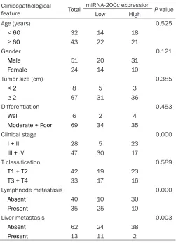
Table 1. Association between miR-200c expression and clinicopathologic features of PDAC patients
Clinicopathological
feature Total
miRNA-200c expression P value
Low High
Age (years) 0.525
< 60 32 14 18
≥ 60 43 22 21
Gender 0.121
Male 51 20 31
Female 24 14 10
Tumor size (cm) 0.385
< 2 8 5 3
≥ 2 67 31 36
Differentiation 0.453
Well 6 2 4
Moderate + Poor 69 34 35
Clinical stage 0.000
I + II 28 5 23
III + IV 47 30 17
T classification 0.589
T1 + T2 42 19 23
T3 + T4 33 17 16
Lymphnode metastasis 0.000
Absent 40 10 30
Present 35 25 10
Liver metastasis 0.003
Absent 62 24 38
Present 13 11 2
primer for miRNA-200c was 5’-GTCGTATCCA- GTGCGTGTCGTGGAGTCGGCAATGCACTGGATAC- GACTCCATC-3’; the sequence of sense primer of miRNA-200c was 5’-GGTAATACTGCCGGGT- AAT-3’; the sequence of antisense primer of miRNA-200c was 5’-CAGTGCGTGTCGTGGAGT- 3’. The Ct value was analyzed using the RFQ-PCR (Applied Biosystems Viia7, USA) analy- sis program. Relative mRNA expression levels were determined by the 2-ΔΔCt method in
com-parison with control cells.
Statistical analysis
All statistical analyses were performed by using the SPSS 18.0 statistical software pack-age. The data were presented as the mean ± SD. The association between miR-200c ex- pression level and clinicopathologic factors of the patients was analyzed using the chi-square test. Survival curves were plotted by the Kaplan-Meier method and compared by
Correlations of miRNA-200c expression with clinicopathologic features of PDAC patients
To further investigate the correlations of miR-200c expression with clinicopathologic fea-tures of PDAC patients. The relative express- ion of miR-200c was divided into two groups based on the mean value (0.33): High miR-200c expression group had miR-miR-200c expres-sion levels more than mean value and low miR-200c expression group had miR-200c expression levels less than mean value. Then, the correlations of miR-200c expression with clinicopathologic features of patients were statistically analyzed. As shown in Table 1, low miR-200c expression was observed to be closely correlated with advanced clinical stage, higher incidence of lymph node metastasis and liver metastasis (Table 1; P < 0.05). How- ever, there were no significant correlations be-tween miR-200c expression and other clinico-pathologic factors including age, gender, tumor
the log-rank test. The significance of different variables with respect to survival was analyzed using the mul- tivariate Cox proportional hazards mo- del. Differences were considered sta-tistically significant when P < 0.05.
Results
Expression of miRNA-200c in human PDAC cells and tissue samples
size, differentiation and T classification (Table 1; P > 0.05).
Correlations of miRNA-200c expression with overall survival of PDAC patients
To further assess the correlation of miR-200c expression with survival of PDAC patients, Kaplan-Meier analyses were performed. As shown in Figure 2, the 5-year overall survival of low miR-200c expression group was sig- nificantly shorter than that of high miR-200c expression group (Figure 2; P < 0.05). Our re-sults demonstrated that downregulation of miR-200c might be correlated with poor sur- vival of PDAC patients.
Univariate and multivariate analyses of prog-nostic variables in PDAC patients
To further determine the prognostic signifi -cance of miR-200c expression for PDAC pa- tients, survival data were obtained for each patient and univariate and multivariate analy-ses were performed (Table 2). Univariate Cox regression analysis showed that clinicopatho-logical variables including clinical stage, lymph node metastasis, liver metastasis, and miR-200c expression were significantly associated with overall survival (Table 2; P < 0.05). Also, to evaluate whether low miR-200c expression (low vs. high) might be as an independent predictor for overall survival of PDAC patients, multivariate Cox regression analyses were
per-formed. Along clinical stage, lymph node me- tastasis, and liver metastasis (Table 2; P < 0.05), miR-200c expression was an indepen-dent molecular biomarker for predicting of the poor overall survival of PDAC patients (RR: 2.314, 95% CI: 1.731-6.378, P = 0.008). Discussion
There are currently no means for the reliable diagnosis of early stages of pancreatic cancer (PDAC) and the curative treatment of late stag-es. Consequently, the vast majority of patients (80%) display an advanced disease that results in a low resection rate leading to a dismal over-all median survival of less than 6 months [1]. Thus, there is an urgent need to discover diag-nostic as well as progdiag-nostic molecular markers together with reliable therapeutics to improve pancreatic cancer management.
The recent discovery of microRNAs (miRNAs or miRs) has revealed a novel mechanism of gene regulation and provided new avenues for cancer research. MiRNAs are small, non-coding RNA molecules, which regulate the gene expression at post-transcription level [11, 12]. MiRNAs are involved in the regulation of vari- ous biological processes including prolifera-tion, apoptosis, differentiation and develop-ment [13]. Cimmino et al reported that miR- 15a and miR-16-1 negatively regulate the antiapoptotic B cell lymphoma 2 gene (Bcl2) causing CLL cells to undergo apoptosis [14]. Li Ma unraveled a mechanism in which miR-10b positively regulates cell migration and in- vasion in a non-metastatic breast cancer cell line in a multi-step process that ultimately leads to activation of RHOC, a pro-metastatic gene [15]. The Slack group introduced a no- vel strategy to efficiently inhibit miR-155, an oncomiR in a murine model of lymphoma, by using a peptide nucleic acid antagomiR atta- ched to a pH-induced transmembrane struc-ture (pHLIP) [16]. These data demonstrated the potential oncogenic or tumor suppressor role of miRNAs. However, the relationship be- tween miRNAs and cancer patient prognosis re- mains largely unknown.
[image:4.612.91.286.73.240.2]Here we reported miR-200c, which was pre- viously shown to function as a tumor suppres-sor and to repress epithelial mesenchymal transition (EMT) and tumor metastasis. For in- stance, increased miR-200c expression leads Figure 2. miR-200c expression is correlated with
to a reversal of EMT in bladder cancer [17]. MiR-200c can also inhibit cancer stem cell self-renewal and attenuate differentiation [18]. MiR-200c were confirmed to be downregulated in human breast cancer stem cells as well as in normal human and murine mammary stem/ progenitor cells. Moreover, miR-200c has a modulatory function in cell division and apopto-sis [19]. However, the association between miR-200c expression and the clinicopathologic features and patient prognosis in PDAC is unknown. In this study, we aimed to explore the association between miR-200c expression and cancer prognosis and clinical pathology. We found that the relative level of miR-200c in PDAC cell lines was significantly lower than that in normal human pancreatic duct epithelial cell line. Also, we compared the expression of miR-200c in PDAC tissues and adjacent non-tumor tissues, and showed that the expression of miR-200c in PDAC tissues was also significantly lower than that in adjacent non-tumor tissues. Then, we investigated the clinicopathologic sig-nificance of miR-200c expression in PDAC. Statistical analyses demonstrated that low miR-200c expression was significant correlated with advanced clinical stage, higher incidence of lymph node metastasis and liver metastasis. These data implied that downregulation of
miR-200 might play critical roles in PDAC progres-sion and development. Furthermore, we found miR-200c expression was observed to be sig-nificantly associated with overall survival of PDAC patients. Kaplan-Meier analysis of overall survival showed that patients with low miR-200c expression tended to have a significantly shorter overall survival than those with high miR-200c expression. Cox proportional haz-ards model proved that low miR-200c was an independent prognostic marker for predict-ing the poor prognosis of PDAC patients. Thus, miR-200c expression could be used as a mo- lecular prognostic factor to identify patients who are more likely to have higher risk of death. Taken together, this study showed that down-regulation miR-200c was associated with tu- mor progression and poor prognosis in PDAC and was identified as an independent poor prognostic factor for PDAC patients.
Disclosure of conflict of interest
None.
Address correspondence to: Chao Ma, Department
of Hepatopancreatobiliary Surgery, The Affiliated
[image:5.612.92.520.84.350.2]Tu-mor Hospital of Zhengzhou University, Zhengzhou 450008, China. E-mail: mjc1566@sina.com Table 2. Univariate analyses of different prognostic factors in PDAC patients
Clinicopathological feature Univariate analysis Multivariate analysis
Risk ratio 95% CI P Risk ratio 95% CI P
Age (years) 1.176 0.353-1.894 0.267
≥ 60 vs. < 60
Gender 1.289 0.543-2.118 0.325
Male vs. Female
Tumor size (cm) 1.867 0.581-2.945 0.241
≥ 2 cm vs. < 2 cm
Differentiation 1.516 0.283-2.906 0.581
Moderate + Poor vs. Well
T classification 2.537 0.683-4.351 0.207
T3 + T4 vs. T1 + T2
Clinical stage 2.417 1.328-6.139 0.009 2.105 1.327-5.14 0.004
III + IV vs. I + II
N classification 3.469 1.374-6.318 0.016 2.438 1.368-7.017 0.009
Present vs. Absent
Liver metastasis 3.336 0.604-5.872 0.014 2.283 0.625-4.673 0.011
Present vs. Absent
miRNA-200c 2.539 1.549-7.327 0.015 2.314 1.731-6.378 0.008
References
[1] Siegel R, Naishadham D, Jemal A. Cancer sta-tistics, 2013. CA Cancer J Clin 2013; 63: 11-30.
[2] Chen Y, Hao J, Ma W, Tang Y, Gao C, Hao X. Improvement in treatment and outcome of pancreatic ductal adenocarcinoma in north China. J Gastrointest Surg 2011; 15: 1026-34. [3] Jones S, Zhang X, Parsons DW, Lin JC, Leary
RJ, Angenendt P, Mankoo P, Carter H, Kamiyama H, Jimeno A, Hong SM, Fu B, Lin MT, Calhoun ES, Kamiyama M, Walter K, Nikolskaya T, Nikolsky Y, Hartigan J, Smith DR, Hidalgo M, Leach SD, Klein AP, Jaffee EM, Goggins M, Maitra A, Iacobuzio-Donahue C, Eshleman JR, Kern SE, Hruban RH, Karchin R, Papadopoulos N, Parmigiani G, Vogelstein B, Velculescu VE and Kinzler KW. Core signaling pathways in hu-man pancreatic cancers revealed by global ge-nomic analyses. Science 2008; 321: 1801-6. [4] Sun C, Rosendahl AH, Ansari D, Andersson R.
Proteome-based biomarkers in pancreatic cancer. World J Gastroenterol 2011; 17: 4845-52.
[5] Macgregor-Das AM, Iacobuzio-Donahue CA. Molecular pathways in pancreatic
carcinogen-esis. J Surg Oncol 2013; 107: 8-14.
[6] Pillai RS. MicroRNA function: multiple mecha-nisms for a tiny RNA? RNA 2005; 11: 1753-61. [7] Calin GA, Croce CM. MicroRNA signatures in
human cancers. Nat Rev Cancer 2006; 6: 857-66.
[8] Papagiannakopoulos T, Kosik KS. MicroRNAs: regulators of oncogenesis and stemness. BMC Med 2008; 6: 15.
[9] Palanichamy JK, Rao DS. miRNA dysregulation in cancer: towards a mechanistic understand-ing. Front Genet 2014; 5: 54.
[10] Hede K. Studies define role of microRNA in
cancer. J Natl Cancer Inst 2005; 97: 1114-5. [11] Srivastava SK, Bhardwaj A, Leavesley SJ,
Grizzle WE, Singh S, Singh AP. MicroRNAs as potential clinical biomarkers: emerging ap-proaches for their detection. Biotech Histo- chem 2013; 88: 373-87.
[12] Bhardwaj A, Arora S, Prajapati VK, Singh S, Singh AP. Cancer “stemness”-regulating mi-croRNAs: role, mechanisms and therapeutic potential. Curr Drug Targets 2013; 14: 1175-84.
[13] Zhang L, Jamaluddin MS, Weakley SM, Yao Q, Chen C. Roles and mechanisms of microRNAs in pancreatic cancer. World J Surg 2011; 35: 1725-31.
[14] Cimmino A, Calin GA, Fabbri M, Iorio MV, Ferracin M, Shimizu M, Wojcik SE, Aqeilan RI, Zupo S, Dono M, Rassenti L, Alder H, Volinia S, Liu CG, Kipps TJ, Negrini M and Croce CM. MiR-15 and miR-16 induce apoptosis by targeting BCL2. Proc Natl Acad Sci U S A 2005; 102: 13944-9.
[15] Ma L, Teruya-Feldstein J, Weinberg RA. Tumour invasion and metastasis initiated by microR-NA-10b in breast cancer. Nature 2007; 449: 682-8.
[16] Cheng CJ, Bahal R, Babar IA, Pincus Z, Barrera F, Liu C, Svoronos A, Braddock DT, Glazer PM, Engelman DM, Saltzman WM and Slack FJ. MicroRNA silencing for cancer therapy target-ed to the tumour microenvironment. Nature 2015; 518: 107-10.
[17] Wiklund ED, Bramsen JB, Hulf T, Dyrskjøt L, Ramanathan R, Hansen TB, Villadsen SB, Gao
S, Ostenfeld MS, Borre M, Peter ME, Ørntoft TF,
Kjems J and Clark SJ. Coordinated epigenetic repression of the miR-200 family and miR-205 in invasive bladder cancer. Int J Cancer 2011; 128: 1327-34.
[18] Lin CH, Jackson AL, Guo J, Linsley PS, Eisenman RN. Myc-regulated microRNAs attenuate em-bryonic stem cell differentiation. EMBO J
2009; 28: 3157-70.