Original Article
Bone morphogenetic protein 9 is a
potential tumor suppressor in osteosarcoma
Jinghao Liang1*, Jing Zhang2*, Fan Chen2, Zhengjun Lian2, Yalu Dong2, Ning Lu2, Yong Jia1
Departments of 1Orthopedics, 2Oncology, General Hospital of Xinjiang Military Command of PLA, Urumqi, P. R.
China. *Equal contributors.
Received September 2, 2016; Accepted November 11, 2016; Epub November 1, 2017; Published November 15, 2017
Abstract: Transforming growth factor-β (TGF-β) is known to promote tumor migration and invasion. Bone morpho-genetic proteins (BMPs) are members of the TGF-β family expressed in a variety of human carcinoma cell lines. Although accumulating evidence has shown that BMP9 plays important roles in the regulation of various cellular processes, the function of BMP9 in clinical osteosarcoma remains to be explored. In this study, BMP9 expression was analyzed in 55 osteosarcoma patient samples and their matching, distant non-cancerous tissues. And the roles of BMP9 in osteosarcoma cell proliferation, apoptosis and cell cycle were also examined. Our results showed that different expression level of BMP9 was detected in all osteosarcoma samples while no expression in normal tissues. Surprisingly, there was a negative association between the expression level of BMP9 and osteosarcoma grade, with low level of BMP9 being found in high histological grade osteosarcoma. Knockdown of BMP9 accelerated the proliferation of MG63, SaOS-2, and U2OS cells. BMP9 overexpression, however, induced cell apoptosis in U2OS cells. Together, these results indicated that BMP9 plays a pivotal role in osteosarcoma. Future studies defining the mechanism of BMP9 effect may lead to novel therapeutic approaches for osteosarcoma.
Keywords: BMP9, osteosarcoma, tumor suppressor
Introduction
Osteosarcoma (OS) is the most common pri-mary malignant bone tumor in children and young adults. Conventional OS is classified into osteoblastic, chondroblastic and fibroblastic OS, according to its histological features [1]. OS is highly aggressive and it metastasizes mostly to the lungs and bones [2]. However, despite the use of aggressive chemotherapeutic treat-ment strategies, the survival of OS patients has shown limited improvement. The prognosis is very poor, particularly in patients with clinically detectable metastasis at diagnosis or relapsed disease [3]. Thus, a novel strategy to efficiently inhibit metastasis is highly desirable.
Bone morphogenetic proteins (BMPs) are mem-bers of the TGF-β superfamily and play a critical role in skeletal development, bone formation and stem cell differentiation [4, 5]. At least 15 different BMPs have been identified in humans and disruptions in BMP signaling result in a variety of skeletal and extra skeletal anomalies [6-8]. BMP9, also known as growth
differentia-characterized member of the BMP family [9]. It has been shown to function as a pleiotropic cytokine and is implicated in the bone morpho-genesis, functional differentiation, glucose homeostasis, iron homeostasis, and angiogen-esis [10-14]. Recently, accumulating evidence shows that BMP9 plays important roles in the regulation of various cellular processes, includ-ing cell proliferation, migration, differentiation and apoptosis [9, 13, 15-19]. However, the expression pattern of BMP9 and the correlation of expression level of BMP9 and other clinico-pathological parameters in clinical osteosarco-ma reosteosarco-main to be explored.
In our study, 55 osteosarcoma patient samples were collected for analysis of the expression level of BMP9 and its correlation with osteosar-coma grade. Then the potential function of BMP9 was further determined in OS cell lines.
Materials and methods Materials
tis-11031 Int J Clin Exp Pathol 2017;10(11):11030-11036 sues were collected from 55 patients with
osteosarcoma who underwent radical surgery at Urumqi General Hospital of Lanzhou Military Area Command of Chinese PLA. None of the patients had received any chemotherapy, radio-therapy or other adjuvant radio-therapy before the operation. This study was approved by The Ethics Committee of Urumqi General Hospital of Lanzhou Military Area Command of Chinese PLA and all patients provided informed con-sent. Tumors were diagnosed and classified according to the American Joint Committee on Cancer breast cancer TNM staging system and the World Health Organization breast cancer histology classifications [20, 21].
Immunohistochemical SP method
All fresh specimens were fixed with formalin and embedded in paraffin according to the standard protocol. Tissue sections were depar-affinized and rehydrated routinely and then subjected to antigen retrieval by placing sli- des in 1× citrate buffer for 15 min at 100°C in a microwave oven. After treatment with 3% H2O2 for 30 minutes, the sections were incu-bated with 20% normal serum for 50 minutes and then with the BMP9 primary antibody (ab35088, Abcam) overnight at 4°C. On the fol-lowing day, the sections were washed with PBS thrice and then processed using an ultrasensi-tive TM S-P kit (Maixin Biotechnology, Fuzhou, China). After the washes in PBS, the color reac-tion was conducted using a 3,3’-diaminobenzi-dine kit (Maixin Biotechnology). The sections were counter-stained with hematoxylin and cov-ered with a coverslip. The stained tissue sec-tions were reviewed and scored independently by two pathologists (Dr. Jiang Wang and Min Liu). The percentage of BMP9 positive cells was rated as follows: -, ≤5% positive tumor cells; +, 5-30% positive cells; ++, 30-55% positive cells; and +++, >55% positive cells.
Cell culture and small interfering RNA knock-downs
The human OS cell lines, MG63, SaOS-2, and U2OS were cultured in DMEM-HG supplement-ed with 10% FBS and 100 U/ml penicillin G/ streptomycin at 37°C in a humidified atmo-sphere of 5% CO2/95% air. Small interfering RNA (siRNA) oligonucleotides for BMP9 (Invi- trogen) were introduced into cells using Lipo- fectamine 2000 transfection reagent (Invi- trogen) according to the manufacturer’s proto-col, and cells were analyzed for 5 days after transfection.
Cell proliferation analysis
The cells were seeded at after 48 hours of transfection and collected in 1D/2D/3D/4D/5D for cell proliferation analysis using CCK-8 ass- ay (YEASEN) following the manufacturer’s instructions. Cell Counting Kit-8 (CCK-8) allows very convenient assays by utilizing Dojindo’s highly water-soluble tetrazolium salt. WST-8 2-(2-methoxy-4-nitrophenyl)-3-(4-nitrophenyl)-5-(2,4-disulfophenyl)-2H-tetrazolium, monoso-dium salt) produces a water-soluble formazan dye upon reduction by dehydrogenase in mito-chondria in the presence of an electron carrier. Color depth is proportional to the cell prolifera-tion. The absorbance value at 450 nm wave-length by enzyme standard instrument reflects the number of living cells indirectly.
Real-time-PCR
Total RNA was extracted from collected sam-ples 48 hours after transfection using RNeasy mini kit (Qiagen). Reverse transcription was performed using PrimeScript® RT Reagent Kit (Perfect Real Time, TaKaRa). For quantitative real-time PCR, reactions were performed using SYBR® Premix ExTaqTM II (Perfect Real Time, TaKaRa) and 7500 Real-Time PCR System (Applied Biosystems). For each sample, the cycle threshold (CT) values were obtained from three replicates. Primer sequences are the fol-lowing: GAPDH forward: 5’-AGGTCGGTGTGAA- CGGATTTG-3’, reverse: 5’-GGGGTCGTTGATGGC- AACA-3’; BMP9 forward: 5’-CTGCCCTTCTTTG- TTGTCTT-3’, reverse: 5’-CCTTACACTCGTAGGC- TTCATA-3’. The relative expression levels of tar-get genes were analyzed using the 2-ΔΔCT method.
Western blot
Cell extracts were prepared at 48 hours after transfection as previously described. Proteins were electrophoresed in 10% SDS-PAGE gels, transferred to nitrocellulose membranes, and incubated overnight at 4°C in PBS buffer con-taining 5% BSA. β-actin (ab8227) expression levels were determined with a monoclonal anti-body to monitor protein loading and retention.
Cell cycle analysis
80% ethanol while vortexing, followed by incu-bation on ice for 60 min. The fixed cells were washed with cold PBS and incubated at 37°C for 30 min in 0.5 ml of PBS containing 10 µg/ml propidium iodine (Sigma) and 5 µg/ml RNase A (New England Biolabs). DNA content was determined by FACS scan analysis (Becton Dickinson).
Statistical analysis
Data were analyzed by SPSS 19.0 statistical software. Measurement data were analyzed by Student’s t-test, while categorical data were
[image:3.612.92.376.72.190.2]We then conducted the association analysis between the expression level of BMP9 protein and clinicopathological parameters from osteo-sarcoma patients (Table 2). The results showed that higher level of BMP9 was found at grade IIA osteosarcoma than that at grade IIB/III (P < 0.05), which suggested that the expression level of BMP9 was negatively correlated with osteosarcoma grade. However, there was no association between expression level of BMP9 and other clinicopathological parameters, such as age, gender, tumor size, TNM stage and sur-vival time (Table 2).
[image:3.612.89.378.273.313.2]Figure 1. Expression of BMP9 in osteosarcoma and its distant noncancerous tissues. The representative images of non-cancerous samples and osteosar-coma samples were shown. Bar, 200 μm.
Table 1. The expression level of BMP9 determined by IHC in osteo-sarcoma samples
Class No. BMP9 grade (No./%)
- + ++ +++
Osteosarcoma 55 0/0.00 6/10.91 29/52.73 20/36.36
analyzed by the chi-square test. P < 0.05 was considered as significant.
Results
Differential expression level of BMP9 protein in osteosar-coma and its distant noncan-cerous tissues
We first detected the expres-sion level of BMP9 by immu-nohistochemical staining in osteosarcoma and the mat- ching distant normal tissue samples from 55 patients. The results of BMP9 staining were scored as none (-), weak (+), moderate (++), and str- ong (+++) according to the assessment of two indepen-dent pathologists. No BMP9 expression was detected in the distant non-cancerous cells (Figure 1). In contrast, BMP9 protein was found in all osteosarcoma samples at varying degrees (Table 1). In most cases, BMP9 protein was expressed moderately or strongly (++, 29/55, 52.73%; +++, 20/55, 36.36%). Over- all, the results demonstrated that BMP9 is overexpressed in osteosarcoma compared with normal tissues
Correlation between BMP9 expression and clinicopatho-logical parameters
Table 2. Association between BMP9 expression and clinicopatho-logical factors from osteosarcoma
Clinicopathological
features Variable N (%) staining (mean ± SD)Intensity of BMP9 P value Age (years) ≥30 26 (47.27) 0.443 ± 0.225 0.845
< 30 29 (52.73) 0.462 ± 0.264
Gender Male 30 (54.55) 0.436 ± 0.216 0.823 Female 25 (45.45) 0.476 ± 0.287
Tumor size (cm) < 5.00 23 (41.82) 0.388 ± 0.259 0.786 ≥5.00 32 (58.18) 0.354 ± 0.341
Histology grade IIA 37 (67.27) 0.628 ± 0.135 0.027 IIB/III 18 (32.73) 0.319 ± 0.204
TNM stage I 24 (43.64) 0.366 ± 0.320 0.758 II 31 (56.36) 0.454 ± 0.386
[image:3.612.92.377.360.544.2]11033 Int J Clin Exp Pathol 2017;10(11):11030-11036
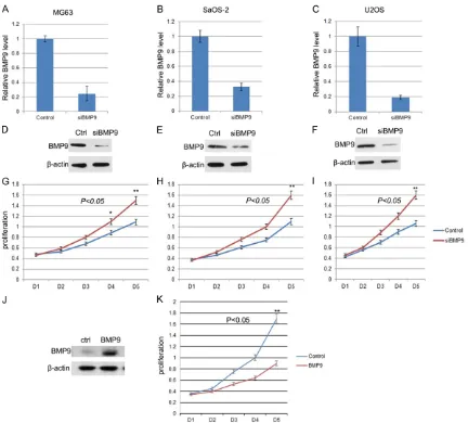
BMP9 knockdown accelerated osteosarcoma cell proliferation
To investigate the role of BMP9 in OS cell prolif-eration, we knock downed the expression of BMP9 in three human OS cell lines, MG63, SaOS-2, and U2OS, using siRNA. The results showed that the RNA level and protein level were both reduced significantly in the three cell lines after RNA interference (Figure 2A-F). And the proliferation rate was accelerated in all three cell lines (Figure 2G-I). We also did a res-cue experiment that we overexpressed BMP9 in BMP9 knockdown cells using a siRNA resis-tant BMP9 expression vector. The BMP9
pro-tein level was checked by Western blot and cell proliferation was analyzed as above. The re- sults were showed in Figure 2J and 2K that BMP9 overexpression was able to decrease the cell proliferation rate. Thus BMP9 negatively regulate cell proliferation in OS cell lines.
Overexpress BMP9 induced cell apoptosis in U2OS cells but did not affect cell cycle
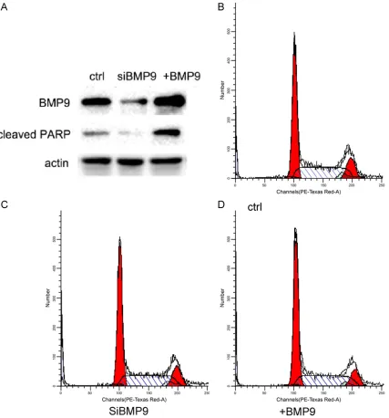
[image:4.612.91.523.69.459.2]induced cleaved PARP level, while knockdown BMP9 decrease the background level of cleaved PARP. These results indicated that BMP9 promoted cell apoptosis. We also com-pared cell cycle profiles between control, BMP9 knockdown and BMP9 overexpressed cells. BMP9 expression level did not affect cell cycle (Figure 3B-D).
Discussion
It is reported that BMP9 positively regulates the osteogenic differentiation of mesenchymal
[image:5.612.96.523.72.530.2]11035 Int J Clin Exp Pathol 2017;10(11):11030-11036 to be positive in all of the osteosarcoma
sam-ples. However, we found a negative association between the expression level of BMP9 and osteosarcoma grade. We found that, compared with low grade tumors, BMP9 expression level was lower in high histological grade osteosar-coma. This result indicated that BMP9 could have a pivotal role during osteosarcoma development.
In the present study, we also investigated the effect of BMP9 on human OS cell lines, MG63, SaOS-2, and U2OS. We observed that BMP9 knockdown had a significant facilitative effect on MG63, SaOS-2, and U2OS cell proliferation. These results are consistent with previous studies that have shown an inhibitory effect of BMP9 on the migration of prostate cancer and breast cancer cells [18, 22]. The response to BMP9, however, is not uniform among all can-cers. BMP9 has been shown to trigger epitheli-al-mesenchymal transition of hepatocellular carcinoma cells [23]. The pro-tumorigenic or anti-tumorigenic effect of BMP9 on different cancer cells might be caused by its complicate interactions with other proteins that exist in dif-ferent cells or difdif-ferent developmental stage. Therefore, the biological effect of BMP9 on dif-ferent cells may depend not only on BMP9 expression level but also on cell type, microen-vironment and the presence of other factors that are not yet defined.
In conclusion, we found that BMP9 was over- expressed in osteosarcoma, and the expres-sion level of BMP9 was negatively correlated with osteosarcoma grade. And BMP9 silencing accelerated the proliferation of MG63, SaOS-2, and U2OS cells. Thus, BMP9 may serve as a biological marker for osteosarcoma grad- ing. Future studies should focus on defining the mechanism of BMP9 function, which may lead to novel therapeutic approaches for osteosarcoma.
Disclosure of conflict of interest
None.
Address correspondence to: Ning Lu, Department of Oncology, General Hospital of Xinjiang Military Command of PLA, 359 Youhao North Road, Urumqi 830000, P. R. China. Tel: +86-991-4991747; E-mail: [email protected]; Yong Jia, Department of Orthopedics, General Hospital of Xinjiang Military Command of PLA, 359 Youhao North Road, Urumqi
830000, P. R. China. Tel: +86-991-4991766; E-mail: [email protected]
References
[1] Bielack S, Jurgens H, Jundt G, Kevric M, Ku- hne T, Reichardt P, Zoubek A, Werner M, Winkelmann W and Kotz R. Osteosarcoma: the COSS experience. Cancer Treat Res 2009; 152: 289-308.
[2] Gill J, Ahluwalia MK, Geller D and Gorlick R. New targets and approaches in osteosarcoma. Pharmacol Ther 2013; 137: 89-99.
[3] Wachtel M and Schafer BW. Targets for cancer therapy in childhood sarcomas. Cancer Treat Rev 2010; 36: 318-327.
[4] Kang Q, Sun MH, Cheng H, Peng Y, Montag AG, Deyrup AT, Jiang W, Luu HH, Luo J, Szatkowski JP, Vanichakarn P, Park JY, Li Y, Haydon RC and He TC. Characterization of the distinct ortho-topic bone-forming activity of 14 BMPs using recombinant adenovirus-mediated gene deliv-ery. Gene Ther 2004; 11: 1312-1320. [5] Luther G, Wagner ER, Zhu G, Kang Q, Luo Q,
Lamplot J, Bi Y, Luo X, Luo J, Teven C, Shi Q, Kim SH, Gao JL, Huang E, Yang K, Rames R, Liu X, Li M, Hu N, Liu H, Su Y, Chen L, He BC, Zuo GW, Deng ZL, Reid RR, Luu HH, Haydon RC and He TC. BMP-9 induced osteogenic differ-entiation of mesenchymal stem cells: molecu-lar mechanism and therapeutic potential. Curr Gene Ther 2011; 11: 229-240.
[6] Hogan BL. Bone morphogenetic proteins: mul-tifunctional regulators of vertebrate develop-ment. Genes Dev 1996; 10: 1580-1594. [7] Wozney JM, Rosen V, Celeste AJ, Mitsock LM,
Whitters MJ, Kriz RW, Hewick RM and Wang EA. Novel regulators of bone formation: mo-lecular clones and activities. Science 1988; 242: 1528-1534.
[8] Zhao GQ. Consequences of knocking out BMP signaling in the mouse. Genesis 2003; 35: 43-56.
[9] Lamplot JD, Qin J, Nan G, Wang J, Liu X, Yin L, Tomal J, Li R, Shui W, Zhang H, Kim SH, Zhang W, Zhang J, Kong Y, Denduluri S, Rogers MR, Pratt A, Haydon RC, Luu HH, Angeles J, Shi LL and He TC. BMP9 signaling in stem cell differ-entiation and osteogenesis. Am J Stem Cells 2013; 2: 1-21.
[10] Duan L, Ye L, Wu R, Wang H, Li X, Li H, Yuan S, Zha H, Sun H, Zhang Y, Chen X, Zhang Y and Zhou L. Inactivation of the phosphatidylinositol 3-kinase/Akt pathway is involved in BMP9-mediated tumor-suppressive effects in gastric cancer cells. J Cell Biochem 2015; 116: 1080-1089.
B. BMP9-induced survival effect in liver tumor cells requires p38MAPK activation. Int J Mol Sci 2015; 16: 20431-20448.
[12] Levet S, Ouarne M, Ciais D, Coutton C, Subileau M, Mallet C, Ricard N, Bidart M, Debillon T, Faravelli F, Rooryck C, Feige JJ, Tillet E and Bailly S. BMP9 and BMP10 are necessary for proper closure of the ductus arteriosus. Proc Natl Acad Sci U S A 2015; 112: E3207-3215. [13] Tillet E and Bailly S. Emerging roles of BMP9
and BMP10 in hereditary hemorrhagic telangi-ectasia. Front Genet 2014; 5: 456.
[14] Wan S, Liu Y, Weng Y, Wang W, Ren W, Fei C, Chen Y, Zhang Z, Wang T, Wang J, Jiang Y, Zhou L, He T and Zhang Y. BMP9 regulates cross-talk between breast cancer cells and bone marrow-derived mesenchymal stem cells. Cell Oncol (Dordr) 2014; 37: 363-375.
[15] Herrera B, Garcia-Alvaro M, Cruz S, Walsh P, Fernandez M, Roncero C, Fabregat I, Sanchez A and Inman GJ. BMP9 is a proliferative and survival factor for human hepatocellular carci-noma cells. PLoS One 2013; 8: e69535. [16] Li R, Zhang W, Cui J, Shui W, Yin L, Wang Y,
Zhang H, Wang N, Wu N, Nan G, Chen X, Wen S, Deng F, Zhang H, Zhou G, Liao Z, Zhang J, Zhang Q, Yan Z, Liu W, Zhang Z, Ye J, Deng Y, Luu HH, Haydon RC, He TC and Deng ZL. Targeting BMP9-promoted human osteosarco-ma growth by inactivation of notch signaling. Curr Cancer Drug Targets 2014; 14: 274-285. [17] Lv Z, Yang D, Li J, Hu M, Luo M, Zhan X, Song P,
Liu C, Bai H, Li B, Yang Y, Chen Y, Shi Q and Weng Y. Bone morphogenetic protein 9 overex-pression reduces osteosarcoma cell migration and invasion. Mol Cells 2013; 36: 119-126. [18] Wang K, Feng H, Ren W, Sun X, Luo J, Tang M,
Zhou L, Weng Y, He TC and Zhang Y. BMP9 in-hibits the proliferation and invasiveness of breast cancer cells MDA-MB-231. J Cancer Res Clin Oncol 2011; 137: 1687-1696.
[19] Yoshimatsu Y, Lee YG, Akatsu Y, Taguchi L, Suzuki HI, Cunha SI, Maruyama K, Suzuki Y, Yamazaki T, Katsura A, Oh SP, Zimmers TA, Lee SJ, Pietras K, Koh GY, Miyazono K and Watabe T. Bone morphogenetic protein-9 inhibits lym-phatic vessel formation via activin receptor-like kinase 1 during development and cancer progression. Proc Natl Acad Sci U S A 2013; 110: 18940-18945.
[20] Benson JR, Weaver DL, Mittra I and Hayashi M. The TNM staging system and breast cancer. Lancet Oncol 2003; 4: 56-60.
[21] Frank GA, Danilova NV, Andreeva I and Nefedova NA. [WHO classification of tumors of the breast, 2012]. Arkh Patol 2013; 75: 53-63. [22] Ye L, Kynaston H and Jiang WG. Bone morpho-genetic protein-9 induces apoptosis in pros-tate cancer cells, the role of prospros-tate apoptosis response-4. Mol Cancer Res 2008; 6: 1594-1606.