Original Article
Expression of toll-like receptor 2, 4 and
related cytokines in intraperitoneally inoculated
Balb/C mice with
Echinococcus multilocularis
Shadike Apaer1,2*, Tuerhongjiang Tuxun1,2,4*, Hai-Zhang Ma3, Heng Zhang1, Hao Zhang1, Jiangduosi Payiziwula2, Pei-Ji Zhao2, Aizimaiti Aihaiti2, Yu-Peng Li2, Tao Li2, Jin-Ming Zhao2,4, Ren-Yong Lin1,4, Hao Wen1,2,4
1State Key Laboratory Incubation Base of Xinjiang Major Diseases Research and Xinjiang Key Laboratory of
Echinococcosis, 2Departments of Liver Transplantation & Laparoscopic Surgery, Digestive and Vascular Surgery
Centre, The First Affiliated Hospital of Xinjiang Medical University, Urumqi, Xinjiang Uyghur Autonomous Region, China; 3Department of General Surgery, Qilu Hospital, Jinan, China; 4WHO Collaborating Center for Prevention and
Care Management of Echinococcosis, First Affiliated Hospital of Xinjiang Medical University and Xinjiang Centers for Disease Control, Urumqi, Xinjiang Uyghur Autonomous Region, China. *Equal contributors.
Received March 30, 2017; Accepted June 1, 2017; Epub July 1, 2017; Published July 15, 2017
Abstract: Immune response pattern between host and Echinococcus multilocularis (E. multilocularis) is considered as a crucial point in development of alveolar echinococcosis (AE). In this study, we are aiming to study the expression patterns of TLR2 and TLR4 with related cytokines and transcription factors in secondary E. multilocularis infected murine model. The murine model of AE was developed by using intraperitoneal inoculation of E. multilocularis pro-toscolexes and albendazole (E. m+ABZ group) or carboxy methyle cellulose (CMC; E. m+CMC group) administration via gastric tube was initiated in the third month and continued for one month. Mice with CMC administration served asnegative controls (C+CMC group). The splenic cells and peritoneal exudates cells (PECs) were prepared and the levels of IFN-γ, IL-10, and IL-5 in splenic cells and PECs culture supernatants were detected using enzyme linked immune-sorbent assay (ELISA). Besides, the mRNA expression levels of TLR2, 4, transcription factors and cytokines were detected by using real-time fluorescent quantitative reverse-transcription polymerase chain reaction (qRT-PCR). The concentration levels of IFN-γ, IL-10, and IL-5 in PECs and splenic cell supernatants were extremely lower, however, significantly elevated after stimulated with Concanavalin A (ConA) for 36 h with higher concentrations in
E. m+CMC group comparing to both E. m+ABZ and C+CMC group. The mRNA levels of TLR2, 4 and GATA3, IFN-γ, IL-10 in splenic cells were significantly increased in E. m+CMC group comparing with other groups. Simultaneously, T-bet mRNA expressions were elevated in E. m+ABZ and C+CMC group compared to E. m+CMC group. In addition, T-bet/GATA3 ratios was higher in E. m+ABZ group compared to E. m+CMC group and were higher in C+CMC group than those in E. m+CMC group. TLR2 mRNA expression in splenic cells showed a positive correlation with IL-10 concentration levels in splenic cell culture supernatants. The present study provides evidence on the possible role of TLR2 in the process of immune tolerance during E. multilocularis infection and suggests albendazole treatment might reverse the immune tolerance situation and improve parasite clearance process.
Keywords: Toll-like receptor, immune modulation, albendazole, echinococcus multilocularis
Introduction
Alveolar echinococcosis (AE) caused by the lar-val stage of tapeworm Echinococcus multilocu-laris (E. multilocularis) continues to be a major public health issue around the world, especially to pastoral and/or semi-pastoral area in China, Central Asia, Middle East, South America and some part of Europe [1]. Radical resection and autologous liver transplantation (ALT) along with long-term albendazole (ABZ)
driven component and Toll-like receptors (TLRs) in various parasitic infections, and thus, en- ables parasite escape from the immune sys-tem’s attack and guarantee long-period surviv-al [7-11]. Our previous studies indicated the involvement of elevated TLR2 and TLR4 in the process of immune tolerance both in cystic and alveolar echinococcosis [7, 12]. However, very little is known as both the alteration and possi-ble role of TLRs in murine model to date. In addition, no related reports, to our knowledge, were found about the possible role of ABZ treat-ments on the TLRs alterations. Therefore, in current study, secondary E. multilocularis infec-tion murine model was developed to further assess the alteration of TLR2, 4 mRNA expres-sions and related cytokines before and after ABZ administration, if any, as well as to study their relationship with relative cytokines. Materials and methods
Biochemicals and drugs
If not otherwise stated, all tissue culture media were purchased from Gibco-BRL (Zurich, Switzerland), biochemical reagents were from Sigma (MO, USA), and ABZ was purchased from GlaxoSmithKline (Tianjin, China). All reagents used for drug analysis were of analytical or research grade.
Mice
Female 8-week-old BALB/C mice were pur-chased from the experimental animal center of Xinjiang Medical University for secondary infec-tion with E. multilocularis and as control ani-mals. All mice were housed in a temperature-controlled, light-cycle room in animal facilities according to the Chinese animal protection guidelines, with food and water ad libitum. Maintenance and isolation of E. multilocularis metacestode Echinococcus
The E. multilocularis metacestode were main-tained by serial transplantation passages through i.p. injection in gerbils. Four to 10 weeks after i.p. injection, gerbils were sacri-ficed, and the parasite tissue was removed from the peritoneal cavity under aseptic condi-tions, placed into 0.9% normal saline (NS), and was washed several times. After grinding the tissue through a sterile 50-lm sieve, 100-fresh-ly prepared acephalic vesicular cysts were
sus-peritoneally into a mouse for secondary infection. Control mice received 100 µL of 0.9% NS.
ABZ administration started at the time point of chronic infection
ABZ suspensions were prepared in carboxy methyle cellulose (CMC) 0.5% (w/v) in water. ABZ suspensions were freshly prepared each week and stored at -20°C for a period of 7 days maximum. The control suspensions containing only CMC were treated identically. Treatments were initiated on the same day, and were repeated daily for 35 consecutive days. Mice were categorized into three groups of 8 in each: (1) E. multilocularis infection treated with ABZ (E. m+ABZ) group; (2) E. multilocularis infection with CMC (E. m+CMC) group; (3) Control+CMC (C+CMC) group.
Cell preparations
Mice were sacrificed 35 days post-treatment with ABZ. Spleen cell suspensions were pre-pared from both infected and control BALB/C mice. The cell suspensions were depleted of erythrocytes by treatment with 0.83% NH4Cl in 0.01 m Tris-HCl (pH 7.2) and subsequently resuspended in RPMI-1640 complete medium containing heat-inactivated 10% fetal calf serum (FCS, Gibco), 2 mm l-glutamine, 0.05 mm 2-mercaptoethanol, 100 U/ml penicillin and 100 μg/ml streptomycin (Gibco). PECs from control and infected mice were collected by peritoneal rinsing with 10 mL RPMI-1640. Cells were subsequently washed twice with HBSS and resuspended in RPMI-1640.
Cell cultures
Spleen cells and PECs were cultured in 48- well round-bottom plates at 2×10⁶/well for 36 hours and the supernatants were collected. The cell cultures were stimulated with Concanavalin A (Con A) (5 μg/ml; Sigma) or were left unstimulated as negative controls. All tests were performed in quadruplicates. Real-time fluorescent quantitative reverse-transcription polymerase chain reaction (qRT-PCR)
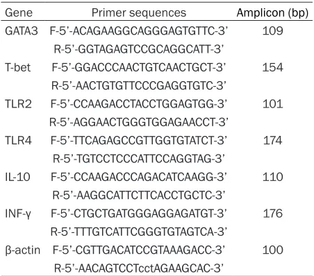
The primers were synthesized by Shenggong Biotech (Shanghai, China) shown as in Table 1. β-actin was analyzed as an internal control and gene expression was normalized to it. The quantitative PCR analyses of the data were performed using SYBR Green program on i-Q 5.0 Real-time PCR system (Bio-Rad, Foster City, CA, USA). The relative amounts of PCR products were determined using the relative standard curve method. mRNA expression level fold changes were calculated as described by the SYBR Green I protocol.
Enzyme-linked immune-sorbent assay (ELISA)
Concentrations of IFN-γ (assay sensitivity was 4 pg/mL), IL-5 (4 pg/mL), and IL-10 (2 pg/mL), were determined from splenic cell superna-tants and PECs’ by ELISA using a commercial mice ELISA kit (eBioscience, San Diego, CA, USA), according to the manufacturer’s instruc-tions. Cytokine concentrations were calculated by using the mean optical density of two wells and comparison with a standard curve.
Statistical analysis
All the quantitative data were expressed as Median [interquartile (IQR)] in the text. Sta- tistical analysis was performed using statis- tical software (SPSS, version 17.0, Chicago, IL, USA). Mann-Whitney (M-W) test was used to determine the differences between groups. Spearman correlation analysis was applied as a test of correlation between two continuous
compared to E. m+CMC mice 5.2 (IQR, 4.4-5.8) g as shown in Figure 1B.
TLR2, TLR4 mRNA expression levels in splenic cells
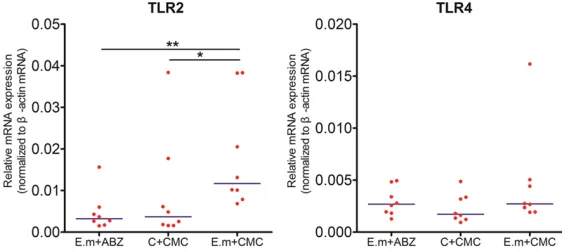
The mRNA expression levels of TLR2 and TLR4 were detected in spleen cells using qRT-PCR technique. TLR2 mRNA levels were significantly elevated in E. m+CMC group when comparing with C+CMC group and E. m+ABZ group (P<0.05, P<0.01 respectively). TLR4 mRNA lev-els were slightly higher in E. m+CMC group compared to both E. m+ABZ and C+CMC group, albeit no statistical differences were found between them (P>0.05) as shown in Figure 2. GATA3, IFN-γ, T-bet and IL-10 mRNA expression levels in splenic cells
[image:3.612.89.315.83.282.2]Th1/Th2 cell related transcription factors and cytokines GATA3, IFN-γ, T-bet and IL-10 mRNA expression levels were measured by using qRT-PCR technique. As shown in Figure 3, relative mRNA expression levels of GATA3 and IFN-γ in E. m+CMC group were found to be elevated compared to E. m+ABZ and C+CMC group, how-ever, without statistical differences (P>0.05, P>0.05 respectively). T-bet mRNA expression levels were increased in E. m+ABZ group com-pared to both E. m+CMC group and C+CMC group, nevertheless, no statistical differences between them (P>0.05). IL-10 mRNA expres-sions were significantly higher in E. m+CMC group than those in E. m+ABZ and C+CMC Table 1. Primer sequence and amplicon of genes
Gene Primer sequences Amplicon (bp)
GATA3 F-5’-ACAGAAGGCAGGGAGTGTTC-3’ 109
R-5’-GGTAGAGTCCGCAGGCATT-3’
T-bet F-5’-GGACCCAACTGTCAACTGCT-3’ 154
R-5’-AACTGTGTTCCCGAGGTGTC-3’
TLR2 F-5’-CCAAGACCTACCTGGAGTGG-3’ 101
R-5’-AGGAACTGGGTGGAGAACCT-3’
TLR4 F-5’-TTCAGAGCCGTTGGTGTATCT-3’ 174
R-5’-TGTCCTCCCATTCCAGGTAG-3’
IL-10 F-5’-CCAAGACCCAGACATCAAGG-3’ 110
R-5’-AAGGCATTCTTCACCTGCTC-3’
INF-γ F-5’-CTGCTGATGGGAGGAGATGT-3’ 176
R-5’-TTTGTCATTCGGGTGTAGTCA-3’
β-actin F-5’-CGTTGACATCCGTAAAGACC-3’ 100
R-5’-AACAGTCCTcctAGAAGCAC-3’ F: Forward; R: Reverse.
variables, and determined by Spearman cor-relation coefficients. Probable values of 0.05 and below were considered to be statistically significant.
Results
Evaluation/Assessing of secondary E. mul -tilocularis experimental model and parasite burden
group with a statistical differences between E. m+CMC and E. m+ABZ groups (P<0.05). We also detected the T-bet/GATA3 ratio and found it significantly elevated in E. m+ABZ group compared to both E. m+CMC and C+CMC group (P<0.05).
Concentration levels of cytokines in PECs supernatants
The concentration levels of cytokines before and after Con A stimulation were detected by using ELISA techniques. Con centrations of
both E. m+ABZ and C+CMC group, however, there were no statistical differences between them (P>0.05) as shown in Figure 4.
Correlation analysis between TLR2 mRNA ex-pression levels and IL-10 concentration levels
[image:4.612.91.374.74.225.2]Spearman correlation coefficients indicated that TLR2 mRNA expressions in splenic cells had a positive correlation with spleen cell IL-10 concentration levels (r=0.4344, P=0.0339), when no correlations were found between spleen cell TLR4 mRNA expressions and IL-10
Figure 1. Assessment of parasite load in intraperitoneal cavity of mice in-fected with E. multilocularis. All mice were sacrificed, and lesion tissues and masses were removed for monitoring parasite burden in the peritoneal cavity (A). Mice treated with ABZ administration resulted in a significantly reduced median parasite weight which was lower when compared to E. m+CMC mice (B).
Figure 2. qRT-PCR analyses of TLR2 and TLR4 mRNA expressions in differ-ent groups. The relative expressions of TLR2 and TLR4 were calculated, and results showed TLR2 mRNA expressions were significantly elevated in E. m+CMC group compared to C+CMC and E. m+ABZ group with statistical dif-ferences (P<0.05, P<0.01 respectively). Relative mRNA expressions of TLR4 were higher in E. m+CMC group comparing with both E. m+ABZ and C+CMC group, albeit no statistical differences were found between them (P>0.05).
P value <0.001 with marker ***; P value <0.01 with marker **; P value <0.05 with marker *.
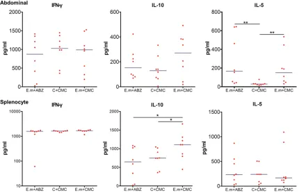
IFN-γ and IL-10 were extremely lower (data not shown), and elevated markedly after stimu-lating with Con A, resulting in the higher level in E. m+CMC group than those in E. m+ABZ and C+CMC group, neverthe-less no statistical differences were found between them (P>0.05). IL-5 concentration levels were also too lower, and interestingly, then elevated markedly with the stimulation of Con A. It is increased signifi-cantly in E. m+CMC and E. m+ABZ group compared to C+CMC group with statistical differences (P<0.01) as shown in Figure 4.
Concentration levels of cyto-kines in splenic cell superna-tants
[image:4.612.91.372.319.445.2]concentrations (r=0.2153, P=0.3124) as shown in Figure 5.
Discussion
Human AE caused by the larval stage of meta-cestode of E. multilocularis is a lethal neglect-ed tropical disease with “cancer-like” infiltrative growth pattern. After successful entering the portal blood stream, E. multilocularis reside itself primarily in liver and develop a granulo-matous lesion with potential to invade adjacent organs and metastasize to remote organs [13]. Early diagnosis and radical resection are chal-lenged due to insidious and asymptomatic growth. Radical resection varies from partial hepatectomy to ALT is optimal method along with long-term ABZ treatment [14, 15].
It is widely accepted that immune interaction between E. multilocularis and host’s immune system was pivotal during disease progression [16, 17]. E. multilocularis has evolved a broad spectrum of ability to actively modulate, even
escape from the host’s immune system for suc-cessful survival [18, 19]. Different subtypes of T helper (Th) cells such as Th1, Th2, Th17 and Th9, as well as Treg cells are involved in the interaction and related with the ability of para-site clearance and/or immune tolerance [4, 20, 21]. Identifying of parasite and/or parasite driv-en compondriv-ents through pathogdriv-en associated molecular patterns (PAMPs) are crucial for achieving an appropriate polarized T helper cell immune response that would enable the para-site to escape from the immune system’s attack and guarantee long-period survival. TLRs are firstly discovered and mostly studied among pattern recognition receptors (PRRs). They take a significant role in antigen recogni-tion and play as powerful immune-stimulant in innate immunity which in turn develops anti-gen-specific acquired immunity as well [22, 23]. The cellular localization of TLRs and respective PAMPs they identify will determine, to a large extent, the nature of the T cell polarization. It is showed that Th2 based responses
orchestrat-Figure 3. Relative mRNA expressions of cytokines and transcription factors in splenic cells. Relative cytokines and transcription factors GATA3, IFN-γ, T-bet, and IL-10 mRNA expression levels were calculated by using qRT-PCR tech-nique. GATA3, IFN-γ and IL-10 mRNA expression levels were elevated in E. m+CMC group compared to both E. m+ABZ and C+CMC group. Relative mRNA expressions of T-bet were found to be increased in E. m+ABZ group com-paring with both E. m+CMC and C+CMC group. Similarly, T-bet/GATA3 was significantly elevated in E. m+ABZ group comparing with both E. m+CMC and C+CMC group. P value <0.001 with marker ***; P value <0.01 with marker **;
[image:5.612.95.522.72.338.2]ed by TLRs signaling pathways are beneficial for the pathogen survival and disease progression [24, 25]. The potent critical role of them in hel-minthic infections such as leishmaniasis,
[image:6.612.92.522.74.352.2]TLR2 and TLR4 have been reported to be expressed during helminthic infection that uphold and maintain Th2 type-immune re- sponses to make “worm favorable” conditions
Figure 4. Concentration levels of cytokines in PECs and spleen cell supernatants. Cytokine concentration levels before and after Con A stimulation were detected by using ELISA technique. After stimulating with Con A, tions of IFN-γ from both PECs and spleen cell supernatants were similar between all three groups. IL-10 concentra-tion levels were elevated after Con A stimulaconcentra-tion and significantly decreased in E. m+ABZ group when comparing with E. m+CMC group. Concentration levels of IL-5 were significantly increased in E. m+CMC and E. m+ABZ group compared to C+CMC group in PECs, however, it was higher than that in Control group in spleen cell supernatants with no statistical differences. P value <0.001 with marker ***; P value <0.01 with marker **; P value <0.05 with marker *.
Figure 5. Correlation analysis between TLR2 mRNA expression levels and IL-10 concentration levels. Spearman correlation coefficients indicated that TLR2 mRNA expressions in splenic cells had a positive correlation with spleen cells IL-10 concentration levels (r=0.4344, P=0.0339). No correla-tions were found between spleen cell TLR4 mRNA expressions and IL-10 con-centrations (r=0.2153, P=0.3124).
[image:6.612.92.376.465.590.2][23, 32]. In our previous study, we observed elevated expression of TLR2/4 both in periph-eral blood and hepatic tissue and their correla-tions to anti-inflammatory cytokines [7, 12]. In current secondary E. multilocularis infection murine model, TLR2, 4 expression in splenic cells and related cytokines in splenic and PECs culture supernatant and their alterations after ABZ administration were detected and dis-cussed. Significantly increased TLR2 mRNA expression levels were observed in E. m+CMC subjects when compared with C+CMC sub- jects. Meanwhile, decreased level of TLR2 was detected after ABZ treatment. Relative TLR4 mRNA expressions were slightly increased in E. m+CMC subjects and decreased after ABZ treatment, however, no statistical differences were found among three groups. Increased TLR2 and TLR4 expressions in intraperitoneally infected mice in our study might be associated with the role of TLRs in the recognition of E. multilocularis PAMPs. Such increased recogni-tion by TLR2, 4 may play a critical role in initiat-ing different immune response that might help the parasite to maintain its survival by inducing immune tolerance.
Besides, the Th1/Th2 subset immune response profile have been studied by detecting Th1 related transcription factor and cytokine T-bet, IFN-γ as well as Th2 related transcription factor and cytokine GATA3, IL-5, meanwhile, immu- ne suppressive cytokine IL-10 was also detect-ed, if any, and its correlation with TLRs were analyzed. The increased levels of IL-10 and IL-5 both in PECs and spleen cell supernatants are shown in infected mice and decreased after ABZ administration, especially in spleen cells with statistical significance. This might be due to enhanced TLR2 and TLR4 recognition skewed the naïve CD4+ T cells towards Th2 and Treg that are favor for parasite survival. However, the levels of IFN-γ have shown no sig-nificance among these groups. Furthermore, supported by our previous findings, expression levels of TLR2 showed a positive correlation with IL-10 levels in splenic cells supernatants [7, 12]. Such an interesting result might be translated into overwhelming anti-inflammatory immune response and over expression of TLR2 and TLR4 in peripheral and regional milieu. [3, 21, 32]. The immune tolerance induced by par-asitic infection may help it to grow under the umbrella with compromised threat by host’s
immune system and thus develop an occupying lesion. However, the anti-helminthic agent ABZ administration may abate the vitality of the parasite, thus consequently, the cross-talk between E. multilocularis and host’s immune system is attenuated and resulted in clearance of parasite or abortion of the lesion.
Conclusions
Collectively, expression levels of TLR2 mRNA significantly increased in intraperitoneally E. multilocularis infected mice and displayed a positive correlation with IL-10 levels that is criti-cal for immune tolerance. On the other hand, decreased level of TLR2 mRNA and IL-10 were observed and indicates that ABZ administra-tion might play a role to reverse the immune tolerant situation and parasite clearance. Acknowledgements
This work was supported by grants from the National Natural Science Foundation of China (No.U1303222; No.81560329).
Disclosure of conflict of interest
None.
Address correspondence to: Hao Wen, State Key Laboratory Incubation Base of Xinjiang Major Diseases Research and Xinjiang Key Laboratory of Echinococcosis, Digestive & Vascular Surgery Cen- ter, The First Affiliated Hospital of Xinjiang Medical University, 137 Liyushan South Road, Xinshi District, Urumqi 830054, Xinjiang Uyghur Autonomous Region, China; Departments of Liver Transplantation & Laparoscopic Surgery, Digestive & Vascular Surgery Center, The First Affiliated Hospital of Xinjiang Medical University, 137 Liyushan South Road, Xinshi District, Urumqi 830054, Xinjiang Uyghur Autonomous Region, China; WHO Co- llaborating Center for Prevention and Care Management of Echinococcosis, First Affiliated Hospital of Xinjiang Medical University and Xinjiang Centers for Disease Control, 137 Liyushan South Road, Xinshi District, Urumqi 830054, Xinjiang Uyghur Autonomous Region, China. Tel: (+86) 991 436 2844; Fax: (+86) 991 436 0051; E-mail: [email protected]
References
ge-notypic diversity, and implications for veteri-nary public health. Vet Parasitol 2014; 202: 69-94.
[2] Wen H, Dong JH, Zhang JH, Duan WD, Zhao JM, Liang YR, Shao YM, Ji XW, Tai QW, Li T, Gu H, Tuxun T, He YB and Huang JF. Ex vivo liver resection and autotransplantation for end-stage alveolar echinococcosis: a case series. Am J Transplant 2016; 16: 615-624.
[3] Vuitton DA and Gottstein B. Echinococcus multilocularis and its intermediate host: a model of parasite-host interplay. J Biomed Bio-technol 2010; 2010: 923193.
[4] Tuxun T, Wang JH, Lin RY, Shan JY, Tai QW, Li T, Zhang JH, Zhao JM and Wen H. Th17/Treg im-balance in patients with liver cystic echinococ-cosis. Parasite Immunol 2012; 34: 520-527. [5] Vuitton DA, Zhang SL, Yang Y, Godot V, Beurton
I, Mantion G and Bresson-Hadni S. Survival strategy of Echinococcus multilocularis in the human host. Parasitol Int 2006; 55 Suppl: S51-55.
[6] Wang J, Lin R, Zhang W, Li L, Gottstein B, Bla-gosklonov O, Lu G, Zhang C, Lu X, Vuitton DA and Wen H. Transcriptional profiles of cyto-kine/chemokine factors of immune cell-hom-ing to the parasitic lesions: a comprehensive one-year course study in the liver of E. multi-locularis-infected mice. PLoS One 2014; 9: e91638.
[7] Shan JY, Ji WZ, Li HT, Tuxun T, Lin RY and Wen H. TLR2 and TLR4 expression in peripheral blood mononuclear cells of patients with chronic cystic echinococcosis and its relation-ship with IL-10. Parasite Immunol 2011; 33: 692-696.
[8] van der Kleij D, Latz E, Brouwers JF, Kruize YC, Schmitz M, Kurt-Jones EA, Espevik T, de Jong EC, Kapsenberg ML, Golenbock DT, Tielens AG and Yazdanbakhsh M. A novel host-parasite lipid cross-talk. Schistosomal lyso-phosphati-dylserine activates toll-like receptor 2 and af-fects immune polarization. J Biol Chem 2002; 277: 48122-48129.
[9] Joshi AD, Raymond T, Coelho AL, Kunkel SL and Hogaboam CM. A systemic granulomatous response to Schistosoma mansoni eggs alters responsiveness of bone-marrow-derived mac-rophages to Toll-like receptor agonists. J Leu-koc Biol 2008; 83: 314-324.
[10] Silvestre R, Silva AM, Cordeiro-da-Silva A and Ouaissi A. The contribution of Toll-like receptor 2 to the innate recognition of a Leishmania in-fantum silent information regulator 2 protein. Immunology 2009; 128: 484-499.
[11] Rodrigues MM, Oliveira AC and Bellio M. The immune response to trypanosoma cruzi: role of toll-like receptors and perspectives for vac-cine development. J Parasitol Res 2012; 2012: 507874.
[12] Tuxun T, Ma HZ, Apaer S, Zhang H, Aierken A, Li YP, Lin RY, Zhao JM, Zhang JH and Wen H. Ex-pression of Toll-like receptors 2 and 4 and re-lated cytokines in patients with hepatic cystic and alveolar echinococcosis. Mediators In-flamm 2015; 2015: 632760.
[13] McManus DP, Gray DJ, Zhang W and Yang Y. Diagnosis, treatment, and management of echinococcosis. BMJ 2012; 344: e3866. [14] Wen H, Dong JH, Zhang JH, Zhao JM, Shao YM,
Duan WD, Liang YR, Ji XW, Tai QW, Aji T and Li T. Ex vivo liver resection followed by autotrans-plantation for end-stage hepatic alveolar echi-nococcosis. Chin Med J (Engl) 2011; 124: 2813-2817.
[15] Wen H, Dong JH, Zhang JH, Duan WD, Zhao JM, Liang YR, Shao YM, Ji XW, Tai QW, Li T, Gu H, Tuxun T, He YB and Huang JF. Ex vivo liver re-section and autotransplantation for end-stage alveolar echinococcosis: a case series. Am J Transplant 2016; 16: 615-624.
[16] McGovern KE and Wilson EH. Role of chemo-kines and trafficking of immune cells in para-sitic infections. Curr Immunol Rev 2013; 9: 157-168.
[17] Vuitton DA. The ambiguous role of immunity in echinococcosis: protection of the host or of the parasite? Acta Trop 2003; 85: 119-132. [18] La X, Zhang F, Li Y, Li J, Guo Y, Zhao H, Pang N,
Ma X, Wen H, Fan H and Ding J. Upregulation of PD-1 on CD4(+)CD25(+) T cells is associated with immunosuppression in liver of mice in-fected with Echinococcus multilocularis. Int Immunopharmacol 2015; 26: 357-366. [19] Harraga S, Godot V, Bresson-Hadni S, Mantion
G and Vuitton DA. Profile of cytokine production within the periparasitic granuloma in human alveolar echinococcosis. Acta Trop 2003; 85: 231-236.
[20] Tuxun T, Apaer S, Ma HZ, Zhang H, Aierken A, Lin RY and Wen H. The potential role of Th9 cell related cytokine and transcription factors in patients with hepatic alveolar echinococcosis. J Immunol Res 2015; 2015: 895416.
[21] Wang J and Gottstein B. Immunoregulation in larval Echinococcus multilocularis infection. Parasite Immunol 2016; 38: 182-192.
[22] Venugopal PG, Nutman TB and Semnani RT. Activation and regulation of toll-like receptors (TLRs) by helminth parasites. Immunol Res 2009; 43: 252-263.
[23] Mukherjee S, Karmakar S and Babu SP. TLR2 and TLR4 mediated host immune responses in major infectious diseases: a review. Braz J Infect Dis 2016; 20: 193-204.
strate-gies for control of inflammation. Crit Rev Immunol 2010; 30: 53-67.
[25] Goodridge HS, Marshall FA, Else KJ, Houston KM, Egan C, Al-Riyami L, Liew FY, Harnett W and Harnett MM. Immunomodulation via novel use of TLR4 by the filarial nematode phosphorylcholine-containing secreted prod-uct, ES-62. J Immunol 2005; 174: 284-293. [26] Faria MS, Reis FC and Lima AP. Toll-like
recep-tors in leishmania infections: guardians or promoters? J Parasitol Res 2012; 2012: 930257.
[27] Gowda DC. TLR-mediated cell signaling by ma-laria GPIs. Trends Parasitol 2007; 23: 596-604.
[28] Ramasawmy R, Cunha-Neto E, Fae KC, Borba SC, Teixeira PC, Ferreira SC, Goldberg AC, Ianni B, Mady C and Kalil J. Heterozygosity for the S180L variant of MAL/TIRAP, a gene express-ing an adaptor protein in the Toll-like receptor pathway, is associated with lower risk of devel-oping chronic Chagas cardiomyopathy. J Infect Dis 2009; 199: 1838-1845.
[29] Babu S and Nutman TB. Immunopathogenesis of lymphatic filarial disease. Semin Immuno-pathol 2012; 34: 847-861.
[30] Brattig NW, Bazzocchi C, Kirschning CJ, Reiling N, Buttner DW, Ceciliani F, Geisinger F, Hoch-rein H, Ernst M, Wagner H, Bandi C and Hoe-rauf A. The major surface protein of Wolbachia endosymbionts in filarial nematodes elicits im-mune responses through TLR2 and TLR4. J Im-munol 2004; 173: 437-445.
[31] Whelan M, Harnett MM, Houston KM, Patel V, Harnett W and Rigley KP. A filarial nematode-secreted product signals dendritic cells to ac-quire a phenotype that drives development of Th2 cells. J Immunol 2000; 164: 6453-6460. [32] Ludwig-Portugall I and Layland LE. TLRs, Treg,