Pengcheng Zhang1*, Qin Luo2*, Tao Hu1, Daxue Yan1
Departments of 1Cardiothoracic Surgery, 2Neonatal Intensive Care Unit, Xiangyang No.1 People’s Hospital, Hubei University of Medicine, Xiangyang, Hubei Province, China. *Equal contributors.
Received November 8, 2016; Accepted January 6, 2017; Epub March 1, 2017; Published March 15, 2017
Abstract: Background: Esophageal squamous cell carcinoma (ESCC) dominates in esophageal carcinomas and remains a serious threat to public health in China with an extremely poor prognosis. Thus, it is still necessary to deeply explore the underlying mechanisms and comprehensive management of ESCC. Materials and methods: 45 ESCC patients and equal amount of healthy volunteers were recruited in our study. QPCR was used to test the expression of miRNAs in the serums. In vitro, after transfection of miR-23b mimics into EC109 cells, the biological function of miR-143 was examined using CCK8 assay, caspase 3 activity assay, wound healing, transwell chamber and Western blot. The correlation between miR-23b and IMP2 was validated using luciferase reporter and Western blot. Results: (1) Serum levels of miR-23b were significantly increased in ESCC patients compared with healthy con -trols (P<0.001) and upregulation level of miR-23b tended to be associated with clinicopathological grade of ESCC patients. (2) Over-expression of miR-23b inhibited proliferation, caspase 3 activity migration and invasion ability of EC109 cells in vitro. (3) Luciferase reporter identified 3’-UTR of IMP2 mRNA harbored the target sequence of miR-23b in EC109 cells, which was also validated by Western blot result. (4) Restoration of IMP2 significantly reversed miR-23b induced anti-proliferation and anti-metastasis of EC109 cells. Conclusion: These findings pointed out that miR-23b might function through targeting IMP2 to suppress ESCC proliferation and metastasis. Our results provide new insights into the function of miR-23b in the development of ESCC and suggest it might represent a potential therapeutic target for ESCC.
Keywords: Esophageal squamous cell carcinoma, microRNA-23b, IMP2, proliferation, metastasis
Introduction
Esophageal carcinoma represents the eighth most common malignancy and the sixth most common worldwide cause of cancer-related death. The fact that about 500000 new cases of esophageal carcinoma were diagnosed annually makes it a global health problem [1, 2]. Esophageal squamous cell carcinoma (ESCC) accounts for approximately 90% of esophageal carcinomas with regard to
histo-logical classification and dominates in Asian
countries [3]. As one of the most aggressive carcinomas of the gastrointestinal tract, ESCC remains a serious threat to public health in China with an extremely poor prognosis [4]. Despite the progress in perioperative, chemo surgical techniques and/or radiotherapy
regi-mens, facilitating appropriate therapeutic strat-egies for ESCC patients and comprehensive management of advanced disease are needed [5].
MicroRNAs (miRNAs) are a family of endoge-nous, small noncoding RNAs. They regulate the
translation or induce degradation of specific
protein coding genes through binding to the
3’-untranslatd regions of the mRNA [6].
pro-gression, prognosis and diagnosis [11]. Several human miRNAs have been shown to be dysreg-ulated in ESCC, including miR-23b, miR-23a, 103a and 223 [12-15]. Although miR-23b has been reported to act as tumor sup-pressor in many diseases [13, 15-17], the func-tions of miR-23b in ESCC were rare unexplored previously. We, therefore, investigated the role of miR-23b in ESCC prognosis and therapeutic monitoring.
IMP2/p62 (insulin-like growth factor 2 mRNA
binding protein, IMP) was originally identified as
an autoantigen in a hepatocellular carcinoma patient [18]. And the autoantibodies against the IMP2/p62 was reported to be elevated in patients with esophageal squamous carcinoma [19]. Here we hypothesize that miR-23b may be involved in esophageal carcinoma through reg-ulating IMP2 expression in ESCC.
In this study, the serum miR-23b concentration was compared between patients with ESCC and healthy people. The relationship between miR-23b and IMP2, as well as their biological
activity, were identified in ESCC cells.
Materials and methods
Participants and samples
26 males and 19 females ESCC patients (aged 45-75 years) and a control group of 45 healthy volunteers were recruited in our study. Serum samples were obtained from patients and vol-unteers. All 45 patients did not receive surgi-cal, preoperative chemotherapy or radiothera-py. The 45 ESCC serum samples consisted of 19 cases of well differentiated, 15 cases of moderately differentiated, 11 cases of poorly differentiated. All of these patients agreed to participate in this study gave written informed consent. The study authorized by the Xiangyang
Hospital, Hubei University of Medicine Ethics
Committee in China. Serum samples obtained as follows: venous blood was extracted, 4000 rpm, 10-minutes centrifugation. The superna-tant was stored at -80°C.
Cell lines and cell culture
ESCC cell lines EC109 was obtained from the Institute of Biochemistry and Cell Biology, Chinese Academy of Sciences (Shanghai, China). Cells were cultured in RPMI-1640
medi-um (Gibco, Carlsbad, CA) supplemented with 10% fetal bovine serum (10% FBS) (Bioind,
Israel) and 1% of 100 U/mL penicillin and strep -tomycin sulfates. All the cell lines were
incubat-ed in a humidifiincubat-ed incubator at 37°C with 5%
CO2.
Cell transfection
Hsa-miR-23b mimics (pGCMV/EGFP) was syn-thesized by GenePharma, and Human IMP2 gene was constructed into pcDNA3.1+HA-emp-ty vector by Life Technologies, while the emppcDNA3.1+HA-emp-ty vector (NC) served as negative control (Both, Shanghai, China). 1×10⁵ per well of EC109 cells
were added to the 24 well-plates. After 24 h, Plasmids were transfected into cells using Lipofectamine 2000 (Invitrogen-Life Techno- logies, Shanghai, China) (DNA/Lipofectamine
2000=1/2.5) according to the manufacturer’s
instructions. Incubated in RPMI-1640 medium with blasticidin (12 µg/mL) or G418 (500 mg/ ml) (Both, Sigma, Shanghai, China) for 15 days, Stable transfection expression of cells were
verified by Western blot and real-time quantita -tive polymerase chain reaction (RT-PCR). Successful clones were pooled and frozen in liquid nitrogen for further experiments.
Cell proliferation assay
Cell proliferation was analyzed by Cell Counting Kit-8 (CCK-8, Dojindo, Japan) assay. EC109 cells with established stable expression (NC, miR-23b mimics, miR-23b mimics+pcDNA3.1+ HA-empty vector, miR-23b mimics+pcDNA3.1+ HA-IMP2) were seeded at a density of 8×10³
cells per well in 96-well plates and allowed to grow for 24 h, 48 h, and 72 h. After incubation, the absorbance at 450 nm was measured using a microtiter plate reader (Thermo Fisher
Scientific, Waltham, MA) and a growth curve
was drawn.
Caspase-3 activity analysis
Caspase-3 colorimetric activity assay kit (Beyotime, Shanghai, China) was used to deter-mine Caspase-3 activity according to the
incubated with Ac-DEVD-AMC (a caspase-3 substrate) at 37°C for 2 h. After incubation, the absorbance at 405 nm was measured using an electroluminescence immunosorbent assay reader.
Cell migration assay
Cell migration ability was determined by wound-healing assay. EC109 cells with established stable expression were plated into 12-well
plates, after grew approximately 90% conflu -ence, the cells layer was scratched gently. After
wash twice with cold PBS to remove float cells,
plates were continue to be incubated in RPMI-1640 medium containing 2% FBS for additional 48 h. Digital camera system (Olympus Corp.,
Tokyo, Japan) was used to acquire five random
images of the scratches of each group at a
magnification of 200×.
Cell invasion assay
Transwell assay was used to determine cell invasion. EC109 cells with established stable expression were incubated in RPMI-1640 medi-um supplemented with 1% FBS, which added in the upper chamber of 24-well plates precoated with diluted Matrigel. The lower chamber was
filled with 20% FBS as a chemoattractant. After
48 h at 37°C, invading cells migrating to the
lower surface were fixed and stained, while
non-invading cells in the upper chamber were
removed by cotton swab. Five random fields in
each chamber were photographed and
count-ed at ×100 magnification.
Isolation of RNA and quantitative polymerase chain reaction analysis
Total RNA from serum and ESCC cells were
extracted using TRIzol (Invitrogen, USA) follow
-ing the manufacturer’s protocols. MiRNA-specific RT primers (RiboBio, Guangzhou,
China) for miR23b and random primer (TaKaRa, Dalian, China) for IMP2 were synthesized. Quantitative polymerase chain reaction (qPCR) was used to measure Reverse-transcribed cDNA with SYBR Green PCR Kit (QIAGEN, Shanghai, China) under the following condi-tions: predenaturation at 95°C for 5 min, dena-turation at 95°C for 10 sec, annealing and extension at 60°C for 30 sec, the followed steps were running for 40 cycles. The relative miRNA and mRNA expression levels were
nor-malized by U6 and GAPDH, respectively.
The reverse transcription primer miR-23b: 5’- GTCGTATCCAGTGCAGGGTCCGAGGTATTCGCAC- TGGATACGACAAATCA-3’; The qPCR primers
miR-23b: 5’-GAGGGTTCCTGGCATGC-3’ (for-ward),5’-GTGCAGGGTCCGAGGT-3’ (reverse).
Western blot
[image:3.612.91.525.71.298.2]Proteins, extracted from serum and ESCC cells, were determined by the bicinchoninic acid (BCA) method (Beyotime, Hangzhou, China). An aliquot of 25 µg of denatured protein from each sample were loaded onto a 10% SDS polyacryl-amide gel. Samples were transferred to nitro-cellulose membrane. The membranes were probed with antibodies against IMP2 (1:1000,
Abcam, England), PCNA (1:1000, Abcam), vimentin (1:500, Abcam), and GAPDH (1:1000, Abcam) overnight at 4°C followed by incubation with secondary antibody (1:2000 dilution; both, Cell Signaling Technology, Boston, MA) for 1 h
at room temperature. The specific proteins
were visualized with Odyssey™ Infrared Imaging
System (Gene Company, Lincoln, NE, USA).
Luminescent reporter gene transfection and luciferase assays
The potential miR-23b binding sites of IMP2 were predicted by using the TargetScan and miRanda database. In the luciferase reporter assay, ESCC cells were added to 96-well plates
at 1×10⁴ cells per well, after 48 h
post-transfec-tion, the dual-luciferase reporter assay system with the luminometer (Promega, Madison, WI,
USA) was used to measure the luciferase activ
-ity. IMP2 mRNA 3’-UTR contained sequences with mutations (MUT) in the putative binding sites of miR-23b. The firefly luciferase activities
were used as an internal control for
transfec-tion efficiency.
Statistical analyses
All experiments were performed at least three independent times. SPSS 17.0 software (SPSS
Inc., Chicago, IL, USA) were used for the statisti -cal analyses. Statisti-cal graphs were drawn using GraphPad 6.0 software. Comparisons between groups were analyzed by analysis of variance or 2-tailed Student t-test. Data were presented as the mean value ± SD. A p-value
<0.05 was considered statistically significant.
Results
Expression level of miRNAs in ESCC
The expressions of miRNAs from serum were investigated by qRT-PCR methodologies in our study. We selected four miRNAs (namely miR-23a, miR-223, miR-23b and miR-103a), which were reported to be aberrant expressed in serum of ESCC patient [12-15]. As shown in Fig-ure 1A, 1B, the plasma level of miR-223 and miR-23a were validated to be higher than healthy group (all panels, P<0.001). In addition,
the results revealed no significant difference in
the expression of miR-103a (P=0.4373) (Figure 1C). However, the expression of miR-23b in
ESCC patients showed significant up-regulation
[image:4.612.91.294.95.478.2]compared with healthy control (P<0.001) (Figure 1D). Furthermore, miR-23b (P<0.01 well differentiated vs moderately differentiated; P<0.001 moderately differentiated vs poorly differentiated, well differentiated vs poorly dif-ferentiated) tended to be higher in the serum of ESCC patients with well differentiated than in those with moderately differentiated or poorly differentiated (Figure 1E). We also analyzed whether miR-23b was correlated with some clinicopathological factors in ESCC patients. Result showed that serum miR-23b level tend-ed to be high in the presence of venous inva-sion (P=0.0486) and advanced P stage (P= 0.0361; Table 1).
Table 1. Association between serum mir-23b levels and clinicopathological factors
Variables Patients (n=45) MiR-23b medianb P-valueb Age
>60 23 (51%) 0.4016 0.1423
≤60 22 (49%) 0.3754
Sex
Male 26 (58%) 0.2610 0.1650
Female 19 (42%) 0.3431 Venous invasiona
v0 27 (60%) 0.1267 0.0486*
v1-3 18 (40%) 0.3911
Lymphatic invasiona
Ly0 21 (47%) 0.1465 0.0712
Ly1-3 24 (53%) 0.3022
Pt-stagea
T0-1 12 (26%) 0.1003 0.0398
T2 9 (20%) 0.1352
T3 21 (47%) 0.2513
T4 3 (7%) 0.4611
PN-stagea
N0 17 (38%) 0.2155 0.1068
N1 19 (42%) 0.1620
N2 4 (9%) 0.3623
N3 5 (11%) 0.4011
Pstagea
I 5 (11%) 0.1121 0.0361*
II 17 (38%) 0.1835
III 20 (44%) 0.2674
IV 3 (7%) 0.5360
aTNM classification. bThe Mann-Whitney U-test and Kruskal
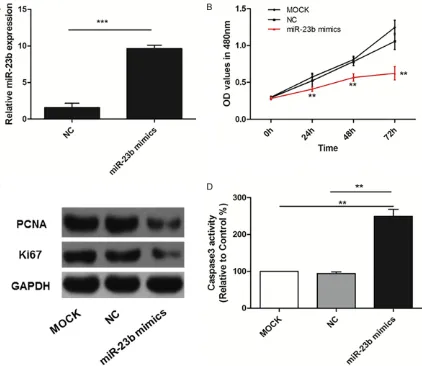
MiR-23b overexpression inhibits the prolifera-tion of EC109 ESCC cells
As shown in the Figure 2A, the expression of
miR-23b was significantly increased in EC109
cells after treatment with miR-23b mimics (P<0.001). Ectopic over-expression of miR-23b could dramatically inhibit proliferation ability of EC109 cells at 24 h, 48 h and 72 h (all panels, P<0.01), no significant difference was found
between MOCK group and NC group (P>0.05) (Figure 2B). In addition, we also found that res-toration of miR-23b decreased the protein expression levels of Ki67 and PCNA (Figure 2C). Meanwhile, Caspase-3 activity was incr-
eased significantly after transfected with
miR-23b mimics (all panels, P<0.01) (Figure 2D).
MiR-23b overexpression inhibits EC109 cells migration
In the wound healing assay, transfected with miR-23b mimics dramatically inhibited the migration ability of EC109 cells compared to the control (P<0.01, Figure 3A, 3B).
MiR-23b overexpression inhibits EC109 cells invasion
In accordance with migration, over-expression of miR-23b resulted in strong invasion inhibito-ry of EC109 cells (all panels, P<0.01) (Figure 4A, 4B). Moreover, the protein expression lev-els of MMP2 in EC109 cells transfected with
[image:5.612.99.521.72.438.2]miR-23b mimics were also decreased signifi -cantly compared with control (Figure 4C).
IMP2 is a direct target of miR-23b
The potential miR-23b binding sites of IMP2 were predicted by three computer-aided algo-rithms including TargetScan, miRanda and PicTar. Figure 5A showed the potential target
sequence of miR-23b harbored in 3’-UTR of
IMP2 mRNA. Luciferase reporter assay was
then performed and result demonstrated that
miR-23b mimics in the wild type (WT) signifi -cantly inhibited the luciferase activity, while the inhibitory effect of miR-23b mimics was
van-ished in the MUT vector (all panels, P<0.05) (Figure 5B). Moreover, qPCR and Western blot analyses revealed that ectopic over-expression of miR-23b dramatically decreased the
[image:6.612.94.524.72.215.2]expres-Figure 3. MiR-23b overexpression inhibited EC109 cells migration. A, B. miR-23b mimics dramatically inhibited the migration ability of EC109 cells compared to the control. **P<0.01 ***P<0.001.
[image:6.612.93.523.263.570.2]sion of IMP2 in EC109 cells (P<0.01) (Figure
[image:7.612.88.518.70.295.2]5C-E). In summary, our results suggest that IMP2 might be a target of miR-23b in EC109 cells.
Figure 5. IMP2 is a direct target of miR-23b. A. Target sequence of miR-23b harbored in 3’-UTR of IMP2 mRNA; B. miR-23b mimics in the wild type (WT) significantly inhibited the luciferase activity, inhibitory effect of miR-23b mim -ics was vanished in the MUT vector; C-E. Over-expression of miR-23b dramatically decreased the expression of IMP2 in EC109 cells. **P<0.01 vs control.
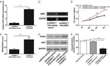
[image:7.612.94.518.364.619.2]IMP2 contributes to miR-23b increased prolif-eration of EC109 cells
PcDNA3.1+HA-IMP2 were transfected in over-expression of miR-23b mimics cells to increase the expression of IMP2 in EC109 cells. qPCR and western blot validated that the IMP2 was increased in the cells transfected with pcDNA3.1+HA-IMP2 compared with the pc- DNA3.1+HA-empty plasmid (P<0.01) (Figure 6A-C).
As shown in Figure 6D, miR-23b contributed to a decrease of EC109 cell proliferation, whereas IMP2 re-introduction reversed the anti-prolifer-ation role of miR-23b. Additionally, Western blot examined that protein expression levels of PCNA and ki-67 in EC109 cells transfected with miR-423-5p mimics were no longer decreased after overexpression of IMP2 (Figure 6E). As for Caspase-3 activity, overexpression IMP2 also decreased caspase-3 activity of cells
transfect-ed with miR-423-5p mimics significantly (all
panels, P<0.01) (Figure 6F).
IMP2 contributes to miR-23b induced migra-tion of EC109 cells
IMP2 re-introduction reversed the inhibitory role of miR-23b in cell migration (all panels, P<0.001) (Figure 7A, 7B).
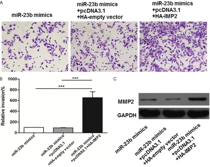
IMP2 contributes to miR-23b induced invasion of EC109 cells
As with migration, over-expression of IMP2 resulted in invasion increasing of EC109 cells
(all panels, P<0.001) (Figure 8A, 8B). Moreover, IMP2 also increased the protein expression lev-els of MMP2 in EC109 cells transfected with
miR-23b mimics significantly compared with
control (Figure 8C).
Discussion
In the present study, we showed that the serum
levels of miR-23b were significantly increased
in ESCC patients compared with healthy con-trols. Further research revealed that the expres-sion of miR-23b tended to be higher in the serum of ESCC patients with well differentiated than in those with moderately differentiated or poorly differentiated. Over-expression of miR-23b inhibited proliferation, migration and inva-sion ability of EC109 cells in vitro. More
impor-tantly, we identified IMP2 as a direct target
gene of miR-23b in EC109 cells. Restoration of
IMP2 significantly reversed miR-23b induced
anti-proliferation and anti-metastasis of EC109 cells.
ESCC dominates in esophageal carcinomas and remains a serious threat to public health in China with an extremely poor prognosis [4, 20]. Despite of the recent rapid promotion in the diagnosis and therapeutic strategies for ESCC patients, prognosis improvement and compre-hensive management of such disease are needed [5]. MiRNAs have been shown to aber-rantly express in a variety of cancers including ESCC [12, 21-23]. Since Tanaka et al. first
[image:8.612.92.521.71.240.2]investigated serum miRNAs and proved the high expression of miR-27a/b in serum to be correlated to poor prognosis [24], Komatsu etc
also identified the plasma miRNA miR-23bas a predictive biomarker for chemoresistance in ESCC [12]. Decreased miR-23b has been reported in ovarian cancer, bladder cancer and Human Gliomas [13, 15, 25]. Among these can-cers, miR-23b might be regarded as an tumor suppressive miRNA and miR-23b overexpres-sion was correlated to restrain cell prolifera-tion, migration and invasion. Which are
consis-tent with our results that miR-23b was signifi -cantly downregulated in ESCC patients and elevated miR-23b expression was oppositely associated with capability of ESCC cell prolifer-ation, caspase 3 activity, migration and inva- sion.
To further understand the mechanism of miR-23b in ESCC, predicting potential binding sites of miR-23b were conducted. We found that
3’-UTR of IMP2 mRNA harbored the target
sequence of miR-23b. Moreover, ectopic
over-expression of IMP2 in EC109 cells transfected with miR-23b mimics led to anticancer capabil-ity silence of miR23b. Previous studies showed that IMP2 expression was increased with tumor size and clinical tumor stage, similar to the results in our study.
These findings pointed out that miR-23b might
function through targeting IMP2 to suppress ESCC proliferation and metastasis.
In conclusion, our current study demonstrates
that serum levels of miR-23b were significantly
increased in ESCC patients compared with healthy controls and upregulation level of miR-23a tended to be associated with
[image:9.612.93.520.73.412.2]clinicopatho-logical grade of ESCC patients. Moreover, find -ings validated that miR-23b might function through targeting IMP2 to suppress ESCC pro-liferation and metastasis. Our results provide new insights into the function of miR-23b in the
development of ESCC and suggest it might rep-resent a potential therapeutic target for ESCC.
Acknowledgements
This study is supported by the National Science Foundation of China (8742543). The authors thank Dr. Xu Shiyang for his kindly providing sta-tistical support in this paper.
Disclosure of conflict of interest
None.
Address correspondence to: Pengcheng Zhang, Department of Cardiothoracic Surgery, Xiangyang No.1 People’s Hospital, Hubei University of Me-dicine, 75 Jiefang Road, Xiangyang 441000, Hubei Province, China. E-mail: zhangpengchenghbxy@163. com
References
[1] Napier KJ, Scheerer M and Misra S. Esopha-geal cancer: a review of epidemiology, patho-genesis, staging workup and treatment mo-dalities. World J Gastrointest Oncol 2014; 6: 112-120.
[2] Jemal A, Bray F, Center MM, Ferlay J, Ward E and Forman D. Global cancer statistics. CA Cancer J Clin 2011; 61: 69-90.
[3] Zhang Y. Epidemiology of esophageal cancer. World J Gastroenterol 2013; 19: 5598-5606. [4] Lin Y, Totsuka Y, He Y, Kikuchi S, Qiao Y, Ueda J,
Wei W, Inoue M and Tanaka H. Epidemiology of esophageal cancer in Japan and China. J Epi-demiol 2013; 23: 233-242.
[5] Sun P, Zhang F, Chen C, Ren C, Bi XW, Yang H, An X, Wang FH and Jiang WQ. Prognostic im-pact of body mass index stratified by smoking status in patients with esophageal squamous cell carcinoma. Onco Targets Ther 2016; 9: 6389-6397.
[6] Ambros V. The functions of animal microRNAs. Nature 2004; 431: 350-355.
[7] Xu W, San Lucas A, Wang Z and Liu Y. Identify-ing microRNA targets in different gene regions. BMC Bioinformatics 2014; 15 Suppl 7: S4. [8] He L, Thomson JM, Hemann MT,
Hernando-Monge E, Mu D, Goodson S, Powers S, Cordon-Cardo C, Lowe SW, Hannon GJ and Hammond SM. A microRNA polycistron as a potential hu-man oncogene. Nature 2005; 435: 828-833. [9] He L, He X, Lim LP, de Stanchina E, Xuan Z,
Li-ang Y, Xue W, Zender L, Magnus J, Ridzon D, Jackson AL, Linsley PS, Chen C, Lowe SW, Cleary MA and Hannon GJ. A microRNA
compo-nent of the p53 tumour suppressor network. Nature 2007; 447: 1130-1134.
[10] Calin GA and Croce CM. MicroRNA signatures in human cancers. Nat Rev Cancer 2006; 6: 857-866.
[11] Bartels CL and Tsongalis GJ. MicroRNAs: novel biomarkers for human cancer. Clin Chem 2009; 55: 623-631.
[12] Komatsu S, Ichikawa D, Kawaguchi T, Takeshi-ta H, Miyamae M, Ohashi T, Okajima W, Imam-ura T, Kiuchi J, Arita T, Konishi H, Shiozaki A, Fujiwara H, Okamoto K and Otsuji E. Plasma microRNA profiles: identification of miR-23a as a novel biomarker for chemoresistance in esophageal squamous cell carcinoma. Onco-target 2016; 7: 62034-62048.
[13] Li W, Liu Z, Chen L, Zhou L and Yao Y. MicroR-NA-23b is an independent prognostic marker and suppresses ovarian cancer progression by targeting runt-related transcription factor-2. FEBS Lett 2014; 588: 1608-1615.
[14] Liang J, Liu X, Xue H, Qiu B, Wei B and Sun K. MicroRNA-103a inhibits gastric cancer cell proliferation, migration and invasion by target-ing c-Myb. Cell Prolif 2015; 48: 78-85.
[15] Majid S, Dar AA, Saini S, Deng G, Chang I, Greene K, Tanaka Y, Dahiya R and Yamamura S. MicroRNA-23b functions as a tumor sup-pressor by regulating Zeb1 in bladder cancer. PLoS One 2013; 8: e67686.
[16] Chen C, Tang Z, Song Q, Yang M, Shi Q and Weng Y. Downregulated microRNA-23b pro-motes BMP9-mediated osteogenesis in C2C12 myoblast cells by targeting Runx2. Mol Med Rep 2016; 13: 2492-2498.
[17] Yan J, Jiang JY, Meng XN, Xiu YL and Zong ZH. MiR-23b targets cyclin G1 and suppresses ovarian cancer tumorigenesis and progres-sion. J Exp Clin Cancer Res 2016; 35: 31. [18] Zhang JY, Chan EK, Peng XX and Tan EM. A
novel cytoplasmic protein with RNA-binding motifs is an autoantigen in human hepatocel-lular carcinoma. J Exp Med 1999; 189: 1101-1110.
[19] Zhou SL, Yue WB, Fan ZM, Du F, Liu BC, Li B, Han XN, Ku JW, Zhao XK, Zhang P, Cui J, Zhou FY, Zhang LQ, Fan XP, Zhou YF, Zhu LL, Liu HY and Wang LD. Autoantibody detection to tu-mor-associated antigens of P53, IMP1, P16, cyclin B1, P62, C-myc, Survivn, and Koc for the screening of high-risk subjects and early detec-tion of esophageal squamous cell carcinoma. Dis Esophagus 2014; 27: 790-797.
expression of microRNA 574-3p as a predictor of postoperative outcome in patients with esophageal squamous cell carcinoma. World J Surg Oncol 2016; 14: 228.
[23] Jin L, Yi J, Gao Y, Han S, He Z, Chen L and Song H. MiR-630 inhibits invasion and metastasis in esophageal squamous cell carcinoma. Acta Biochim Biophys Sin (Shanghai) 2016; 48: 810-819.