Original Article
Evaluation of quantitative real-time polymerase chain
reaction method in detecting
Her-2
gene status of
immunohistochemically scored 2+ invasive breast
carcinoma patients in Yunnan province of China
Wanpu Wang1,2*, Huihua Zhang1,2*, Lilin Luo1,2, Shuang Qiu1,2, Jingwen Hu3, Guanglong Liu4, Long Yang1,2, Shuaiyao Lu3, Tianxing Chen1,2
1Department of Pathology, The First People’s Hospital of Yunnan Province, Kunming, Yunnan, China; 2Affiliated
Hospital of Kunming University of Science and Technology, Kunming, Yunnan, China; 3Institute of Medical Biology,
Chinese Academy of Medical Sciences, Kunming, Yunnan, China; 4Department of Medical, Kunming University of
Science and Technology, Kunming, Yunnan, China. *Equal contributors.
Received March 10, 2017; Accepted May 15, 2017; Epub June 1, 2017; Published June 15, 2017
Abstract: Human epidermal growth factor receptor-2 (Her-2) is a significant prognostic factor and the most impor -tant protein in breast carcinoma target therapy. We aimed to evaluate Her-2 gene amplification status detected by quantitative real-time polymerase chain reaction (Q-PCR) with fluorescent in situ hybridization (FISH) as a golden standard, and searched the optimum threshold of Q-PCR when it can be equivalent to FISH. A total of 108 immuno -histochemistry (IHC) 2+ invasive breast carcinoma cases from Yunnan province of China were enrolled in this study, assessed Her-2 gene status by FISH and Q-PCR and investigated some clinicopathological variables association with Her-2 amplification results of these two methods individually. A significant correlation of Her-2 FISH results and Q-PCR amplification status with differentiation of lymphatic metastasis (P = 0.001, P = 0.005) was observed and metastasis ratio increased with the rising of mRNA expression. When the cutoff value set at 2.60, compared with FISH, Q-PCR had the same great sensitivity (96.59%), specificity (75%), negative predictive value (94.44%) and positive predictive value (83.33%). This study showed that Q-PCR (cutoff = 2.60) had an extremely good consistency (kappa = 0.739) with FISH and could assess Her-2 status instead of FISH in some cases.
Keywords: Invasive breast carcinoma, Her-2 gene amplification assessment, Q-PCR, FISH, Yunnan province of China
Introduction
Breast carcinoma is the most common malig-nant tumor (accounting for 15% of total female tumors) and ranks first in morbidity and mor-tality among Chinese female malignant tumors on one time. Per the current incidence trend of breast carcinoma, by 2030, the incidence cases and death toll will reach to 2.64 and 1.7 million, respectively [1, 2]. Recently, the mor-bidity of breast carcinoma remains high (first), but the mortality is declining (fifth) [2, 3]. Be- tween 2005 and 2011, the 5-year relative sur-vival was found to be 89% [4]. The increasing of survival is not only benefited from the estab -lishment of the female population-wide screen-ing and early detection system but also profited
from the development of molecular biology technology in recent years and the improve-ment of comprehensive diagnosis and treat-ment standardization.
therapy and chemotherapy, but can benefit from the treatment of the targeted drugs-re- combinant DNA derived humanized monoclo -nal antibodies-Trastuzumab (Herceptin) [8, 9], lapatinib (Tykerb), and pertuzumab (Perjeta) [10]. At present, IHC and FISH are performed to detect Her-2 status per ASCO/CAPs, predicting prognosis and guiding the breast carcinoma targeted drugs (especially Herceptin) [11]. As the first line screening method to assess Her-2 protein expression, IHC is widely applied by clinic pathological laboratory since its low cost and simple operation. IHC membrane specificity staining was semi quantitatively scored as 0/1+ (negative), 2+ (equivocal) and 3+ (positive), led to subjective differences in interpretation [12, 13]. The Chinese Human Epidermal Growth Factor Receptor 2 Testing Guideline in Breast carcinoma indicated that IHC score of 2+ cases should perform ISH to ensure Her-2 gene status [14-16]. Although being regarded as the golden standard [17], FISH is operational complexity, time-consuming and costliness [11]. The staining slide should be timely read; otherwise fluorescence quench -ing may lead to inaccurate interpretation. The Guideline also point out that whether the cases with dual-probe Her-2/CEP17 ratio ≥ 2.0 but average Her-2 copy number/cell < 4.0 should be regarded as FISH positive is controversial, whether the cases with dual-probe Her-2/ CEP17 ratio < 2.0 but average Her-2 copy num-ber/cell ≥ 4.0 and < 6.0 should be regarded as FISH positive is indeterminate. In addition, some samples fixed not in time also could not obtain reliable results by FISH [11, 18].
On account of high accuracy, sensitivity, reli-ability and streli-ability, quantitative real-time po- lymerase chain reaction (Q-PCR) is widely ap- plied to assess copy number variation (CNV) in many other types of carcinoma cells including colorectal carcinoma, ovarian carcinoma, and melanoma, especially in formalin-fixed paraf-fin-embedded (FFPE) tissues and fine needle aspiration cytology (FNAC) samples [19-24]. The quantification of Her-2 gene CNV by Q-PCR is always targeting at short fragmented sequ- ences which exists in FFPE samples.
Based on these, take the FISH as a golden standard, we screened 108 patients of breast invasive ductal carcinoma from Yunnan Pro- vince of China to evaluate Q-PCR when it ap-
plied to test Her-2 gene amplification. Under certain conditions, due to its low cost and sim-ple operation, Q-PCR is designed to be as an alternative method when FISH is unable to interpret. When the expression value is bigger than cutoff value, the result is objectively jud- ged as positive and conversely as negative. Materials and methods
Patient material
Tissue samples of 108 invasive breast carci-noma females aged 27-78 (47.4±11.5) who underwent modified radical mastectomy were screened during October 2014 to June 2016 at the Department of Pathology in the First People’s Hospital of Yunnan Province of China. Anonymous use of redundant tissue for rese- arch purposes is part of the treatment agree-ment with patients. And the study was appro- ved by the Ethics Committee of the First People’s Hospital of Yunnan Province. All pa- tients were confirmed for invasive breast car-cinoma first time and had not received any treatment like chemotherapy, radiotherapy or biotherapy before surgery. All specimens were fixed in 10% formalin, embedded in paraffin, and then sliced into 2-3 microns’ slides which in the following with IHC stain scores at 2+. Immunohistochemistry (IHC)
IHC result is interpreted per the percentage of positive cells whose membrane presented clear brown or tan. We selected IHC ambiguity cases scored 2+ referred to The Chinese Human Epidermal Growth Factor Receptor 2 Testing Guideline in Breast carcinoma: more than 10% of the invasive carcinoma cells pre -sented incomplete and/or weak to moderate membrane staining or 10% or less invasive car -cinoma cells presented strong and circumfer-ential membrane staining. Every slide was in- terpreted by two independent pathologists. Fluorescent in situ hybridization (FISH)
Three microns’ thickness slides were baked overnight, dewaxed, and boiled for 23 min at 100±5°C. Digested with pepsin for 13 to 15
number/cell < 4.0 were treated as none Her-2 gene amplification; the samples with red/green ratios < 2.0 and average Her-2 copy number/ cell ≥ 4 but < 6.0 or with red/green ratios ≥ 2.0 but average Her-2 copy number/cell < 4.0 were indeterminate; the samples with many clustered red signals were directly treated as Her-2 gene amplification without calculation.
Quantitative real-time polymerase chain reac-tion (Q-PCR)
[image:3.612.89.377.90.433.2]DNA was extracted from paraffin-embedded tissue (PPFE) with DNA Rapid Extraction Kit (centrifugal column type) (TIANGEN, China). The tissue block was sliced up into 5-8 10-μM-thick pieces, lysis and dissolved, and kept the middle layer of water phase into a new centrifuge tube Table 1. Primers for Her-2 and β-actin
Gene Sense primer sequence Anti-sense primer sequence
β-actin GCGCGGCTACAGCTTCA CTTAATGTCACGCACGATTTCC Her-2 GAAGGACATCTTCCACAAGAACAA CGAGAGCGGTTGGTGTCTATC
min after airing and sufficient -ly washed in 2 × SSC. De- hydrated in 70%, 90%, 100% ethanol gradient solutions and added probe after air-dried (the following steps need to be operated away from light). Hybridization was performed in a hybridizer (Thermo Brite, USA) at 85°C for 5 min, followed by 16 hours at 37°C. After washing in 2 × SSC and 0.1% in NP-40/2 × SSC at 37°C, re- spectively, the slides were dried in the dark, dehydrated in 70% ethanol, and added DAPI solution for the further evaluation under a fluores -cence microscope after seal-ing coverslip.
In distinct invasion tumor region, total GSP Her-2 sig-nals (red) and CEP17 sigsig-nals (green) were counted respec-tively within 60 tumor cells nuclear to calculated the ratio of red signals to green sig-nals. The samples with red/ green ratios ≥ 2.0 or with red/ green ratios < 2.0 but aver- age Her-2 copy number/cell ≥ 6.0 were treated as Her-2 gene amplification; the sam -ples with red/green ratios < 2.0 and average Her-2 copy Figure 1. Example specimen of breast infiltrating ductal carcinoma IHC 2+
after centrifugal. Added 2 times volume of an- hydrous ethanol, 3 min standing after incorpo-rating thoroughly, the liquid was injected into an adsorption column CR2. After centrifugal, it was washed with buffer GD, rinsed with buffer PW, idling centrifugal for 2 min, dried at room temperature for 2-5 min. Put the CR2 adsorp-tion column into a clean centrifuge tube and add 30 μl elution buffer or ddH2O preheated to 65°C TE, placed for 2-5 min at room tempera-ture, and then centrifuged for 2 min at 12,000 rpm (~13,400 × g) to collect DNA. The purity
and the concordance percentages and correla-tions (Spearman’s rho) by Chi square test with GraphPad Prism5 statistical software and PASW statistics software. For Q-PCR, sensitivi -ty, specifici-ty, positive (PPV) and negative pre -dictive value (NPV) were calculated with FISH as a golden standard.
Results
[image:4.612.91.370.108.294.2]The mean age of the enrolled patients is 47.4-years old, which is similar to the relevant study Table 2. Correlation of Her-2 FISH results with age, location,
tumor size and lymphatic metastasis in 108 breast carcinoma patients
Clinicopathological
parameters Patients
Her-2
Positive (%) Negative (%) χ2, P
Age
< 40 years 22 16 (72.7) 6 (27.3%) χ2 = 0.769 ≥ 40 years 86 72 (83.7) 14 (16.3%) P = 0.38
Location
Left 33 27 (81.8%) 6 (18.2%) χ2 = 0
Right 75 61 (81.3%) 14 (18.7%) P = 1 Tumor size
< 5 cm 96 82 (85.4%) 14 (14.6%) χ2 = 6.675 ≥ 5 cm 12 6 (50%) 6 (50%) *P = 0.01
Lymphatic metastasis
Positive 60 56 (93.3%) 4 (6.7%) χ2 = 10.862
Negative 48 32 (66.7%) 16 (33.3%) **P = 0.001
* and **significant p values are shown bold.
Table 3. Correlation of her-2 Q-PCR results with age, location, tumor size and lymphatic metastasis in 108 breast carcinoma patients
Clinicopathological
parameters Patients
Her-2 CNV
≥ 2 (%) < 2 (%) χ2, P Age
< 40 years 22 18 (81.8%) 4 (18.2%) χ2 = 0.718 ≥ 40 years 86 78 (90.7%) 8 (9.3%) P = 0.474 Location
Left 33 31 (93.9%) 2 (6.1%) χ2 = 0.511 Right 75 65 (86.7%) 10 (13.3%) P = 0.610 Tumor size
< 5 cm 96 86 (89.5%) 10 (10.4%) χ2 = 0.494 ≥ 5 cm 12 10 (83.3%) 2 (16.7%) P = 0.630 Lymphatic metastasis
Positive 60 60 (100%) 0 (0%) χ2 = 2.897 Negative 48 36 (75%) 12 (25%) **P = 0.005
**significant p values are shown bold.
and integrity of the DNA were evaluated by calculating the O.D. 260:280 ratios.
The Q-PCR mixtures were pre-pared per the Bio-Rad SsoFast Evagreen protocol (Singapore). The primers for Her-2 and β-actin are shown in Table 1. Amplifications were performed in a Bio-Rad CFX Connect Real-Time System (California, USA) under the following proce-dures: 95°C for 10 min and followed by 39 amplification cycles at 95°C for 10 sec and 57°C for 30 sec. Then 65°C for 31 sec and followed by 60 amplification cycles at 65°C for 5 sec with +0.5°C/cycle, rising 0.5°C/s. Data were analyzed by the CFX manager 3.0 soft -ware. The housekeeping gene β-actin was used for the quan -tification of Her-2 CNV status in this study. Her-2 CNV was cal-culated using a comparative threshold cycle (Ct) with the following formula: CNV = 2-ΔΔCT,
where ΔΔCt = (CtHer-2
-Ctβ-actin) sample-(CtHer-2
-Ctβ-ac-tin) normal. The gene copy number ratio of the unknown sample ≥ 2 was defined as amplified. The gene copy num -ber ratio of the unknown sam-ple < 2 was defined as non-amplified [23, 25, 26].
Statistics
[image:4.612.92.369.364.550.2][2], much younger than western countries. There are 22 patients younger than 40 years old and 86 patients older than or equal to the age of 40. The tumors of 75 cases located in the right, and 33 cases located in the left. There were 60 cases with lymph node sis, and 48 cases without lymph node metasta-sis. Nighty-six cases tumors were smaller than 5 cm, and 12 cases were bigger than 5 cm. All samples immunohistochemical results were scored at 2+ (Figure 1A) and then were ana-lyzed for FISH and Q-PCR to determine Her-2 gene amplification. In total 108 cases, there are 88 cases of FISH positive shown in Figure 1B and 1C with positive rate of 81.5% and 20 cases of FISH negative shown in Figure 1D with negative rate of 18.5%.
There was no significant statistical difference of Her-2 FISH amplification respect to different age groups (P = 0.38) and tumor location (P = 1). But, there was significant statistical differ -ence (χ2 = 6.675, P = 0.01) for Her-2 FISH amplification between different tumor sized. The positive rate of bigger size was 4.2 times (95% CI 2.084~8.466) of the smaller group. There was also a significant statistical differ -ence (χ2 = 10.862, P = 0.001) for Her-2 FISH amplification between lymphatic metastasis group and non-lymphatic metastasis group. The former positive rate was 7 times (95% CI 2.154~22.749) of the latter (Table 2).
The gene copies number ratios of the samples ≥ 2 were defined as amplified, and < 2 were defined as non-amplified. There was no signifi -cant statistical difference for Her-2 Q-PCR am-
plified and non-amplified respect to age (P = 0.474), tumor location (P = 0.610) and tumor size (P = 0.630). However, there was a signifi -cant statistical difference (P = 0.005) for Her-2 Q-PCR amplified and non-amplified between lymphatic metastasis positive group and nega-tive group. The former posinega-tive rate was 1.333 times (95% CI 1.132~1.57) of the latter (Table 3).
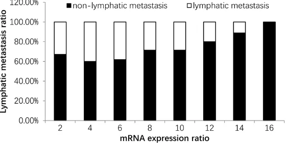
It is worth to noting that there was a signifi-cant statistical difference (P < 0.01) of mRNA expression between lymphatic metastasis specimen and non-lymphatic metastasis speci-men (Figure 2). And the more the amount of mRNA expression, the higher the metastasis cases proportion. When expressing quantity reached 16 times of the control, metastasis proportion reached 100% (Figure 3).
There were 60 cases in lymphatic metastasis group of which included 1 case with FISH and Q-PCR negative, 55 cases with double-positive and 4 cases with FISH negative but Q-PCR positive. Both results coincidence rate was 93.3%. There were 48 cases in non-lym -phatic metastasis group of which included 12 cases with FISH and Q-PCR double-negative, 32 cases with double-positive and 4 cases with FISH negative but Q-PCR positive. Both results coincidence rate was 91.7% (Figure 4).
To find the correlation between Q-PCR and FISH results, after repeated testing, we setup Her-2 Q-PCR cutoff value at 2.60. An mRNA ratio greater than or equal to 2.60 was taken as positive, and less than 2.60 was taken as negative. With FISH method as the gold stan -dard, the sensitivity of Q-PCR was 96.59%, specificity was 75%, positive predictive value was 94.44%, and negative predictive value was 83.33%. So, concluded that the Q-PCR method is highly consistent with the FISH (kappa = 0.739) (Figure 5).
Discussion
Human epidermal growth factor receptor-2 (Her-2) gene amplification and/or mRNA and/or protein over-expression cause excessive prolif-erated cells forming tumors. Some research claimed that Her-2 molecular positive expres-sion ratio in breast carcinoma is between 20% and 30%, which is 3-4 times higher than the other carcinomas, especially higher in infiltrat -Figure 2. There was a significant statistical differ
ing ductal carcinoma with high malignancy. The target drugs such as Herceptin can precisely kill malignant cells with Her-2 positive status instead of affecting normal cells survival. Breast carcinoma cells with HER-2 protein over-expression and/or Her-2 gene amplification are sensitive to Herceptin. Moreover, Her-2 molecu-lar status is a significant prognosticator of breast carcinoma in clinic. IHC and FISH are universally accepted for these two molecular states [27-29]. Because of the efficient-cost and simple operation, IHC is much preferred by many hospital laboratories, meanwhile, its results might be more subjective per the artifi -cially interpretation system. Besides, the inap-propriate tissues processing can lead to ambig-uous results sometimes [30]. As a protein tar-geted detection technology, IHC cannot report the true amplification status of Her-2 gene pre-cisely. Nucleic acid as a target detection tech-nology, FISH is used to assess Her-2 gene CNV, especially for indeterminate specimens with an IHC score of 2+ according to National Com- prehensive Carcinoma Network (NCCN) Guide- lines. Contrast with IHC, FISH result is much closer to the true amplification status of Her-2 gene status in breast carcinoma cells with
bet-sitivity, accuracy and reliability, especially the fragmented DNA in the delay fixed specimens [32]. Tianjie Pu (2015) et al. showed that the Q-PCR results did not change with a cold isch-emia time of up to 12 hours. Thus, to the DFF (the delay to formalin fixation) issues, Q-PCR might be a surrogate for the Her-2 detection. As a result, some researchers suggested that Q-PCR could be a stable and reliable alterna-tive method for the evaluation of Her-2 gene CNV in breast carcinoma especially for samples that were not promptly placed in fixative. In summary, Q-PCR has advantages in sensitivity, accuracy and reliability, easy operating, high throughput time-saving. Meanwhile, the only disadvantage of Q-PCR utilization in breast infil -trating ductal carcinoma is that it cannot locate the target area in situ, especially in some het-erogeneous tumors and can be solved by laser capture micro dissection method [32].
[image:6.612.94.374.77.217.2]Previous investigations showed that breast carcinoma in different regions, different races have differences. For ethnic minorities, eco -nomically and traffic underdeveloped and geo -graphical restricted in Yunnan province, can lead to the particularity of the incidence of Figure 3. Nighty-four cases of mRNA expression quantity were 2 times in the
control group, the lymphatic metastasis for 58 cases (61.7%). Sixty cases of mRNA expression quantity were 4 times in the control group, the lymphatic metastasis for 36 cases (60%). Forty-two cases of mRNA expression quantity were 6 times in the control group, the lymphatic metastasis for 26 cases (61.9%). Twenty-eight cases of mRNA expression quantity were 8 times in the control group, the lymphatic metastasis for 20 cases (71.4%). Twenty-eight cases of mRNA expression quantity were 10 times in the control group, the lymphatic metastasis for 20 cases (71.4%). Twenty cases of mRNA expres -sion quantity were 12 times in the control group, the lymphatic metastasis for 16 cases (80%). Eighteen cases of mRNA expression quantity were 14 times in the control group, the lymphatic metastasis for 16 cases (88.9%). Six cases of mRNA expression quantity were 16 times in the control group, the lymphatic metastasis for 6 cases (100%).
ter stability, accuracy, sensi-tivity and repeatability. How- ever, fluorescence signals must be observed and count-ed within 20 or even more invasive carcinoma cells in time which is time-consum-ing. Fluorescence quenching caused by delay or exposure in white light for a long time, this could ultimately lead to erroneous interpretation. For this reason, together with costly apparatus, FISH is not widely used in many primary hospitals. It is worth to be noted that the ASCO/CAP guideline and other studies have cautioned that approxi-mately 20-26% of current Her-2 test results might be inaccurate when detected by FISH [31].
sen-breast carcinoma. In conclusion, Undergoing the repeated tests, we set the reasonable cut-off value at 2.60 (kappa = 0.739) within the Q-PCR detection data of 108 females. Take the FISH as golden standard, when interpret the result per this value, Q-PCR method has a great sensitivity (96.59%), specificity (75%), negative predictive value (94.44%) and positive predic -tive value (83.33%), which can be used as a potential alternative of FISH to test Her-2 gene status. Moreover, the correlation of Her-2 FISH results and Q-PCR results are respectively com-pared with clinic pathological variables in 108 breast carcinoma patients in our study. And we
cated that in some cases, Q-PCR can be a reli-able and streli-able alternative method for the eval-uation of Her-2 status in immunohistochemical scored 2+ breast carcinoma in Yunnan Province of China, even wider area.
Acknowledgements
This study was supported by Applied Found-ation Research Project of Yunnan Province 2014FZ069.
Disclosure of conflict of interest
[image:7.612.94.523.78.254.2]None.
Figure 4. Summary of FISH and Q-PCR results for FFPE tissue samples from 108 breast carcinomas. Samples numbered 1 to 60 are lymphatic metastasis cases and samples numbered 61 to number 108 are non-lymphatic metastasis cases. Ratios of samples clustered amplification of FISH were determined at 30. Each Q-PCR copy num -ber value represents the Her-2 status for each sample. The dashed horizontal line indicates the Q-PCR and FISH threshold cutoff of 2 copies for a sample to be deemed Her-2 positive. The FISH values were expressed as the copy number (Her-2/CEP17 ratio). There were 7 cases numbered 2, 3, 4, 5 (metastasis group), 61, 70, 74, and 76 (non-metastasis group) were positive by Q-PCR but negative by FISH.
Figure 5. When the threshold value was set to 2.60, kappa value reached maximum (kappa = 0.739), which meant that Q-PCR method was highly con-sistent with the FISH method.
[image:7.612.96.371.359.496.2]-Address correspondence to: Shuaiyao Lu, Institute of Medical Biology, Chinese Academy of Medical Sciences, 379 Jiaoling Road, Kunming, Yunnan, China. Tel: +86-15096625297; Fax: +86-871-684-00646; E-mail: lushuaiyao-km@163.com; Tianxing Chen, Department of Pathology, The First People’s Hospital in Yunnan Province, Kunming, Yunnan, China; Affiliated Hospital of Kunming University of Science and Technology, 157 Jinbi Road, Kunming, Yunnan, China. Tel: 13708858871; Fax: +86-871-63638441; E-mail: 82898626@qq.com
References
[1] Akarolo-Anthony SN, Ogundiran TO and Ade-bamowo CA. Emerging breast cancer epidem-ic: evidence from Africa. Breast Cancer Res 2010; 12 Suppl 4: S8.
[2] Balekouzou A, Yin P, Pamatika CM, Bishwajit G, Nambei SW, Djeintote M, Ouansaba BE, Shu C, Yin M, Fu Z, Qing T, Yan M, Chen Y, Li H, Xu Z and Koffi B. Epidemiology of breast cancer: retrospective study in the central African re-public. BMC Public Health 2016; 16: 1230. [3] Shaukat U, Ismail M and Mehmood N. Epide
-miology, major risk factors and genetic predis-position for breast cancer in the Pakistani population. Asian Pac J Cancer Prev 2013; 14: 5625-5629.
[4] Rojas K and Stuckey A. Breast cancer epide- miology and risk factors. Clin Obstet Gynecol 2016; 59: 651-672.
[5] Slamon DJ, Clark GM, Wong SG, Levin WJ, Ull -rich A and McGuire WL. Human breast cancer: correlation of relapse and survival with amplifi -cation of the HER-2/neu oncogene. Science 1987; 235: 177-182.
[6] Goud KI, Dayakar S, Vijayalaxmi K, Babu SJ and Reddy PV. Evaluation of HER-2/neu status in breast cancer specimens using immunohis-tochemistry (IHC) & fluorescence in-situ hybrid -ization (FISH) assay. Indian J Med Res 2012; 135: 312-317.
[7] Gheybi MK, Baradaran A, Mohajeri MR, Ost-ovar A, Hajalikhani P and Farrokhi S. Validity of immunohistochemistry method in predicting HER-2 gene status and association of clinico-pathological variables with it in invasive breast cancer patients. APMIS 2016; 124: 365-371. [8] Romond EH, Perez EA, Bryant J, Suman VJ,
Geyer CJ, Davidson NE, Tan-Chiu E, Martino S, Paik S, Kaufman PA, Swain SM, Pisansky TM, Fehrenbacher L, Kutteh LA, Vogel VG, Visscher DW, Yothers G, Jenkins RB, Brown AM, Dakhil SR, Mamounas EP, Lingle WL, Klein PM, Ingle JN and Wolmark N. Trastuzumab plus adjuvant chemotherapy for operable HER2-positive breast cancer. N Engl J Med 2005; 353: 1673-1684.
[9] Piccart-Gebhart MJ, Procter M, Leyland-Jones B, Goldhirsch A, Untch M, Smith I, Gianni L, Baselga J, Bell R, Jackisch C, Cameron D, Dow-sett M, Barrios CH, Steger G, Huang CS, Ander-sson M, Inbar M, Lichinitser M, Lang I, Nitz U, Iwata H, Thomssen C, Lohrisch C, Suter TM, Ruschoff J, Suto T, Greatorex V, Ward C, Straeh-le C, McFadden E, Dolci MS and Gelber RD. Trastuzumab after adjuvant chemotherapy in HER2-positive breast cancer. N Engl J Med 2005; 353: 1659-1672.
[10] Jelovac D and Emens LA. HER2-directed thera-py for metastatic breast cancer. ONCOL (Wil-liston Park) 2013; 27: 166-175.
[11] Wolff AC, Hammond ME, Hicks DG, Dowsett M, McShane LM, Allison KH, Allred DC, Bartlett JM, Bilous M, Fitzgibbons P, Hanna W, Jenkins RB, Mangu PB, Paik S, Perez EA, Press MF, Spears PA, Vance GH, Viale G and Hayes DF. Recommendations for human epidermal growth factor receptor 2 testing in breast can-cer: American society of clinical oncology/col-lege of American pathologists clinical practice guideline update. J Clin Oncol 2013; 31: 3997-4013.
[12] Gancberg D, Jarvinen T, di Leo A, Rouas G, Cardoso F, Paesmans M, Verhest A, Piccart MJ, Isola J and Larsimont D. Evaluation of HER-2/ NEU protein expression in breast cancer by im -munohistochemistry: an interlaboratory study assessing the reproducibility of HER-2/NEU testing. Breast Cancer Res Tr 2002; 74: 113-120.
[13] Portier BP, Wang Z, Downs-Kelly E, Rowe JJ, Patil D, Lanigan C, Budd GT, Hicks DG, Rimm DL and Tubbs RR. Delay to formalin fixation ‘cold ischemia time’: effect on ERBB2 detec-tion by in-situ hybridizadetec-tion and immunohisto -chemistry. Mod Pathol 2013; 26: 1-9.
[14] Bethune GC, Veldhuijzen VZD, MacIntosh RF, Rayson D, Younis T, Thompson K and Barnes PJ. Impact of the 2013 American society of clinical oncology/college of American patholo-gists guideline recommendations for human epidermal growth factor receptor 2 (HER2) testing of invasive breast carcinoma: a focus on tumours assessed as ‘equivocal’ for HER2 gene amplification by fluorescence in-situ hy -bridization. Histopathology 2015; 67: 880-887.
[15] Rakha EA, Starczynski J, Lee AH and Ellis IO. The updated ASCO/CAP guideline recommen-dations for HER2 testing in the management of invasive breast cancer: a critical review of their implications for routine practice. Histopa-thology 2014; 64: 609-615.
immunohistochemistry-equivo-cal (2+) invasive breast cancer. J Thorac Dis 2014; 6: 896-904.
[17] Yoon N, Do IG and Cho EY. Analysis of HER2 status in breast carcinoma by fully automated HER2 fluorescence in situ hybridization (FISH): comparison of two immunohistochemical tests and manual FISH. APMIS 2014; 122: 755-760. [18] Moatamed NA, Nanjangud G, Pucci R, Lowe A,
Shintaku IP, Shapourifar-Tehrani S, Rao N, Lu DY and Apple SK. Effect of ischemic time, fixa -tion time, and fixative type on HER2/neu im -munohistochemical and fluorescence in situ hybridization results in breast cancer. Am J Clin Pathol 2011; 136: 754-761.
[19] Wang TL, Maierhofer C, Speicher MR, Lengauer C, Vogelstein B, Kinzler KW and Velculescu VE. Digital karyotyping. Proc Natl Acad Sci U S A 2002; 99: 16156-16161.
[20] Wang TL, Diaz LJ, Romans K, Bardelli A, Saha S, Galizia G, Choti M, Donehower R, Parmigiani G, Shih I, Iacobuzio-Donahue C, Kinzler KW, Vogelstein B, Lengauer C and Velculescu VE. Digital karyotyping identifies thymidylate syn -thase amplification as a mechanism of resis -tance to 5-fluorouracil in metastatic colorectal cancer patients. Proc Natl Acad Sci U S A 2004; 101: 3089-3094.
[21] Salani R, Davidson B, Fiegl M, Marth C, Muller-Holzner E, Gastl G, Huang HY, Hsiao JC, Lin HS, Wang TL, Lin BL and Shih I. Measurement of cyclin E genomic copy number and strand length in cell-free DNA distinguish malignant versus benign effusions. Clin Cancer Res 2007; 13: 5805-5809.
[22] Yu J, Miller R, Zhang W, Sharma M, Holtschlag V, Watson MA and McLeod HL. Copy-number analysis of topoisomerase and thymidylate synthase genes in frozen and FFPE DNAs of colorectal cancers. Pharmacogenomics 2008; 9: 1459-1466.
[23] Moore DA, Saldanha G, Ehdode A, Potter L, Dyall L, Bury D and Pringle JH. Accurate detec-tion of copy number changes in DNA extracted from formalin-fixed, paraffin-embedded mela -noma tissue using duplex ratio tests. J Mol Diagn 2013; 15: 687-694.
[24] Rodriguez C, Suciu V, Poterie A, Lacroix L, Mi -ran I, Boichard A, Delaloge S, Deneuve J, Azou -lay S, Mathieu MC, Valent A, Michiels S, Arne-dos M and Vielh P. Concordance between HER-2 status determined by qPCR in fine nee -dle aspiration cytology (FNAC) samples com -pared with IHC and FISH in core needle biopsy (CNB) or surgical specimens in breast cancer patients. Mol Oncol 2016; 10: 1430-1436.
[25] Abdul Murad NA, Razak ZA, Hussain RM, Syed Hussain SN, Ko Ching Huat C, Che Md Ali SA, Abdullah N, Muhammad R, Ibrahim N, Jamal R. Quantification of Her-2/Neu gene in breast cancer patients using real time-polymerase chain reaction (Q-PCR) and correlation with im-munohistochemistry findings. Asian Pac J Can -cer Prev 2013; 14: 1655-1659.
[26] O’Malley FP, Parkes R, Latta E, Tjan S, Zadro T, Mueller R, Arneson N, Blackstein M and Andru-lis I. Comparison of HER2/neu status assessed by quantitative polymerase chain reaction and immunohistochemistry. Am J Clin Pathol 2001; 115: 504-511.
[27] Moelans CB, de Weger RA, Van der Wall E and van Diest PJ. Current technologies for HER2 testing in breast cancer. Crit Rev Oncol Hema-tol 2011; 80: 380-392.
[28] Rosa FE, Silveira SM, Silveira CG, Bergamo NA, Neto FA, Domingues MA, Soares FA, Caldeira JR and Rogatto SR. Quantitative real-time RT-PCR and chromogenic in situ hybridization: precise methods to detect HER-2 status in breast carcinoma. BMC Cancer 2009; 9: 90. [29] Tse C, Brault D, Gligorov J, Antoine M,
Neu-mann R, Lotz JP and Capeau J. Evaluation of the quantitative analytical methods real-time PCR for HER-2 gene quantification and ELISA of serum HER-2 protein and comparison with fluorescence in situ hybridization and immuno -histochemistry for determining HER-2 status in breast cancer patients. Clin Chem 2005; 51: 1093-1101.
[30] Dobson L, Conway C, Hanley A, Johnson A, Costello S, O’Grady A, Connolly Y, Magee H, O’Shea D, Jeffers M and Kay E. Image analysis as an adjunct to manual HER-2 immunohisto-chemical review: a diagnostic tool to standard-ize interpretation. Histopathology 2010; 57: 27-38.
[31] Qian XL, Wen HY, Yang YL, Gu F, Guo XJ, Liu FF, Zhang L, Zhang XM and Fu L. Assessment of dual-probe Her-2 fluorescent in situ hybridiza -tion in breast cancer by the 2013 ASCO/CAP guidelines produces more equivocal results than that by the 2007 ASCO/CAP guidelines. Breast Cancer Res Tr 2016; 159: 31-39. [32] Pu T, Guo P, Qiu Y, Chen S, Yang L, Sun L, Ye F