Original Article
Expression of p16 predicts poor outcome for patients
with gastrointestinal stromal tumors
Nu Ri Jang, Joon Hyuk Choi, Mi Jin Gu
Department of Pathology, Yeungnam University College of Medicine, Republic of Korea
Received March 13, 2017; Accepted April 20, 2017; Epub June 1, 2017; Published June 15, 2017
Abstract: Gastrointestinal stromal tumors are the most common mesenchymal tumors found in the gastrointestinal tract. Their biological behavior is still predicted by a consensus scheme proposed by the U.S. National Institutes of
Health. In this study, we investigated the prognostic significance of p16 protein expression in gastrointestinal stro
-mal tumors. Expression of p16 protein was observed in 42.4% (92/217) of tumors and was significantly associated
with a high mitotic count, tumor necrosis, recurrence or metastasis, and a higher-risk group. Patients with p16-expressing gastrointestinal stromal tumors showed a shorter overall survival and disease-free survival than those without p16 expression; however, p16 expression was not an independent prognostic factor. The risk of malignant behavior and the presence of recurrence or metastasis were independent prognostic factors. Expression of p16 protein predicts poor outcome and can be a useful marker to predict relapse or metastasis and aggressive behavior in gastrointestinal stromal tumors.
Keywords: Gastrointestinal stromal tumor, p16, risk, survival, immunohistochemistry
Introduction
Gastrointestinal stromal tumors (GISTs) are the most common mesenchymal neoplasms of the gastrointestinal tract and characterized by oncogenic mutation in the KIT (80-85%) and platelet-derived growth factor receptor alpha (PDGRFA; 5-7%) gene [1, 2]. The majority of GISTs occur in adults andrespond to targeted tyrosine kinase therapy. However, approximate-ly 10-15% of GISTs are KIT/PDGFRA wild-type (WT) GISTs, which are less sensitive to tyrosine kinase inhibitors. The KIT/PDGFRA WT GISTs are heterogeneous tumors and include succi-nate dehydrogenase (SDH)-deficient, neurofi -bromatosis 1-associated, BRAF mutant, and quadruple WT GISTs [3].
GISTs show a wide range of biological behav-iors, which are predicted by tumor size and mitotic counts [2]. However, it is hard to predict the biological behavior based on histological findings alone.
The p16 gene is a tumor suppressor that inhib-its cell cycling by arresting cells in the G1-S phase. This genetic alteration results in loss of
p16 protein expression in many human cancers [4, 5]. In contrast, p16 overexpression was observed in breast cancer and premalignant lesions, breast ductal intraepithelial neoplasia, carcinoma in situ of the cervix, and prostatic intraepithelial neoplasia [4, 6-9]. The prognos-tic significance of p16 expression status has been reported in GISTs, but the results were quite inconsistent in studies [10-14]. The aim of this study was to investigate the expression sta-tus of p16 in GISTs and to assess its clinical and pathological significance.
Materials and methods
Patient characteristics
so assessed: tumor location, tumor size, mitotic count, tumor cell type, necrosis, mucosal ulceration, and recurrence or metastasis. The risk of malignant behavior was classified according to the system proposed by Miettinen and Lasota (the so-called AFIP criteria) [15] and further classi-fied as low, moderate, or high risk. Overall survival (OS) was defined as the time from surgical resection to death or the last follow-up. The follow-up period ended in October 2016 (OS range: 0-215 months). This study was approved by our institu-tional Human Ethics Review Board. Tissue microarray construction Two to five 2-mm cores were obtained from the most representative tumor area of each block and arrayed in a new recipient block. Thus, 11 tissue microarray blocks were constructed. Four to five cores comprising breast carcinoma, thyroid papillary carcino-ma, normal gastric mucosa, palatine tonsil, and uterine leiomyoma were used as control tissues.
Immunohistochemistry
[image:2.612.95.522.72.235.2]Immunohistochemistry for p16 (clo- ne E6H4, mouse monoclonal anti-body, prediluted; Ventana, Tucson, AZ, USA) was performed after on-board heat-induced epitope retrieval in standard pH CC1 buffer (37°C, 32 Figure 1. Immunohistochemical study for p16 in gastrointestinal stromal tumor. A. Expression, B. No expression.
Table 1. Correlation between clinicopathologic factors and p16 expression in gastrointestinal stromal tumors
Clinicopathologic
factors No.
p16 expression P No Expression
Sex 0.646
Male 114 64 (56.1%) 50 (43.9%) Female 103 61 (59.2%) 42 (40.8%)
Location 0.485
Stomach 146 89 (61.0%) 57 (39.0%) Small intestine 66 34 (51.5%) 32 (48.5%) Colorectum 3 1 (33.3%) 2 (66.7%) Extra-gastrointestinal 2 1 (50.0%) 1 (50.0%)
Cell type 0.917
Spindle 199 114 (57.3%) 85 (42.7%) Epithelioid 7 4 (57.1%) 3 (42.9%) Mixed 11 7 (63.6%) 4 (36.4%)
Mitosis < 0.001
≤ 5/50 HPF 138 92 (66.7%) 46 (33.7%) > 5/50 HPF 79 33 (41.8%) 46 (58.2%)
Risk <0.001
Low 128 87 (68.0%) 41 (32.0%) Intermediate 40 21 (52.5%) 19 (47.5%) High 49 17 (34.7%) 32 (65.3%)
Necrosis 0.002
No 182 113 (62.1%) 69 (37.9%) Yes 35 12 (34.3%) 23 (65.7%)
Mucosal invasion 0.607
No 193 110 (57.0%) 83 (43.0%) Yes 24 15 (62.5%) 9 (37.5%)
Recurrence or metastasis < 0.001
[image:2.612.91.346.300.701.2]min) on the automated Benchmark® platform
(Ventana Medical Systems). The staining was visualized using the UltraView™ universal DAB
detection kit (Automated BenchMark®, Ven-
tana), which included a hydrogen peroxide sub-strate and a 3, 3’-diaminobenzidine chromogen
[image:3.612.91.388.72.293.2]variables with significant results in univariate analysis were analyzed in multivariate analysis. Hazard ratios (HRs) and associated 95% confi -dence intervals (CIs) were calculated for each variable. Statistical significance was accepted for p values < 0.05.
Figure 2. Survival curves of overall survival for p16 expression versus no p16 expression. The p16-expressing gastrointestinal tumors showed a shorter over-all survival rate (P < 0.001).
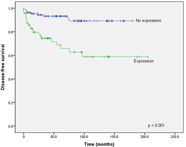
Figure 3. Survival curves of disease-free survival for p16 expression versus no p16 expression. The p16-expressing gastrointestinal tumors showed a shorter disease-free survival rate (P < 0.001).
solution. The slides were subsequently counterstai- ned with hematoxylin. Interpretation of immuno-histochemistry
Slides were assessed by an investigator who was blind-ed to the patients’ clinico-pathological information. We defined p16 expression as more than 20% of total tumor cells showing nucle-ar staining with or without a cytoplasmic reaction. Lym- phocytes and background stromal cells served as the positive controls.
Statistical analysis
Comparisons were perfor- med using SPSS version 23.0 (SPSS Inc., Chicago, IL, USA). The χ2 test and
[image:3.612.91.389.353.589.2]Results
Clinicopathological characteristics
A total of 126 males and 120 female patients with median age of 58.5 years (range: 22-88 years) were included in this study. The median tumor size was 4.79 cm (range: 1-23 cm). Expression of CD117 and DOG1 was found in 222 (98.2%) cases. SDHB-negative GISTs were detected in only two gastric WT GISTs (one in a 56-year-old female and the other in a 15-year-old male patient). The SDHB-negative GISTs
revealed diffuse strong positive staining on CD117 and DOG1.
Comparison between expression of p16 and clinicopathological factors
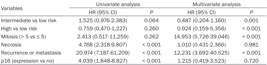
Expression of p16 was found in 42.4% (92/217) of GISTs (Figure 1). The p16 expression was sig-nificantly associated with GISTs with a higher mitotic count (> 5/50 high-power fields [HPF]), tumor necrosis, recurrence or metastasis, and a higher-risk group with respect to aggressive behavior (Table 1). Patients with p16-express-ing GISTs showed shorter OS (P < 0.001) (Figure 2) and DFS (P < 0.001) (Figure 3) than those without p16 expression. On multivariate analy-sis, risk of malignant behavior and recurrence or metastasis were independent prognostic factors. The intermediate-risk group (P = 0.001, HR 10.370, CI 2.611-41.187) and high-risk group (P < 0.001, HR 13.459, CI 3.555-50.952) showed shorter survival than the low-risk group. Patients without recurrence or metastasis had better survival than those with recurrence or metastasis (P < 0.016, HR 0.369, CI 0.164-0.831) (Table 2).
Discussion
Most clinicopathological studies have
[image:4.612.91.523.97.203.2]demon-are the most important prognostic indicators of GISTs. However, they do not always reliably pre-dict patient outcomes. The clinical behavior of GIST varies, and some small and mitotically inactive GISTs show aggressive behaviors [16]. A reliable method to predict the prognosis of GIST is necessary for clinical management. Alteration of cell-cycle regulatory proteins has been implicated in the pathogenesis and tumor progression of various kinds of human cancers. Loss of p16 expression has been reported to be associated with progression to malignant disease [17]. However, p16 overexpression was found in some tumors, and it was associated with the aggressiveness of disease subtypes [6-8]. Although there have been extensive stud-ies of p16 expression in GISTs, discrepancstud-ies still exist with respect to its prognostic value [18]. Loss of p16 expression has been previ-ously reported as a negative prognostic factor in GISTs. Schneider-Stock et al. [19] did not find any correlation between p16 gene alteration and clinicopathologic variables, but p16 loss was associated with a poor prognosis and p16 expression was higher in the benign GISTs. Huang et al. [20] also demonstrated that com-plete loss of p16 expression preferentially affected intermediate- and high-risk groups, and they suggested that p16 deregulation might be involved in early tumorigenesis. Several other studies have confirmed this cor -relation and its implication for poor prognosis [21-23]. However, Haller et al. [24] demonstrat-ed that loss of chromosomal region 9p21 ldemonstrat-ed to reduced mRNA and p16 expression in GISTs. Steigen et al. [25] also showed that patients with p16-expressing GISTs had a significantly worse OS than those without p16 expression. In addition, p16-expressing GISTs tended to have a larger size and a higher mitotic count (> 5/50 HPF) compared with those not expressing
Table 2. Univariate and multivariate analyses of clinicopathologic factors affecting the survival of patients with gastrointestinal stromal tumors
Variables Univariate analysis Multivariate analysis
HR (95% CI) P HR (95% CI) P
Intermediate vs low risk 1.525 (0.976-2.383) 0.064 0.487 (0.204-1.160) 0.001 High vs low risk 0.759 (0,470-1.227) 0.260 0.924 (0.159-5.356) < 0.001
p16. Our study showed similar results, although p16 expression was not correlated with tumor size. These results were also confirmed in another study by Schmieder et al. [26], who revealed that p16-expressing GISTs tended to develop more recurrence or metastasis and showed a worse disease-specific survival and DFS compared with those not expressing p16. They also suggested that p16 expression might be an indicator for high-risk GIST. Our study showed nearly identical results with Schmie- der’s study in that p16-expressing GISTs were significantly associated with a higher-risk group and had a tendency of more recurrence or metastasis and worse OS and DFS. Regarding these contradictory results, although loss of p16 expression biologically contributes to ma- lignancy, other oncogenic changes such as loss of RB or TP53 and aberrant activation of cyclin D1 may lead to increased proliferation and dys-regulation of the cell cycle [14].
Prognostic factors in GISTs have been widely studied, and tumor size and mitotic count have been accepted as reliable factors. Other fac-tors such as anatomic location, cellular atypia, and tumor necrosis have been shown to be independent prognostic factors in some stud-ies [25]. However, it is still difficult to predict the risk of developing recurrence or metasta-sis, a higher mitotic count, and a higher risk, especially in small biopsied GISTs. Our study showed that p16 expression was a highly pre-dictive factor for the presence of recurrence of metastasis and being in a higher-risk group for patients with GISTs.
In summary, p16 expression in GISTs was
sig-nificantly associated with a higher mitotic count, tumor necrosis, recurrence or metasta-sis, and a higher-risk group with respect to aggressive behavior. Furthermore, p16-expre- ssing GISTs revealed shorter OS and DFS com-pared with those without expression. The expression of p16 can be a highly predictive marker to predict recurrence or metastasis and aggressive behavior in GISTs.
Disclosure of conflict of interest
None.
Address correspondence to: Dr. Mi Jin Gu, Depart- ment of Pathology, College of Medicine, Yeungnam University, 170, Hyeonchung-ro, Nam-gu, Daegu, Re-
public of Korea. Tel: 8253-640 6756; Fax: M8253-622-8432; E-mail: mjgu@yu.ac.kr
References
[1] Miettinen M and Lasota J. Gastrointestinal stromal tumors: pathology and prognosis at different sites. Semin Diagn Pathol 2006; 23: 70-83.
[2] Fletcher CD, Berman JJ, Corless C, Gorstein F, Lasota J, Longley BJ, Miettinen M, O’Leary TJ, Remotti H, Rubin BP, Shmookler B, Sobin LH and Weiss SW. Diagnosis of gastrointestinal stromal tumors: A consensus approach. Hum Pathol 2002; 33: 459-465.
[3] Lasota J, Felisiak-Golabek A, Wasag B, Kowalik A, Zieba S, Chlopek M, Wang ZF, Coates T, Kop-czynski J, Gozdz S, Sarlomo-Rikala M and Miet-tinen M. Frequency and clinicopathologic
pro-file of PIK3CA mutant GISTs: molecular genetic
study of 529 cases. Mod Pathol 2016; 29: 275-282.
[4] Witkiewicz AK, Knudsen KE, Dicker AP and Knudsen ES. The meaning of p16(ink4a)
ex-pression in tumors: functional significance,
clinical associations and future developments. Cell Cycle 2011; 10: 2497-2503.
[5] Haller F, Gunawan B, von Heydebreck A, Schwager S, Schulten HJ, Wolf-Salgo J, Langer C, Ramadori G, Sultmann H and Fuzesi L. Prog-nostic role of E2F1 and members of the CDK-N2A network in gastrointestinal stromal tu-mors. Clin Cancer Res 2005; 11: 6589-6597. [6] Bechert C, Kim JY, Tramm T and Tavassoli FA.
Co-expression of p16 and p53 characterizes aggressive subtypes of ductal intraepithelial neoplasia. Virchows Arch 2016; 469: 659-667. [7] Lebok P, Roming M, Kluth M, Koop C, Ozden C, Taskin B, Hussein K, Lebeau A, Witzel I, Wolber L, Geist S, Paluchowski P, Wilke C, Heilenkotter U, Muller V, Schmalfeldt B, Simon R, Sauter G, Terracciano L, Krech RH, von der Assen A and Burandt E. P16 overexpression and 9p21 dele-tion are linked to unfavorable tumor pheno-type in breast cancer. Oncotarget 2016; 7: 81322-81331.
[8] Pare R, Shin JS and Lee CS. Increased expres-sion of senescence markers p14(ARF) and p16(INK4a) in breast cancer is associated with an increased risk of disease recurrence and poor survival outcome. Histopathology 2016; 69: 479-491.
[10] Tetikkurt US, Ozaydin IY, Ceylan S, Gurbuz Y, Erdogan N and Oz F. Predicting malignant po-tential of gastrointestinal stromal tumors: Role of p16 and E2F1 expression. Appl Immunohis-tochem Mol Morphol 2010; 18: 338-343. [11] Huang HY, Huang WW, Lin CN, Eng HL, Li SH, Li
CF, Lu D, Yu SC and Hsiung CY. Immunohisto-chemical expression of p16INK4A, Ki-67, and Mcm2 proteins in gastrointestinal stromal tu-mors: prognostic implications and correlations
with risk stratification of NIH consensus crite -ria. Ann Surg Oncol 2006; 13: 1633-1644. [12] Jung SH, Suh KS, Kang DY, Kang DW, Kim YB
and Kim ES. Expression of DOG1, PDGFRA, and p16 in gastrointestinal stromal tumors. Gut Liver 2011; 5: 171-180.
[13] Schneider-Stock R, Boltze C, Lasota J, Peters B, Corless CL, Ruemmele P, Terracciano L, Pross M, Insabato L, Di Vizio D, Iesalnieks I, Dirnhofer S, Hartmann A, Heinrich M, Miettin-en M, Roessner A and Tornillo L. Loss of p16
protein defines high-risk patients with gastroin -testinal stromal tumors: a tissue microarray study. Clin Cancer Res 2005; 11: 638-645. [14] Schmieder M, Wolf S, Danner B, Stoehr S,
Juchems MS, Wuerl P, Henne-Bruns D, Knipps-child U, Hasel C and Kramer K. P16 Expression differentiates high-risk gastrointestinal stro-mal tumor and predicts poor outcome. Neopla-sia 2008; 10: 1154-1162.
[15] Miettinen M and Lasota J. Gastrointestinal stromal tumors: pathology and prognosis at different sites. Semin Diagn Pathol 2006; 23: 70-83.
[16] Miettinen M, Sobin LH and Lasota J. Gastroin-testinal stromal tumors of the stomach: a clini-copathologic, immunohistochemical, and mo-lecular genetic study of 1765 cases with long-term follow-up. Am J Surg Pathol 2005; 29: 52-68.
[17] Michaloglou C, Vredeveld LC, Soengas MS, De-noyelle C, Kuilman T, van der Horst CM, Majoor DM, Shay JW, Mooi WJ and Peeper DS. BRAFE600-associated senescence-like cell cy-cle arrest of human naevi. Nature 2005; 436: 720-724.
[18] Schneider-Stock R, Boltze C, Lasota J, Miettin-en M, Peters B, Pross M, Roessner A and Gun-ther T. High prognostic value of p16INK4 alter-ations in gastrointestinal stromal tumors. J Clin Oncol 2003; 21: 1688-1697.
[19] Schneider-Stock R, Boltze C, Lasota J, Peters B, Corless CL, Ruemmele P, Terracciano L, Pross M, Insabato L, Di Vizio D, Iesalnieks I, Dirnhofer S, Hartmann A, Heinrich M, Miettin-en M, Roessner A and Tornillo L. Loss of p16
protein defines high-risk patients with gastroin -testinal stromal tumors: a tissue microarray study. Clin Cancer Res 2005; 11: 638-645. [20] Huang HY, Huang WW, Lin CN, Eng HL, Li SH, Li
CF, Lu D, Yu SC and Hsiung CY. Immunohisto-chemical expression of p16INK4A, Ki-67, and Mcm2 proteins in gastrointestinal stromal tu-mors: prognostic implications and correlations
with risk stratification of NIH consensus crite -ria. Ann Surg Oncol 2006; 13: 1633-1644. [21] Sabah M, Cummins R, Leader M and Kay E.
Altered expression of cell cycle regulatory pro-teins in gastrointestinal stromal tumors: mark-ers with potential prognostic implications. Hum Pathol 2006; 37: 648-655.
[22] Haller F, Gunawan B, von Heydebreck A, Schwager S, Schulten HJ, Wolf-Salgo J, Langer C, Ramadori G, Sultmann H and Fuzesi L. Prog-nostic role of E2F1 and members of the CDK-N2A network in gastrointestinal stromal tu-mors. Clin Cancer Res 2005; 11: 6589-6597. [23] Ricci R, Arena V, Castri F, Martini M, Maggiano
N, Murazio M, Pacelli F, Potenza AE, Vecchio FM and Larocca LM. Role of p16/INK4a in gas-trointestinal stromal tumor progression. Am J Clin Pathol 2004; 122: 35-43.
[24] Haller F, Agaimy A, Cameron S, Beyer M, Gu-nawan B, Happel N, Langer C, Ramadori G, von Heydebreck A and Fuzesi L. Expression of p16INK4A in gastrointestinal stromal tumours (GISTs): two different forms exist that indepen-dently correlate with poor prognosis. Histopa-thology 2010; 56: 305-318.
[25] Steigen SE, Bjerkehagen B, Haugland HK, Nor-drum IS, Loberg EM, Isaksen V, Eide TJ and Nielsen TO. Diagnostic and prognostic markers for gastrointestinal stromal tumors in Norway. Mod Pathol 2008; 21: 46-53.