Original Article
Correlation between
AEG-1
gene polymorphisms
and osteosarcoma susceptibility
Wenlong Zhang1,2*, Na Ge3*, Lele Dong2, Jianqiang Lian2, Jinlei Sun2, Yanping Zheng1
1Department of Orthopedics, Qilu Hospital of Shandong University, Jinan, Shandong, China; 2Department of
Orthopedics, First Affiliated Hospital, Baotou Medical College, Baotou, Inner Mongolia, China; 3Institute of
Nutri-tion and Food Health, School of Public Health, Baotou Medical College, Baotou, Inner Mongolia, China. *Equal
contributors andco-first authors.
Received December 2, 2016; Accepted December 22, 2016; Epub June 1, 2017; Published June 15, 2017
Abstract: Aim: The objective of this study was to explore the relationship between single nucleotide polymorphisms (SNPs) of the astrocyte elevated gene-1 (AEG-1) gene (rs16896059 and rs1311) and osteosarcoma (OS) suscep-tibility in Chinese Han population. Methods: The case and control groups were evaluated by Hardy-Weinberg equi-librium (HWE). Genotype and allele frequencies of the two polymorphisms were obtained by direct calculation. Differences of the genotyes and alleles of these polymorphisms were assessed by Chi-square test. Relative risk
of OS was represented by odds ratios (ORs) with 95% confidence intervals (CIs). Results: Frequencies of AA geno
-type and A allele of rs16896059 SNP between OS patients and healthy individuals were significantly different
(P<0.05). AA genotype and A allele may be protective against OS onset respectively (OR=0.258, 95%
CI=0.068-0.986; OR=0.628, 95% CI=0.401-0.985). However, there was no significant differences of the genotype and allele
distributions of AEG-1 gene rs1311 polymorphism between case and control groups (P>0.05). Conclusion: AEG-1 gene rs16896059 polymorphism act as a protect factor for the occurrence of OS. But rs1311 had no significant
association with the OS risk.
Keywords: Osteosarcoma, AEG-1, polymorphisms, susceptibility
Introduction
Osteosarcoma (OS) is a most common primary malignancy in bone tissues, particularly among children and adolescents. But there is a second incidence peak among individuals who is older than 60 years [1, 2]. Although the incidence of OS is not high, it has a rising trend recent years. Meanwhile the mortality of it is obviously high, and with the easy metastasis and recurrence the OS prognosis is very poor. Exploration of the OS etiology is essential, so as to provide a theoretical basis for the therapy method. Until now, etiology of OS remains unknown. But it was considered that OS is a complex disease. Development and progression of this disease is caused by an interaction of environmental fac-tors and genetic susceptibility [3-5]. Recent evi-dence indicated that oncogenes, suppressor genes and DNA-repair genes may play critical roles in the determination of individual suscep-tibility to cancers. Polymorphisms in these
genes possibly alter their expression and func-tion, may increase or decrease carcinogen acti-vation or detoxication and modulate DNA repair. As an important oncogene, astrocyte elevated gene-1 (AEG-1, also known as MTDH and Lyric),
was originally identified as an HIV-inducible
gene in primary human fetal astrocytes [6].
AEG-1 gene is widely over expressed in many malignant tumors [7-16]. AEG-1 participates in tumorigenesis, including cellular transforma-tion, apoptosis inhibitransforma-tion, invasion, metastasis, angiogenesis and resistance to chemothera-peutic agents [17-24]. Up-regulation of AEG-1 promotes the growth, invasion and metastasis of cancers via several signaling pathways such
as PI3K/AKT, NF-κB and MAPK pathways [25].
DNA extraction kit (Tiagen Biotech Co., Ltd, China) according to the manufacturer’s protocol.
PCR primers of AEG-1 gene polymorphisms were designed by Primer Premier 5.0 and syn-thesize by Sangon Biotech Co., Ltd. PCR primer sequences were as follows: forward, 5’-CGT GAT AAG GTG CTG ACT GAT TC-3’; and reverse, 5’-CAG GAA ATG ATG CGG TTG TAA G-3’. PCR
amplification was performed in a total volume
of 25 µl, containing 100 ng genomic DNA, 2.5 µl of 10 × PCR buffer, 1.5 mM MgCl2, 0.15 mM dNTPs, 25 pM of each primer, and 1 U of Taq DNA polymerase. PCR reaction conditions was as follows: an initial denaturation step at 94°C for 5 min, followed by 35 cycles of 30 s at 94°C,
30 s at 60°C, 30 s at 72°C and a final elonga -tion at 72°C for 10 min. PCR products were sequenced by Sangon Biotech (Shanghai) Co., Ltd.
Statistical analysis
Hardy-Weinberg equilibrium (HWE) was used to detect the representativeness of the cases and controls. Differences of clinical features, geno-types and alleles between case and control
groups were assessed by χ2 test. Odds ratios
(ORs) and 95% confidence intervals (95% CIs)
were calculated by Chi-squared test. All statisti-cal analyses were performed by PASW 18.0 statistical software. P-value less than 0.05
were considerate as statistically significant.
Results
General characteristics of subjects
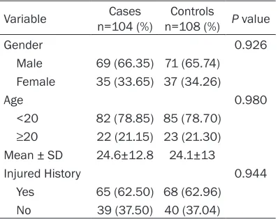
General characteristics of the participants were shown in Table 1. Our data indicated that
there were no significant differences between
OS patients and cancer-free controls in age, gender and injured history.
Association of AEG-1 gene rs16896059 and rs1311 polymorphisms with OS risk
Genotype distributions of rs16896059 and rs1311 SNPs in case and control groups were all in accordance with HWE test.
The results of genotype distribution showed that AA genotype frequency of rs16896059
SNP was significantly higher in controls (P= In this study, we attempted to further
investi-gate the potential role of AGE-1 gene polymor-phisms in OS development. Single nucleotide polymorphism (SNP), the most common
poly-morphism, is defined as genetic variation in a DNA sequence. Identification of SNPs in the
host can potentially facilitate the evaluation of the susceptibility of cancer and predict pro-gression of disease or response to treatment. Here, we investigated rs16896059 and rs1311 SNPs in AEG-1 gene to determine whether a
particular SNP may influence susceptibility to
OS development. Materials and methods
Study population
A total of 104 OS patients (aged 10-41 years old) and 108 healthy controls (aged 13-45 years old) were enrolled in this study. OS patients were diagnosed by histopathological examination at Qilu Hospital of Shandong University from 2005 to 2015. Healthy controls were recruited from the healthy check-up cen-ter of the same hospital during the same peri-od. Control subjects were matched with patients in terms of age, gender and injured history. This study was approved by Review Boards of Qilu Hospital of Shandong University. All the participants were unrelated and signed the written informed consent.
Genomic DNA extraction and genotyping method
[image:2.612.90.289.96.253.2]Gnomic DNA was extracted from 2 ml whole peripheral venous blood using the TIANamp
Table 1. Comparison of the clinical features between case and control groups
Variable n=104 (%)Cases n=108 (%)Controls P value
Gender 0.926
Male 69 (66.35) 71 (65.74)
Female 35 (33.65) 37 (34.26)
Age 0.980
<20 82 (78.85) 85 (78.70)
≥20 22 (21.15) 23 (21.30) Mean ± SD 24.6±12.8 24.1±13
Injured History 0.944
ed in a variety of tumor biological behaviors
[28]. AEG-1 is first find in the study of HIV-1
infection in human astrocytes [29, 30]. AEG-1 is a single-pass transmembrane protein, and mainly presented in nucleolus. This protein is an extremely conservative protein in mammals. Soon after, it was found that AEG-1 expressed in almost all tissues. The expression level of AEG-1 is up-regulated in various cancer cells. Besides, AEG-1 over-expressed in metastatic breast cancer and promoted the adhesion of mammary tumor cells. AEG-1 protein is neces-sary for the proliferation, metastasis invasion and prognosis of tumors, and even the angio-genesis in tumor tissues [31, 32]. AEG-1 emerge as a potential regulator for many ma- lignant tumors, and mediate by complex
signal-ing pathway includsignal-ing PI3K/AKT, NF-κB and
MAPK pathways [25]. AEG-1 protein also relates to the metastasis, invasion and drug tolerance in OS patients [33, 34].
AEG-1 protein is encoded by AEG-1 gene which is located in chromosome 8q22.1. As we all know, polymorphisms in the genes may alter the expression and function of the correspond-ing protein, and then lead to disorders of the 0.036, Table 2), indicating a statistical
associa-tion with the onset of OS (OR=0.258, 95% CI=0.068-0.986). Meanwhile, rs1311 geno-types had no obvious association with the occurrence of OS (P>0.05). As the Table 3
shown, we observed a statistically significant
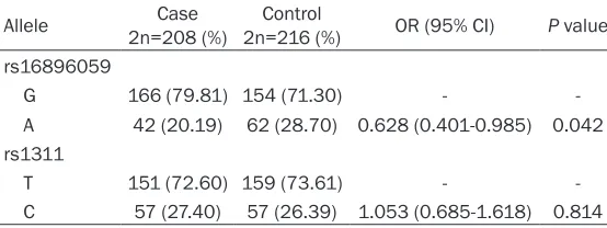
difference of rs16896059 A allele frequency between case and control groups (P=0.042). A allele appears to be protective against OS development (OR=0.628, 95% CI=0.401-0.985). However, C allele distribution of rs1311
polymorphism appears to be insignificant
between case and control groups (P>0.05), suggesting that the rs1311 variant may not relate to the OS occurrence.
Discussion
OS is a most common life-threatening malig-nancy originated from mesenchymal tissues. This tumor often occurs in children, adoles-cents and young adults. Besides, OS incidence is highly in males than that in females [26, 27]. With a high tendency of metastasis and recur-rence, OS has a poor prognosis. Although the incidence of OS is not high, the mortality of it is high, and the morbidity and mortality has a
ris-Table 2. Genotype frequency of AEG-1 gene polymorphisms in the osteosarcoma (OS) cases and cancer-free controls
Genotype n=104 (%)Case n=108 (%)Control OR (95% CI) P value rs16896059
GG 65 (62.5) 56 (51.85) -
GA 36 (34.62) 42 (38.89) 0.738 (0.417-1.307) 0.297 AA 3 (2.88) 10 (9.26) 0.258 (0.068-0.986) 0.036 rs1311
TT 57 (54.81) 58 (53.70) -
[image:3.612.91.372.96.228.2]CT 37 (35.58) 43 (39.81) 0.876 (0.494-1.551) 0.649 CC 10 (9.62) 7 (6.48) 1.454 (0.518-4.083) 0.476
Table 3. Association between OS risk and allele frequencies of
AEG-1 gene polymorphisms
Allele 2n=208 (%)Case 2n=216 (%)Control OR (95% CI) P value rs16896059
G 166 (79.81) 154 (71.30) -
A 42 (20.19) 62 (28.70) 0.628 (0.401-0.985) 0.042 rs1311
T 151 (72.60) 159 (73.61) -
C 57 (27.40) 57 (26.39) 1.053 (0.685-1.618) 0.814
ing trend recent years. More- over, current therapy method for OS is not effective. There- fore, exploration of the OS pathogenesis is necessary. Despite many studies investi-gate the etiology of OS, but the mechanism of this disease remains unclear. Cumulative studies suggested that OS is a complex disease caused by the combined effects of genet-ic and environmental factors. Among these factors genetic background is an important determinant for OS develop- ment.
In order to certify the patho-genesis of OS, an extensive functional genomics research is needed which will discover new and unknown polymorphic loci for OS etiology. Recent studies indicate that AEG-1
[image:3.612.91.368.275.379.2][2] Mirabello L, Troisi RJ and Savage SA. Osteosar-coma incidence and survival rates from 1973 to 2004: data from the surveillance, epidemi-ology, and end results program. Cancer 2009; 115: 1531-1543.
[3] Powers M, Zhang W, Lopez-Terrada D, Czerniak BA and Lazar AJ. The molecular pathology of sarcomas. Cancer Biomark 2010; 9: 475-491. [4] Bovee JV and Hogendoorn PC. Molecular pa -thology of sarcomas: concepts and clinical
im-plications. Virchows Arch 2010; 456: 193-199.
[5] de Alava E. Molecular pathology in sarcomas. Clin Transl Oncol 2007; 9: 130-144.
[6] Su ZZ, Chen Y, Kang DC, Chao W, Simm M, Vol
-sky DJ and Fisher PB. Customized rapid sub -traction hybridization (RaSH) gene microarrays identify overlapping expression changes in hu-man fetal astrocytes resulting from huhu-man
im-munodeficiency virus-1 infection or tumor ne -crosis factor-alpha treatment. Gene 2003; 306: 67-78.
[7] Zhao Y, Moran MS, Yang Q, Liu Q, Yuan C, Hong S and Kong B. Metadherin regulates radiore-sistance in cervical cancer cells. Oncol Rep 2012; 27: 1520-1526.
[8] Kim DH, Mohapatra G, Bollen A, Waldman FM and Feuerstein BG. Chromosomal abnormali -ties in glioblastoma multiforme tumors and glioma cell lines detected by comparative ge-nomic hybridization. Int J Cancer 1995; 60: 812-819.
[9] Poon TC, Wong N, Lai PB, Rattray M, Johnson PJ and Sung JJ. A tumor progression model for hepatocellular carcinoma: bioinformatic analy-sis of genomic data. Gastroenterology 2006; 131: 1262-1270.
[10] Wang Y, Klijn JG, Zhang Y, Sieuwerts AM, Look
MP, Yang F, Talantov D, Timmermans M, Mei -jer-van Gelder ME, Yu J, Jatkoe T, Berns EM,
Atkins D and Foekens JA. Gene-expression pro
-files to predict distant metastasis of
lymph-node-negative primary breast cancer. Lancet 2005; 365: 671-679.
[11] van’t Veer LJ, Dai H, van de Vijver MJ, He YD,
Hart AA, Mao M, Peterse HL, van der Kooy K, Marton MJ, Witteveen AT, Schreiber GJ, Kerk-hoven RM, Roberts C, Linsley PS, Bernards R
and Friend SH. Gene expression profiling pre -dicts clinical outcome of breast cancer. Nature 2002; 415: 530-536.
[12] van de Vijver MJ, He YD, van’t Veer LJ, Dai H, Hart AA, Voskuil DW, Schreiber GJ, Peterse JL,
Roberts C, Marton MJ, Parrish M, Atsma D,
Wit-teveen A, Glas A, Delahaye L, van der Velde T, Bartelink H, Rodenhuis S, Rutgers ET, Friend
SH and Bernards R. A gene-expression signa-ture as a predictor of survival in breast cancer. N Engl J Med 2002; 347: 1999-2009.
organism. SNP, the most common polymor-phism, play a crucial role in the occurrence and development of multiple diseases. As an impor-tant regulator of cancer progression, polymor-phisms in AEG-1 gene might lead to many can-cers. However, there are few researches focus on the association of AEG-1 gene polymor-phisms and cancer risk.
To our knowledge, this is the first study which
investigated the impact of AEG-1 gene polymor-phism in risk of OS. We selected rs16896059 and rs1311 two SNPs of AEG-1 gene to explore the association with OS risk. Then we found that AA genotype and A allele of rs16896059 polymorphism were frequently observed in con-trols. The results indicated that AA genotype and A allele decreased the OS risk approxi-mately 0.258 and 0.628 fold. That was partly according to the results in ovarian cancer [35]. But GA genotype of rs16896059 and all of the genotypes of rs1311 polymorphism had no
sig-nificant association with the occurrence of OS.
Because the incidence of OS is low, so the sample size of this study is small. Besides, there only one ethnicity involves in our study. Although the participants of this study had a
good representativeness, and no significant
differences of clinical features between case and control groups, the results of this study
was still insufficient to certify the pathogenesis
of OS. Thus well designed studies with large sample size and more ethnicity are necessary in the future, so as to obtain more exact evi-dence to certify the pathogenesis of OS. Acknowledgements
This study received funding from the Depart- ment of Education, Inner Mongolia, China (NJZY16208).
Disclosure of conflict of interest None.
Address correspondence to: Yanping Zheng, De- partment of Orthopedics, Qilu Hospital of Shandong University, Jinan 250012, Shandong, China. E-mail: [email protected]
References
[25] Sarkar D, Emdad L, Lee SG, Yoo BK, Su ZZ and
Fisher PB. Astrocyte elevated gene-1: far more
than just a gene regulated in astrocytes. Can-cer Res 2009; 69: 8529-8535.
[26] Stiller CA, Bielack SS, Jundt G and
Steliarova-Foucher E. Bone tumours in European children
and adolescents, 1978-1997. Report from the automated childhood cancer information sys-tem project. Eur J Cancer 2006; 42: 2124-2135.
[27] Haddox CL, Han G, Anijar L, Binitie O, Letson GD, Bui MM and Reed DR. Osteosarcoma in pediatric patients and young adults: a single institution retrospective review of presenta-tion, therapy, and outcome. Sarcoma 2014; 2014: 402509.
[28] Motalleb G, Gholipour N and Samaei NM. As-sociation of the human astrocyte elevated gene-1 promoter variants with susceptibility to hepatocellular carcinoma. Med Oncol 2014; 31: 916.
[29] Su ZZ, Kang DC, Chen Y, Pekarskaya O, Chao
W, Volsky DJ and Fisher PB. Identification and
cloning of human astrocyte genes displaying
elevated expression after infection with HIV-1 or exposure to HIV-1 envelope glycoprotein by
rapid subtraction hybridization, RaSH. Onco-gene 2002; 21: 3592-3602.
[30] Su ZZ, Kang DC, Chen Y, Pekarskaya O, Chao
W, Volsky DJ and Fisher PB. Identification of
gene products suppressed by human
immuno-deficiency virus type 1 infection or gp120 ex -posure of primary human astrocytes by rapid subtraction hybridization. J Neurovirol 2003; 9: 372-389.
[31] Yang C, Zheng S, Liu Q, Liu T, Lu M, Dai F, Gao
X, Sheyhidin I and Lu X. Metadherin is required for the proliferation, migration, and invasion of esophageal squamous cell carcinoma and its meta-analysis. Transl Res 2015; 166: 614-626.e2.
[32] Liu Y, Kong X, Li X, Li B and Yang Q. Knockdown of metadherin inhibits angiogenesis in breast cancer. Int J Oncol 2015; 46: 2459-2466. [33] Liu B, Wu Y and Peng D. Astrocyte elevated
gene-1 regulates osteosarcoma cell invasion and chemoresistance via endothelin-1/endo-thelin A receptor signaling. Oncol Lett 2013; 5: 505-510.
[34] Tang J, Shen L, Yang Q and Zhang C. Overex-pression of metadherin mediates metastasis of osteosarcoma by regulating epithelial-mes-enchymal transition. Cell Prolif 2014; 47: 427-434.
[35] Yuan C, Li X, Yan S, Yang Q, Liu X and Kong B. The MTDH (-470G>A) polymorphism is associ-ated with ovarian cancer susceptibility. PLoS One 2012; 7: e51561.
[13] Long M, Dong K, Gao P, Wang X, Liu L, Yang S,
Lin F, Wei J and Zhang H. Overexpression of
astrocyte-elevated gene-1 is associated with cervical carcinoma progression and angiogen-esis. Oncol Rep 2013; 30: 1414-1422. [14] Liu L, Wu J, Ying Z, Chen B, Han A, Liang Y,
Song L, Yuan J, Li J and Li M. Astrocyte elevated gene-1 upregulates matrix metalloprotein-ase-9 and induces human glioma invasion. Cancer Res 2010; 70: 3750-3759.
[15] Yoo BK, Emdad L, Su ZZ, Villanueva A, Chiang
DY, Mukhopadhyay ND, Mills AS, Waxman S,
Fisher RA, Llovet JM, Fisher PB and Sarkar D.
Astrocyte elevated gene-1 regulates hepatocel-lular carcinoma development and progression. J Clin Invest 2009; 119: 465-477.
[16] Yu C, Chen K, Zheng H, Guo X, Jia W, Li M, Zeng M, Li J and Song L. Overexpression of astrocyte elevated gene-1 (AEG-1) is associated with esophageal squamous cell carcinoma (ESCC) progression and pathogenesis. Carcinogene-sis 2009; 30: 894-901.
[17] Emdad L, Lee SG, Su ZZ, Jeon HY, Boukerche
H, Sarkar D and Fisher PB. Astrocyte elevated
gene-1 (AEG-1) functions as an oncogene and regulates angiogenesis. Proc Natl Acad Sci U S A 2009; 106: 21300-21305.
[18] Emdad L, Sarkar D, Lee SG, Su ZZ, Yoo BK,
Dash R, Yacoub A, Fuller CE, Shah K, Dent P, Bruce JN and Fisher PB. Astrocyte elevated
gene-1: a novel target for human glioma thera-py. Mol Cancer Ther 2010; 9: 79-88.
[19] Jiang T, Zhu A, Zhu Y and Piao D. Clinical impli-cations of AEG-1 in liver metastasis of colorec-tal cancer. Med Oncol 2012; 29: 2858-2863. [20] Ke ZF, Mao X, Zeng C, He S, Li S and Wang LT.
AEG-1 expression characteristics in human non-small cell lung cancer and its relationship with apoptosis. Med Oncol 2013; 30: 383. [21] Li C, Liu J, Lu R, Yu G, Wang X, Zhao Y, Song H,
Lin P, Sun X, Yu X, Zhang Y, Ning X and Geng J. AEG -1 overexpression: a novel indicator for peritoneal dissemination and lymph node me-tastasis in epithelial ovarian cancers. Int J Gy-necol Cancer 2011; 21: 602-608.
[22] Song H, Li C, Lu R, Zhang Y and Geng J. Expres-sion of astrocyte elevated gene-1: a novel marker of the pathogenesis, progression, and poor prognosis for endometrial cancer. Int J Gy-necol Cancer 2010; 20: 1188-1196.
[23] Sun W, Fan YZ, Xi H, Lu XS, Ye C and Zhang JT.
Astrocyte elevated gene-1 overexpression in human primary gallbladder carcinomas: an un-favorable and independent prognostic factor. Oncol Rep 2011; 26: 1133-1142.
[24] Yoo BK, Chen D, Su ZZ, Gredler R, Yoo J, Shah