Original Article
MicroRNA-375 confers cisplatin resistance by regulating
KRAS expression in non-small cell lung cancer cell
Yong Li*, Pei Wang*, Xiao-Hui Li, Hao-Jie Wang, Gong-Ning Shi
Department of Cardio-Thoracic Surgery, Huaihe Hospital of Henan University, Kaifeng 475001, Henan Province, China. *Equal contributors and co-first authors.
Received September 16, 2015; Accepted October 24, 2015; Epub March 1, 2016; Published March 15, 2016
Abstract: Cisplatin is a classic chemotherapeutic drug that has been widely used for non small cell lung cancer (NSCLC) treatment, but NSCLC cells are often resistant to primary or acquired cisplatin therapy. Several studies have shown that miR-375 is significantly elevated in tumor and suggests that this miRNA might play a role in this re-sistance. In this study, we investigated this possibility and the possible mechanism underlying this role. We showed that forced expression of miR-375 significantly inhibited apoptosis, enhanced cell proliferation and increased the resistance of tumor cells to cisplatin in NSCLC H1975 cells. Furthermore, knock-down of miR-375 reversed these effects on H1975 cells and increased the sensitivity of H1975 to cisplatin chemotherapy. Finally, we showed that miR-375 targeted the KRAS gene, and directly regulated the expression of KRAS mRNA. These results demonstrate that miR-375 may play an important role in the cisplatin resistance of NSCLC.
Keywords: miR-375, NSCLC, cisplatin, KRAS
Introduction
Non small cell lung cancer (NSCLC) is one of the most common types of lung cancers. Despite advances in surgery, chemotherapy and molecular targeted therapy, the mortality rate has only decreased slightly over the past years [1]. Among classic chemotherapeutic drugs, cisplatin is one of the widely used che-motherapies for the adjuvant and metastatic therapy of NSCLC. However, primary or acquired resistance to platinum-based chemotherapy is a major clinical obstacle to the successful treatment of NSCLC [2]. Tremendous efforts have been taken to understand molecular and cellular mechanisms for the action of cisplatin over the past decades, but it remains unclear how these NSCLC cells become resistant to cis-platin. Thus, a better understanding of molecu-lar events underlying this drug resistance is important and necessary for achieving effec-tive therapy against this type of cancer.
MicroRNAs (miRNA) are a class of small RNA molecules that regulate the translation and degradation of mRNAs. MiRNAs bind to
The relationship between miR-375 dys-regula-tion and human cancer resistance has attract-ed increasing attention. However, the associa-tion of miR-375 with the sensitivity of NSCLC cells to cisplatin has yet to be explored. The present study aims to provide insights into the association of miR-375 expression with cispla-tin resistance in NSCLC. As a result, inhibition of this miR-375 expression led to hypersensitiv-ity of NSCLC cell H1975 to cisplatin, whereas further over-expression of miR-375 promoted cell proliferation. Also, we found that KRAS ge- ne is a direct target of miR-375. These results suggest that miR-375 might be one of the key cellular components important for the develop-ment of NSCLC cisplatin resistance. Our study also implies that targeting miR-375 may over-come the resistance of NSCLC to cisplatin and thus serve as a potential target for the develop-ment of cancer therapy specifically against cis-platin resistant cancer cells.
Materials and methods
Cell culture and infection
Non-small cell lung cancer cell line H1975 was cultured in RPMI1640 medium with 10% fe- tal bovine serum (FBS). Pre-375 and miR-375-antisense oligonucleotide (ASO) lentiviral vector were synthesized by Nanjing genscript co., LTD, the nonsense sequence lentiviral vec-tor (NC) as a negative control, lentiviral vecvec-tor- vector-U6 snRNA as an endogenous control. H1975 cells were transfected with each group. We used puromycin to screen stable cell lines. TaqMan quantitative real-time PCR analysis of miR-375 expression
miRNA was isolated from the cell lines with miRNA isolation Kit (Omega), reverse scribed using TaqMan® miRNA reverse tran-scription kit, and subjected to real-time PCR using TaqMan® miRNA Assay kit [8]. Real-time PCR was performed using Step One Plus Real-Time PCR System (Applied Biosystems, USA) by standardized protocol. To normalize the expres-sion levels of miR-375, U6 was used as a refer-ence. The relative amount of miR-375 to inter-nal control U6 was calculated by using 2-ΔΔCT. Cell proliferation assay and cell apoptosis as-say
To determine the biological effect of miR-375 on cell proliferation, cell counting CCK-8 kit was
used following manufacturer’s protocol [9]. 1 × 10⁴ cells per well were plated into 96 well pl- ates and each day harvested a 96 well plates, a total of observation for 7 days. Then, 10 μl of CCK-8 solution was added to each well, and cells were incubated for 2 h at 37°C. Absorban- ce at 450 nm was read on a microplate reader. All experiments were performed for 3 times. Apoptosis was detected using a DNA fragmen-tation ELISA kit (Roche, Indianapolis, IN, USA). Cisplatin sensitivity assays
Each group of cells was harvested by trypsin-ization. To adjust the cell suspension to 1 × 10⁴ cells/ml, 100 μl per well were plated into 96 well plates in quadruplicate. The total of cispla-tin concentration were set up to 7 different groups, each group was 0, 5, 10, 15, 20 25, and 30 (μM). Then, 10 μl of CCK-8 solution was added to each well, and cells were incubated for 2 h at 37°C. Absorbance at 450 nm was read on a microplate reader.
TaqMan quantitative real-time PCR for mRNA expression
Total RNA from cells was isolated using mRNA isolation Kit (Omega) according to the instruc-tions supplied by the manufacturer. Reverse transcription (invitrogen) was performed with (2 μg) of total RNA. Real-time quantitative RT-PCR was performed using TaqMan human miRNA assay kit. The relative amount of KRAS to inter-nal control (β-actin) mRNA was calculated by using 2-ΔΔCT. Each experiment was performed in triplicate.
Western blotting
Luciferase reporter assay
The 3’-UTR of KRAS containing miR-375 bind-ing site was amplified usbind-ing the primers: (Forward) 5’-CCGCTCGAGATCCCAGTAATGGAAT- GAAG-3’; (Reverse): 5’-ATAAGAATGCGGCCGC- CATCACTTATTATTGCCTATGT-3’. As a negative control, the mutated binding site of the 3’-UTR sequence (using the reverse complement of the binding site) was amplified using the prim-ers: (Forward) 5’-CCGCTCGAGTGAAGGTAATAT- TGTGAAGCTATTGTC-3’; (Reverse): 5’-ATAAGA- ATGCGGCCGCCA TCACTTATTATTGCCTATGT-3’. Products were reclaimed from agarose gel elec-trophoresis, and cloned into the luciferase reporter PsiCHECK vector (Promega). All con-structs were verified by sequencing. Each group cell was seeded in 5 × 10⁵ cells per 6-well
plates and transfected plasmid when cells reached 70% confluence, luciferase reporters using lipofectamine™ 2000 (Invitrogen) follow-ing the instructions. Cells were harvested 48 h after transfection, being analyzed for lucifera- se activity using the Dual-Luciferase Reporter Assay system (Promega).
Animal experiment
[image:3.629.101.530.80.391.2]Experiments involving animals were performed according to the Guide for the Care and Use of Laboratory Animals and the institutional ethical guidelines of Huaihe Hospital of He- nan University for animal experiments. Scram- ble-transfected and miR-375-overexpressing H1975 cells (6 × 10⁶ cells) were inoculated s.c. into the dorsal flanks of BALB/c nude mice
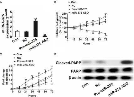
Figure 1. miR-375 increases cell proliferation and anti-apoptotic in H1975 cancer cells. A. Analysis of TaqMan qRT-PCR showed that a decrease or increase after H1975 cells infected with miR-375 ASO and pre-miR-375 lentiviral vector, the nonsense sequence lentiviral vector (NC) as negative control. All data are shown as mean ± SD, **P <
0.01 compared to control cells. B. miR-375 increased H1975 cells proliferation. Cells were infected with miR-375 ASO and pre-miR-375 lentiviral vector, and were plated into 96 well plates and harvested at different times. All data were shown as mean ± SD, *P < 0.05, **P < 0.01 compared to control cells. C. Impact of miR-375 on cell
apopto-sis. Apoptosis was analyzed by cell flow cytometry. The cells undergoing apoptosis were Annexin-V-PE-positive and 7-AAD-negative. All data are shown as mean ± SD, *P < 0.05, **P < 0.01 compared to control cells. D. Western blot
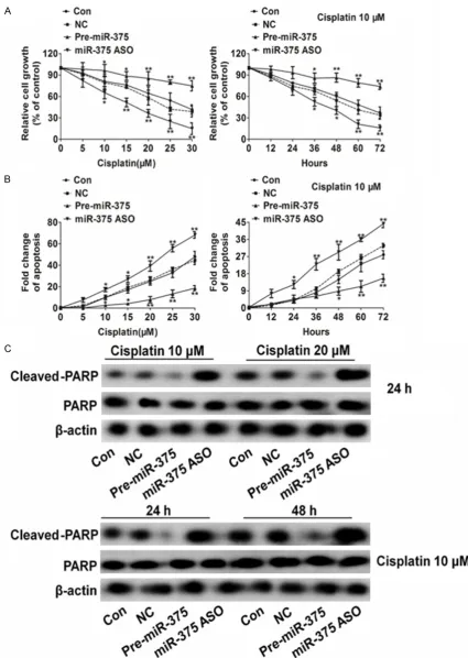
and were average ± SD. values. *P < 0.05, **P < 0.01, ***P < 0.001 compared to control cells. B. miR-375
down-expression promoted cell apoptosis in H1975 cells after treatment with cisplatin. Data were collected from three independent experiments and were average ± SD. values. *P <0.05, **P < 0.01, ***P < 0.001 compared to control
[image:5.629.102.529.152.663.2]cells. C. Western blot analysis showed that cleaved PARP was higher in miR-375 ASO lentiviral vector transfection cells than in NC transfection H1975 cells.
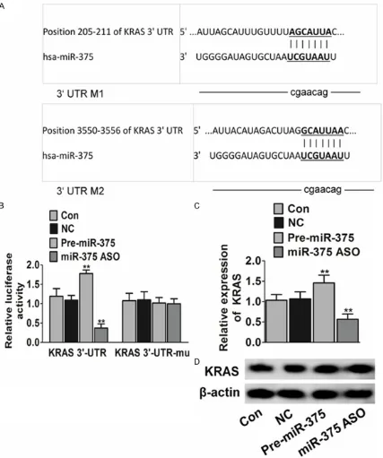
Figure 3. miR-375 regulates the expression of mRNA level of KRAS. A. Sequence of the miR-375 binding sites within the human KRAS 3’-UTR and a schematic of the reporter construct showing the KRAS 3’-UTR sequence and the mutated KRAS 3’-UTR sequence. B. Luciferase activity of the KRAS reporter in the presence of pre-miR-375 lentiviral vector or miR-375-ASO (**P < 0.01). Data were collected from three independent experiments and were average
(female, Nu/Nu, six week old). The tumor vol-ume was measured for 25 days. All mice were killed, s.c. tumors were resected, and weights were recorded.
Statistical analysis
Statistical analysis was performed using SPSS 18.0. Data’s were presented as the mean ± standard deviation. Statistical analyses were done by analysis of variance (ANOVA) or Student’s test. P value < 0.05 was considered statistically significant.
Results
miR-375 increases cell proliferation
In order to investigate the function of miR-375 in NSCLC cells, we tested miR-375 expression
in H1975 cell line using TaqMan real-time PCR after infection 48 h. The result showed that a decrease or increase after infected with miR-375 antisense oligonucleotide (ASO) and pre-miR-375 lentiviral vector (Figure 1A). We observed a significantly increased in prolifera-tion after infecprolifera-tion of pre-miR-375 lentiviral vector, In contrast, miR-375 ASO lentiviral vec-tor significantly decreased H1975 cell prolifera-tion (Figure 1B). These results indicate that H1975 cell proliferation ability can be signifi-cantly enhanced by the increase of miR-375 expression.
We further investigated the effect of miR-375 on cell apoptosis. The results showed that increase of miR-375 expression could decrease apoptosis dramatically. In contrast, knockdown of miR-375 could increase the apoptosis of H1975 cancer cells infected with miR-375 ASO, pre-miR-375 lentiviral vector and the nonsense sequence lentiviral vector (NC) by using qRT-PCR. Data were collected from three independent experiments and were average ± SD. values. **P < 0.01 compared to control cells. D. Analysis of expression levels of endogenous KRAS protein in H1975
[image:6.629.101.527.141.436.2]cells infected with miR-375 ASO, pre-miR-375 lentiviral vector and negative control (NC) by western blot.
H1975 cells (Figure 1C). Meanwhile, the expres-sion of cleavage PARP was significantly up-expression in miR-375 knockdown H1975 ce- lls, while slightly down-expression of cleavage PARP in miR-375-overexpression H1975 cells as shown in western blot analysis (Figure 1D). These results suggest that miR-375 could func-tion as a strong anti-apoptotic factor in human H1975 cancer cells.
Effect of miR-375 on the sensitivity of H1975 cells to cisplatin chemotherapy
Next, we investigated whether miR-375 could affect the sensitivity of H1975 cells to cispla- tin chemotherapy. Compared with the control group (H1975 cells), up-regulation of miR-375 reduced H1975 cells sensitivity to cisplatin, while down-regulation of miR-375 significantly increase killing ability of cisplatin by dose-dependent and time-dose-dependent (Figure 2A). This data indicates that miR-375 is one key player in cisplatin resistance of NSCLC cancer cell H1975. We also investigated the effect of miR-375 on the apoptosis of H1975 cells by DNA fragmentation assay and PARP cleavage assay. Similarly, pre-miR-375 lentiviral vector transfection inhibited the apoptosis in H1975 cells which induced by cisplatin (Figure 2B). Meanwhile, in the presence of cisplatin, the expression of cleavage PARP was significantly slight in H1975 cells transfected with pre-miR-375 lentiviral vector when compared to nonsense sequence lentiviral vector transfec-tion (Figure 2C). These results indicate that miR-375 can protect H1975 cancer cells sur-vival by anti-apoptosis.
KRAS 3’-UTR is a target for miR-375
We then searched for the potential target of miR-375 by TargetScan, PicTar and miRanda-mirSVR [10]. These programs predicted KRAS as one of the prime targets of miR-375. To test whether or not KRAS is a target of miR-375, the plasmid pMIR-reportor containing the wild-type 3’-UTR region of KRAS downstream of the lucif-erase coding region (Figure 3A, KRAS_WT) was constructed. HEK-293T cells were co-transfect-ed with reporter plasmid (KRAS_WT) and scramble. We performed a luciferase reporter assay and observed a significant decrease or increase in luciferase activity in the presence of pre-miR-375 or miR-ASO [11]. In addition, to validate whether KRAS is a direct target of
miR-375, we mutated the miR-375 binding site that was located in the 3’-UTR of KRAS, and this mutant reporter was no longer responsive to this miRNA (Figure 3B). These results indicate that KRAS is a direct target of miR-375. At last, we determined the effect of miR-375 on the expression of KRAS in H1975 cells. The results showed that pre-miR-21 increases the expression of KRAS mRNA. Moreover, miR-375-ASO decreased the expression of KRAS mRNA. Those results suggest that miR-375 presents a positive regulator of KRAS (Figure 3C). It may be partly involved in regulation of the expression of these proteins at transcrip-tional levels. Furthermore, we found that miR-375 had no significant effect on the expression of KRAS (Figure 3D). To our knowledge, it is the first time to verify that miR-21 can indirectly tar-get KRAS.
miR-375 confers H1975 cells cisplatin resis-tance in vivo
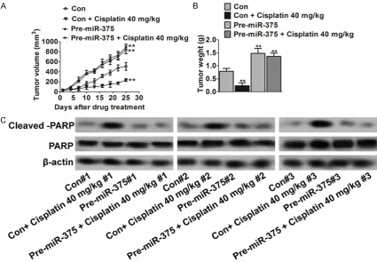
To determine whether miR-375 conferred resis-tance to cisplatin-induced apoptosis in vitro translates to drug resistance in vivo, we used a NSCLC xenograft model for this study. Briefly, H1975 cells expressing the control vector or miR-375 were subcutaneously inoculated into athymic nude mice. Mice were randomly divid-ed into four groups 7 days after innoculation. One group was treated with cisplatin (40 mg/kg per day) administered by i.p. injection and the other with the vehicle. Tumor growth and thera-peutic response were monitored during the course of cisplatin treatment.
growth of miR-375-expressing cells than that of control cells. To determine whether miR-375- mediated resistance to apoptosis in vitro was associated with decreased cisplatin effect in vivo, western blot assays were performed to examine the cleaved-PARP in tumors. The results showed that pre-miR-375 tumors have slightly cleaved -PARP than control tumors in the absence of cisplatin (Figure 4C). In addi-tion, cisplatin treatment did not increase clea- ved -PARP in miR-375 tumors (Figure 4C), indi-cating that miR-375 tumors were more resis-tant to cisplatin-mediated cell apoptosis than control tumors. Taken together, these in vitro and in vivo results reveal a critical role of miR-375 in drug resistance of NSCLC.
Discussion
The dismal outcome of non-small cell lung can-cer cell (NSCLC) patients highlights the need for novel prognostic biomarkers. MiRNAs con-trol the expression levels of particular genes. Therefore, dys-regulation of miRNAs, which occurs in certain diseases, such as cancers, may be attributed to aberrant gene expression [12]. The involvement of microRNAs in cancer and their potential as biomarkers of diagnosis and prognosis are becoming increasingly app- reciated. Many studies have reported signifi-cant associations between miRNA expression profiles and important clinical features of tumors, as well as patient survival rates. MiR-375 has been identified as oncogenes or tumor suppressor genes which has the potential to the development and growth of many human malignancies, including lung cancer, hepatocel-lular cancer, glioma and colorectal cancer [13]. A number of studies also indicate that miR-375 plays a very important role in tumor cell prolif-eration, apoptosis and invasion. However, the limited information concerning the role of miR-375 in NSCLC is available [14]. In consistence with these studies, here we showed that miR-375 can promote cell proliferation and inhibit apoptosis in NSCLC line H1975 cells.
Cisplatin resistances have been the bottleneck in improving the efficacy of chemotherapy. MiR-375 has been considered to function as an oncogenic molecule and may play a role in this drug resistance. Several reports suggest that miR-375 is one of the key miRNAs which plays a broad role in sensitivity to chemotherapeutic agents [15]. A study showed that epigenetic
silencing of miR-375 induces trastuzumab resistance in HER2-positive breast cancer by targeting IGF1R [16]. Recently, one report sh- owed that miR-375 is up-regulated in acquired paclitaxel resistance in cervical cancer and might be a therapeutic target in paclitaxel-resistant cervical cancer [17]. Therefore, those studies provide part of molecular mechanisms for how miR-375 contributes to drug resistance. Our discovery of miR-375 as a mediator of drug response in NSCLC provides a potential the- rapeutic target. Inhibition of miR-375 expres-sion could increase NSCLC cancer chemo- sensitivity.
The KRAS gene is necessary for the mainte-nance of tumor progression. Mismatch binding initiates subsequent events, including cleavage and excision of the error-containing strand fol-lowed by new synthesis and ligation [18]. KRAS gene mutation increased the risk of a wide vari-ety of cancers, especially in colon cancer. MiR-375 appeared to directly target the 3’-UTR of KRAS mRNA, and correlated to cisplatin drug resistance in NSCLC. We further validated this in H1975 cancer cells. We found that up-regula-tion of miR-375 could markedly increase the expression of KRAS. Hence, we propose that miR-375 may increase cisplatin resistance by regulating the expression of KRAS. These results verify that miR-375 could regulate KRAS expression, leading to the increase of cisplatin resistance. Our study provides a new piece of evidence supporting the involvement of KRAS in miR-375-caused cisplatin resistance.
In conclusion, we have shown that miR-375 plays a significant role in proliferation, apopto-sis, invasion and the response to cisplatin and radiotherapy in H1975 cancer cells. Moreover, this role might be attributed to miR-375 target-ed KRAS and influencing cisplatin chemothera-py sensitivity. Our finding suggests miR-375 can be a potentially useful marker for predic-tion of the clinical response to cisplatin therapy, and miR-375 may be a potential target for NSCLC therapy.
Disclosure of conflict of interest
None.
475001, Henan Province, China. E-mail: liyong1025- [email protected]
References
[1] Vijayvergia N, Shah PC, Denlinger CS. Survivorship in Non-Small Cell Lung Cancer: Challenges Faced and Steps Forward. J Natl Compr Canc Netw 2015; 13: 1151-161. [2] Maione P, Sacco PC, Sgambato A, Casaluce F,
Rossi A, Gridelli C. Overcoming resistance to targeted therapies in NSCLC: current ap-proaches and clinical application. Ther Adv Med Oncol 2015; 7: 263-273.
[3] Cong D, He M, Chen S, Liu X, Liu X, Sun H. Expression profiles of pivotal microRNAs and targets in thyroid papillary carcinoma: an anal-ysis of The Cancer Genome Atlas. Onco Targets Ther 2015; 8: 2271-2277.
[4] Jin H, Wang W. MicroRNA-539 suppresses os-teosarcoma cell invasion and migration in vitro and targeting Matrix metallopeptidase-8. Int J Clin Exp Pathol 2015; 8: 8075-8082.
[5] Wei C, Luo Q, Sun X, Li D, Song H, Li X, Song J, Hua K, Fang L. microRNA-497 induces cell apoptosis by negatively regulating Bcl-2 pro-tein expression at the posttranscriptional level in human breast cancer. Int J Clin Exp Pathol 2015; 8: 7729-7739.
[6] Tao J, Ji J, Li X, Ding N, Wu H, Liu Y, Wang XW, Calvisi DF, Song G, Chen X. Distinct anti-onco-genic effect of various microRNAs in different mouse models of liver cancer. Oncotarget 2015; 6: 6977-6988.
[7] Lili Song, Shikai Liu, Saitian Zeng, Liang Zhang, Xia Li. miR-375 Modulates Radiosensitivity of HR-HPV-Positive Cervical Cancer Cells by Targeting UBE3A through the p53 Pathway. Med Sci Monit 2015; 21: 2210-2217.
[8] Kang Kang, Xiaoying Zhang, Hongtao Liu, Zhiwei Wang, Jiasheng Zhong, Zhenting Huang, Xiao Peng, Yan Zeng, Yuna Wang, Yi Yang, Jun Luo, Deming Gou. A Novel Real-Time PCR Assay of microRNAs Using S-Poly(T), a Specific Oligo(dT) Reverse Transcription Primer with Excellent Sensitivity and Specificity. PLoS One 2012; 7: e48536.
[9] Liu Z, Xu Y, Long J, Guo K, Ge C, Du R. microR-NA-218 suppresses the proliferation, invasion and promotes apoptosis of pancreatic cancer cells by targeting HMGB1. Chin J Cancer Res 2015; 27: 247-257.
[10] He S, Zeng S, Zhou ZW, He ZX, Zhou SF. Hsa-microRNA-181a is a regulator of a number of cancer genes and a biomarker for endometrial carcinoma in patients: a bioinformatic and clinical study and the therapeutic implication. Drug Des Devel Ther 2015; 9: 1103-75. [11] Andruska N, Mao C, Cherian M, Zhang C,
Shapiro DJ. Evaluation of a Luciferase-based Reporter Assay as a Screen for Inhibitors of Estrogen-ERα-induced Proliferation of Breast Cancer Cells. J Biomol Screen 2012; 17: 921-932.
[12] Lujambio A, Lowe SW. The microcosmos of cancer. Nature 2012; 482: 347-355.
[13] Calin GA, Sevignani C, Dumitru CD, Hyslop T, Noch E, Yendamuri S, Shimizu M, Rattan S, Bullrich F, Negrini M, Croce CM. Human mi-croRNA genes are frequently located at fragile sites and genomic regions involved in cancers. Proc Natl Acad Sci U S A 2004; 101: 2999-3004.
[14] Zhang B, Pan X, Cobb GP, Anderson TA. microR-NAs as oncogenes and tumor suppressors. Dev Biol 2007; 302: 1-12.
[15] Garzon R, Marcucci G, Croce CM. Targeting mi-croRNAs in cancer: rationale, strategies and challenges. Nat Rev Drug Discov 2010; 9: 775-789.
[16] Ye XM, Zhu HY, Bai WD, Wang T, Wang L, Chen Y, Yang AG, Jia LT. Epigenetic silencing of miR-375 induces trastuzumab resistance in HER2-positive breast cancer by targeting IGF1R. BMC Cancer 2014; 14: 134.
[17] Shen Y, Wang P, Li Y, Ye F, Wang F, Wan X, Cheng X, Lu W, Xie X. miR-375 is upregulated in acquired paclitaxel resistance in cervical cancer. Br J Cancer 2013; 109: 92-99.