Original Article
Role of miR-24 in myocardial
fibrosis induced by angiotensin II
Qiufang Lian1, Xianli Wang1, Jianjun Mu2, Fuqiang Liu2, Zhao Wu1
1Department of Cardiology, Xianyang Hospital of Yan’an University, Xianyang 712000, China; 2Department of
Cardiovascular Medicine, First Affiliated Hospital of Medical College, Xi’an Jiaotong University, Xi’an 710061, China
Received November 28, 2015; Accepted January 26, 2016; Epub March 1, 2016; Published March 15, 2016 Abstract: Objective: To investigate the role and mechanism of miR-24 in angiotensin II induced myocardial fibrosis, by miR-24 expression and interference recombinant lentiviral transduction in primary cardiac fibroblasts. Method: The primary cardiac fibroblasts were stimulated by angiotensin II to simulate in vitro myocardial fibrosis. Cardiac fibroblasts were transduced with previously constructed miR-24 expression interference recombinant lentivirus. Cell proliferation, apoptosis and gene expression levels of AGTR-1, β-arrestin-1, GNAQ and PKC-delta were detected to identify the target of miR-24. PKC-delta expression recombinant lentivirus was constructed to investigate the effect of miR-24 on AGTR1-Gq-PKC signaling pathway. Results: MiR-24 facilitated expression of AGTR-1 and β-arrestin-1 and cell apoptosis, and inhibited cell proliferation and the synthesis of hydroxyproline, exhibiting a negative synergis-tic effect with PKC-delta. Conclusion: miR-24 negatively regulated PKC-delta by working on AGTR1 and furthermore inhibited myocardial fibrosis induced by angiotensin II.
Keywords: miR-24, angiotensin II, myocardial fibrosis
Introduction
In normal myocardial tissues, collagen fiber synthesis and metabolic imbalance can cause accumulation of collagen fibers and lead to myocardial fibrosis. Such lesions are closely associated with arrhythmia, cardiac dysfunc-tion and sudden cardiac death [1]. Myocardial fibrosis is a complex process, involving multiple regulatory systems including AngII-BK system, ET-NO system, platelet-derived growth factor (PDGF) and thromboxane [2, 3]. Renin-angio- tensin system (RAS) is a major regulator of renal and cardiovascular functions, playing an important role in myocardial fibrosis and remo- deling process [4]. The physiological function of angiotensin II (AngII) is mediated by its type I receptor (AGTR1) and G protein coupled recep-tors. In addition, AngII can exert a growth fac-tor-like effect, inducing myocardial cell hyper-trophy and stimulating cardiac fibroblast prolif-eration [5, 6].
Micro RNA (miRNA) is an endogenous non-cod-ing snon-cod-ingle-stranded small molecule RNA, exten-sively present in the eukaryotes. miRNA can
inhibit the expression level of target genes through completely or incompletely comple-mentary binding with the 3’-UTR of target miRNA [7]. miRNA participates in various physi-ological and pathphysi-ological processes, such as organism development, cell proliferation, differ-entiation and apoptosis. miR-24, a recently identified miRNA molecule, has been found to be associated with tumor and highly expressed in a variety of malignant tumors (gastric cancer, pancreatic cancer, liver cancer and colorectal cancer, etc.) [8-10]. As a tumor marker, miR-24 participates in signal transduction and apopto-sis regulation in tumor cells; however, the role in myocardial fibrosis has not been reported [11-13].
MiR-24 and RL
Materials and methods
Cell strains and cell culture
Rat cardiac fibroblasts (CFs) were separated from male SD rat heart. After verified by Vimentin and factor VII staining, CFs were cul-tured in DMEM complete media containing 1% antibiotics (Penicillin⁄Streptomycin, Gibco) and 10% FBS (Gibco) at 37°C with 5% CO2 in an incubator with saturated humidity. 24 h after cell passaging, cell confluence reached about 70%. Serum-free media was used to synchro-nize cells and after 24 h media was changed. Recombinant lentivirus or drug was added to the designated group and cells were cultured for another 48 h before further detection. Detection of AGTR1 and β-arrestin-1 mRNA level
CFs were digested, centrifuged and collected. Total RNA was extracted using TRIzol reagent (Qiagen). Reverse transcription and SYBR green I quantitative fluorescence PCR (Eppendorf) were performed to detect the mRNA level of AGTR1 and β-arrestin-1. Primer sequences are listed in Table 1. PCR conditions were as fol-lows: 94°C 4 min, 94°C 20 sec, 60°C 30 sec, 72°C 30 sec, 35 cycles. Each sample was repeated in triplicates.
Detection of protein level of AGTR1, β-arrestin-1, GNAQ and PKC-delta
CFs were digested, centrifuged and washed with PBS once. 0.1 ml RIPA lysis buffer was added to 106 cells. Cells were vortexed, resus-pended and placed on ice for 5 min. Cell lysis was centrifuged at 12000 g, 4°C for 5 min. The supernatant was used for SDS-PAGE and Wes- tern blot. The film was scanned and the inten-sity of target bands was analyzed using UVP
Detection of cell apoptosis by flow cytometry CFs were digested and washed with PBS twice. 1~5×10⁴ cells were collected, added into 200 μl Annexin V-FITC binding buffer (BD) and incu-bated at room temperature for 10 min without light. The supernatant was discarded after cen-trifugation. 190 μl Annexin V-FITC binding buf-fer was added to resuspend cells. Cells were mixed with 10 μl propidium iodide and placed on ice without light for flow cytometry detec- tion.
Preparation of PKC-delta recombinant lentivi-rus
The synthesized PKC-delta gene was cloned into a shuttle plasmid pLV5 for ligation. E.coli competent cells were transformed with the plasmid. Positive clones were selected, sequ- enced and named pLV5/PKC-delta. Plasmids were extracted using high purity and endotoxin-free plasmid midi-extraction kit (NucleoBond Xtra Midi Plus, Macherey-Nagel) and mixted with packaging plasmid PG-p1-VSVG, PG-P2-REV and PG-P3-RRE (Tronolab), respectively, in a certain ratio to transduce 293T cells. 293T cells were cultured for 72 h and the culture supernatant was collected. The supernatnat was centrifuged, filtered, and precipitated in PEG8000. The pellete was resuspended and ultracentrifuged using CsCl and dialyzed to purify virus. 293T cells were seeded in a 96-well plate at a concentratin of 1×10⁵/well and cultu-red in an incubator. After 24 h, the purified virus was diluted in ten-fold DMEM and 100 µl virus dilution was added to each well (10-2-10-6). Control group was also set up. Cells were cultu-red for 24-72 h in the incubator and observed under inverted light microscope to count positi-ve cells. The virus transduing unit (TU/ml) was calculated with dilution factor.
Detection of CFs proliferation by MTT assay after recombinant lentiviral transduction CFs were seeded in a 96-cell plate at a density of 1×10⁴/well and after 24 h, serum-free media was used to synchronize cells for 24 h. Following media change, cells were treated by groups and cultured for another 48 h. 10 μl 5 mg/ml MTT solution (Sigma) was added into each well and cells were cultured for 4 h. The media was discarded and 100 μl DMSO was added in each well. The plate was shaken at a low rate for 10 min at room temperature. The absorbance at 490 nm wavelength was detected using a spec-trophotometer. DMSO blank control was
includ-Table 1. Quantitative fluorescence PCR prim-ers
Primers Sequence (5’-3’)
[image:2.612.91.290.95.190.2]Detection of hydroxyproline level in CFs
The hydroxyproline level in CFs was detected using a double antibody sandwich ELISA kit. CFs were treated following aforementioned
methods. Cell culture supernatant was collect-ed. Cells were digested, resuspended with 100 μl PBS and subjected to repeated freeze-thaw cycles. Cells were centrifuged. The supernatant was collected and mixed with culture
[image:3.612.91.524.70.615.2]MiR-24 and RL
natant. ELISA was performed following instruc-tion of the kit and the hydroxyproline level in each sample was calculated using standard curve.
Statistical analysis
Triplicates were set up for each experiment. Data were analyzed using SPSS 10.0 software. The mean values between two samples were compared using t-test. P<0.05 was considered significant difference.
tin-1 protein expression levels were promoted and inhibited, compared with recombinant len-tivirus control group. Protein expression level of AGTR1 (B subtype), GNAQ and PKC-delta was barely influenced by miR-24 (P>0.05).
MiR-24 promoting cell apoptosis
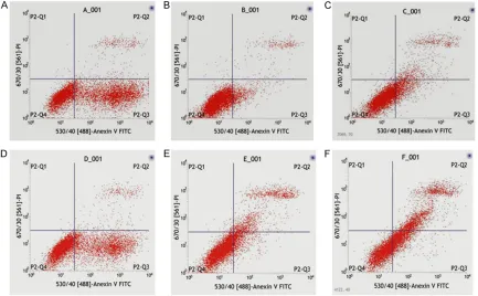
Flow cytometry results (Figure 4) showed that the cell survival rate in control, AngII, AngII + empty vector, AngII + MiR-24 expression, AngII + MiR-24 inter-ference and AngII + AngII
[image:4.612.91.378.72.237.2]inhibi-Figure 2. mRNA expression of AGTR1 and β-arrestin-1 (1-8 present Control Group; AngII group; AngII + empty vector group; AngII + MiR-24 expression group; AngII + empty vector + Rottlerin group; AngII + MiR-24 expression + Rottlerin group; AngII + 24 expression + empty vector group; AngII + MiR-24 expression + PKC delta expression group, respectively).
Figure 3.The protein detection level of AGTR1 (subtype A), AGTR1 (subtype B), β-arrestin-1, GNAQ and PKC-delta. (1-6 present control group, AngII group, AngII + empty vector group, AngII + MiR-24 expression group, AngII + MiR-24 interference and AngII + AngII inhibitor group, respectively).
Results
miR-24 promoting AGTR1 and β-arrestin-1 expression
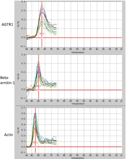
The melting curve of quantita-tive fluorescence PCR demon-strated a good specificity of all primers of AGTR1, β- arrestin-1 and actin gene (Figure 1). The standard curve showed that all three PCR sys-tems had an amplification effi-ciency higher than 95%. The relative quantitative result showed that AngII significantly promoted gene transcription of AGTR1 and β-arrestin-1 in CFs (P<0.05), while AngII inhibitor exhibited a signifi-cant effect only on AGTR1 gene transcription. The re- combinant lentivirus control failed to exhibit an effect on AngII induced AGTR1 and β-arrestin-1 gene transcrip-tion in CFs (P>0.05). Under the effect of miR-24 expres-sion and interference recom-binant lentivirus, gene tran-scription of AGTR1 and β- arrestin-1 were partially pro-moted and inhibited, respec-tively (Figure 2).
[image:4.612.92.374.320.498.2]tor groups was (46.82±0.10)%, (87.47±0.03)%, (76.59±0.03)%, (61.33±0.03)%, (72.37±0.37)% and (46.75±0.09)%, respectively. AngII signifi-cantly facilitated CFs survival (P<0.05) while AngII inhibitor completely offset this effect. Recom- binant lentivirus control had a certain effect on CFs survival following AngII stimulation (P<0.05). Compared with recombinant lentivi-rus control, CFs survival rate was reduced
under the effect of miR-24 expression recombi-nant lentivirus (P<0.05), while miR-24 interfer-ence recombinant lentivirus failed to exert a significant effect on CFs survival (P>0.05). CFs transduction with Lenti-PKC-delta
The titer of Lenti-PKC-delta was 5×10⁸ TU/ml after packaging and purification. The negative control of recombinant lentivirus without
[image:5.612.91.524.73.341.2]exog-Figure 4.CFs apoptosis detection results. (1-6 present control group, AngII group, AngII + empty vector group, AngII + MiR-24 expression group, AngII + MiR-24 interference and AngII + AngII inhibitor group, respectively.
[image:5.612.90.522.396.572.2]MiR-24 and RL
Figure 7.A: ELISA standard curve; B: Hydroxyproline levels.
Figure 6. MTT Assay Result of CFs following Recombinant Lentiviral Trans-duction (1-8 present Control Group; AngII group; AngII + empty vector group; AngII + MiR-24 expression group; AngII + empty vector + Rottlerin group; AngII + MiR-24 expression + Rottlerin group; AngII + MiR-24 expression + empty vector group; AngII + MiR-24 expression + PKC delta expression group, re-spectively).
enous genes was prepared using the same method by Weisiteng Biotechnology Center, with a titer of 1×10⁸ TU/ml. The two types of recombinant lentivirus were used to transduce CFs at a dose of MOI=15. After 96 h, the
trans-the effect of miR-24 expression recombinant lentivirus, hydroxyproline level was partially inhibited, and PKC delta inhibitor Rottlerine exhibited the same effect. MiR-24 expression exhibited a positive synergistic effect with
duction rate of Lenti-PKC-delta reached above 80%, as shown in Figure 5. CFs proliforation following recombinant lentiviral trans-duction
MTT results at 48 h following recombinant lentiviral trans-duction and drug treatment are shown in Figure 6. AngII significantly promoted CFs proliferation. Recombinant lentivirus control had no effect on CFs proliferation following AngII stimulation. Under the effect of miR-24 expression recombinant len-tivirus, cell proliferation was partially inhibited, and the effect was comparable with that of PKC delta inhibitor Rottlerin. MiR-24 expression exhibited a positive synergis-tic effect with Rottlerin, while the effects of miR-24 sion and PKC delta expres-sion offset each other. Changes of CFs hydroxypro-line level following recombi-nant lentiviral transduction
ELISA standard curve was constructed by the OD value of standard, as shown in
[image:6.612.90.379.348.621.2]Rottlerin, while the effects of miR-24 expres-sion and PKC delta expresexpres-sion offset each other.
Change of AGTR1 and β-arrestin-1 protein level in CFs after recombinant lentiviral transduc-tion
Western blot and intensity analysis (Figure 8) showed that AngII significantly promoted AGTR1 (A subtype) and β-arrestin-1 expression in CFs, while recombinant lentivirus control had no sig-nificant effect on the two protein expression in CFs after AngII stimulation. Under the effect of miR-24 expression recombinant lentivirus, the expression levels of both proteins were elevat-ed. PKC delta inhibitor Rottlerin reduced the levels of the two proteins. MiR-24 expression exhibited a positive synergistic effect with PKC delta and a negative synergistic effect with Rottlerin. The change of protein expression level of AGTR1 (B subtype) was relatively small (P>0.05).
Discussion
Based on the recent progress of miRNAs in the research field of biosynthesis, mechanism of action and disease association, great break-through has been made in the association be- tween miRNAs and tumor occurrence and development [14, 15]. The increase or reduc-tion of miRNAs expression level can lead to changes in genetic information, thereby result-ing in certain pathophysiological status. PKC is an important intracellular mediator of signal transduction. The catalytic activity of PKC is activated only when binding with membrane so that PKC can participate in intracellular and
thesis. Further studies found that miR-24 faci-litated cell apoptosis and also promoted expression of AGTR1 (A subtype) and β- arrestin-1, while exhibiting subtle influence on AGTR1 (B subtype), GNAQ and PKC-delta. To investigate the effect of miR-24 on AGTR1-Gq-PKC signaling pathway, recombinant lentivirus expressing PKC-delta was constructed. Working synergistically with recombinant lentivirus expressing miR-24, PKC-delta antagonized the inhibitory effect of miR-24 on cell proliferation and hydroxyproline synthesis, which was com-parable with the effect of PKC-delta inhibitor Rottlerin. The results showed that AngII indu-ced myocardial fibrosis was blocked by the inhi-bitory effect of miR-24 on PKC-delta.
Regulation of PKC is an effective means of intervention in the treatment of human heart failure. Recent clinical trials showed that sys-temic application of PKC inhibitors exhibited good tolerability, but drugs with tissue specific-ity of in vivo distribution are still waiting to be developed. The role and mechanism of miR-24 in angiotensin II induced myocardial fibrosis enrich our understanding on myocardial fibro-sis and provide more possibilities for re- search of treatment target, drug development and even gene therapy.
Disclosure of conflict of interest
None.
[image:7.612.92.374.74.178.2]Address correspondence to: Qiufang Lian, Depart- ment of Cardiology, Xianyang Hospital of Yan’an University, No. 38, Wenlin Road, Xianyang 712000, China. Tel: +8629-33785491; E-mail: wjylqf@163. com
Figure 8.The protein detection level of AGTR1 (subtype A), AGTR1 (subtype B), β-arrestin-1, GNAQ and PKC-delta. (1-8 present Control Group; AngII group; AngII + empty vector group; AngII + MiR-24 expression group; AngII + empty vector + Rottlerin group; AngII + MiR-24 expression + Rottlerin group; AngII + MiR-24 expression + empty vector group; AngII + MiR-24 expression + PKC delta expression group, respectively).
extracellular signal transdu- ction and regulation of ce- ll metabolism, differentiation and proliferation. It has been demonstrated that PKC par-ticipates in the process of myocardial remodeling [16]. AngII can bind with AGTR1 and activate PKC-delta, pro-moting fibroblast prolifera-tion, adherence and migration [17, 18].
syn-MiR-24 and RL
References
[1] Weber KT. Are myocardial fibrosis and diastolic dysfunction reversible in hypertensive heart disease? Congest Heart Fail 2005; 11: 322-324.
[2] Yang L, Zou XJ, Gao X, Chen H, Luo JL, Wang ZH, Liang QS, Yang GT. Sodium tanshinone IIA sulfonate attenuates angiotensin II-induced collagen type I expression in cardiac fibro-blasts in vitro. Exp Mol Med 2009; 41: 508-516.
[3] Diez C, Nestler M, Friedrich U, Vieth M, Stolte M, Hu K, Hoppe J, Simm A. Down-regulation of Akt/PKB in senescent cardiac fibroblasts im-pairs PDGF-induced cell proliferation. Cardio- vasc Res 2001; 49: 731-740.
[4] Wang XP, Zhang R, Wu K, Wu L, Dong Y. Angiotensin II mediates acinar cell apoptosis during the development of rat pancreatic fibro-sis by AT1R. Pancreas 2004; 29: 264-270. [5] Torsoni MA, Carvalheira JB, Calegari VC,
Bezerra RM, Saad MJ, Gontijo JA, Velloso LA. Angiotensin II (AngII) induces the expression of suppressor of cytokine signaling (SOCS)-3 in rat hypothalamus-a mechanism for desensiti-zation of AngII signaling. J Endocrinol 2004; 181: 117-128.
[6] Takayanagi T, Kawai T, Forrester SJ, Obama T, Tsuji T, Fukuda Y, Elliott KJ, Tilley DG, Davisson RL, Park JY, Eguchi S. Role of epidermal growth factor receptor and endoplasmic reticulum stress in vascular remodeling induced by an-giotensin II. Hypertension 2015; 65: 1349-1355.
[7] Sevignani C, Calin GA, Siracusa LD, Croce CM. Mammalian microRNAs: a small world for fine-tuning gene expression. Mamm Genome 2006; 17: 189-202.
[8] Slotta-Huspenina J, Berg D, Bauer K, Wolff C, Malinowsky K, Bauer L, Drecoll E, Bettstetter M, Feith M, Walch A, Höfler H, Becker KF, Langer R. Evidence of prognostic relevant ex-pression profiles of heat-shock proteins and glucose-regulated proteins in oesophageal ad- enocarcinomas. PLoS One 2012; 7: 41420. [9] Zhu HT, Dong QZ, Sheng YY, Wei JW, Wang G,
Zhou HJ, Ren N, Jia HL, Ye QH, Qin LX. MicroRNA-29a-5p is a novel predictor for early recurrence of hepatitis B virus-related hepa- tocellular carcinoma after surgical resection. PLoS One 2012; 7: 52393.
[10] Hayes CN, Akamatsu S, Tsuge M, Miki D, Akiyama R, Abe H, Ochi H, Hiraga N, Imamura M, Takahashi S, Aikata H, Kawaoka T, Kawakami Y, Ohishi W, Chayama K. Hepatitis B virus-specific mi-RNAs and Argonaute2 play a role in the viral life cycle. PLoS One 2012; 7: 47490.
[11] Liu H, Li P, Li B, Sun P, Zhang J, Wang B, Jia B. RKIP suppresses gastric cancer cell proliferati-on and invasiproliferati-on and enhances apoptosis regu-lated by microRNA-224. Tumour Biol 2014; 35: 10095-10103.
[12] Goto Y, Nishikawa R, Kojima S, Chiyomaru T, Enokida H, Inoguchi S, Kinoshita T, Fuse M, Sakamoto S, Nakagawa M, Naya Y, Ichikawa T, Seki N. Tumour-suppressive microRNA-224 in-hibits cancer cell migration and invasion via targeting oncogenic TPD52 in prostate cancer. FEBS Lett 2014; 588: 1973-1982.
[13] Yuan K, Xie K, Fox J, Zeng H, Gao H, Huang C, Wu M. Decreased levels of miR-224 and the passenger strand of miR-221 increase MBD2, suppressing maspin and promoting colorectal tumor growth and metastasis in mice. Gas- troenterology 2013; 145: 853-864.
[14] Tominaga N, Yoshioka Y, Ochiya T. A novel plat-form for cancer therapy using extracellular ve-sicles. Adv Drug Deliv Rev 2015; 95: 50-5. [15] Cui L, Zhang X, Ye G, Zheng T, Song H, Deng H,
Xiao B, Xia T, Yu X, Le Y, Guo J. Gastric juice MicroRNAs as potential biomarkers for the screening of gastric cancer. Cancer 2013; 119: 1618-1626.
[16] Lee MW, Severson DL. Signal transduction in vascular smooth muscle: diacylglycerol second messengers and PKC action. Am J Physiol 1994; 267: C659-678.
[17] Hou M, Pantev E, Möller S, Erlinge D, Edvinsson L. Angiotensin II type 1 receptors stimulate pro-tein synthesis in human cardiac fibroblasts via a Ca2+-sensitive PKC-dependent tyrosine
kina-se pathway. Acta Physiol Scand 2000; 168: 301-309.