Original Article
Local blockage of EMMPRIN impedes pressure ulcers
healing in a rat model
Xi-Lan Zhao*, Xiao Luo*, Ze-Xin Wang, Guo-Li Yang, Ji-Zhong Liu, Ya-Qiong Liu, Ming Li, Min Chen, Yong-Mei Xia, Jun-Jie Liu, Shu-Ping Qiu, Xiao-Qing Gong
Department of Nursing, Fuling Central Hospital, Chongqing 408099, China. *Equal contributors. Received March 3, 2015; Accepted May 18, 2015; Epub June 1, 2015; Published June 15, 2015
Abstract: Excessive extracellular matrix degradation caused by the hyperfunction of matrix metalloproteinases (MMPs) has been implicated in the failure of pressure ulcers healing. EMMPRIN, as a widely expressed protein, has emerged as an important regulator of MMP activity. We hypothesize that EMMPRIN affects the process of pressure ulcer healing by modulating MMP activity. In the rat pressure ulcer model, the expression of EMMPRIN in ulcers detected by Western blot was elevated compared with that observed in normal tissue. To investigate the role of EMMPRIN in regulating ulcer healing, specific antibodies against EMMPRIN were used via direct administration on the pressure ulcer. Local blockage of EMMPRIN resulted in a poor ulcer healing process compared with control ulcers, which was the opposite of our expectation. Furthermore, inhibiting EMMPRIN minimally impacted MMP activ-ity. However, the collagen content in the pressure ulcer was reduced in the EMMPRIN treated group. Angiogenesis and the expression of angiogenic factors in pressure ulcers were also reduced by EMMPRIN local blockage. The results in the present study indicate a novel effect of EMMPRIN in the regulation of pressure ulcer healing by control-ling the collagen contents and angiogenesis rather than MMPs activity.
Keywords: Pressure ulcer, EMMPRIN, CD147, matrix metalloproteinases, collagen, angiogenesis
Introduction
Pressure ulcers are common in the aging and paralyzed populations. Conventional treat-ments of pressure ulcers focus on prevention, and the external interventions focus on the wound-healing process [1]. However, as chronic wounds, pressure ulcers show a wide complex-ity in the healing process. A successful wound
healing process involves inflammation, cell pro -liferation, extracellular matrix (ECM) deposition and remodeling [2]. The degradation of the extracellular matrix (ECM) relay affects the matrix metalloproteinases (MMPs), which are secreted by various cells types as proenzymes and are activated by membrane type MMPs or by serine proteases. However, excessive extra-cellular matrix degradation caused by the hyperfunction of MMPs has been implicated in the failure of pressure ulcer healing [3]. Therefore, alleviating the detrimental effects of MMPs may promote the healing process of pressure ulcers.
Extracellular matrix metalloproteinase inducer
expressed membrane protein [4]. It was initially found on the surface of tumor cells and is expressed on numerous cell types and serves as a cell surface receptor for multiple ligands, such as cyclophilin A, monocarboxylate trans-porter, integrins, and EMMPRIN itself. Binding to these various partners enables EMMPRIN to induce MMPs and mediate various functions [5]. Given the role of EMMPRIN in regulating MMP expression, we hypothesize that EMMPRIN could affect the process of pressure ulcer heal-ing by modulatheal-ing MMP activity. In the present study, we addressed whether EMMPRIN is expressed in pressure ulcers and whether anti-EMMPRIN intervention promotes wound heal-ing in a rat model of chronic skin pressure ulcers.
Materials and methods
Animal model establishment
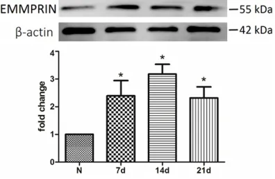
Figure 1. EMMPRIN expression was increased in pressure ulcers. Western blot analysis showed that the expression of EMMPRIN was detectable both in normal and pressure ulcer tissue and was signifi -cantly higher in pressure ulcers. N = 3, *P < 0.05 compared with normal tissue.
Health (NIH Publication No. 85-23, revised 1996). A total of48 male Sprague-Dawley rats (10-11 weeks old; 150-200 g; purchased from Chongqing, China) were used. Animals were randomized to an EMMPRIN antibody-treated group and a physiological saline-treated (con-trol) group. The magnet compression model that was previously reported was used [6].
Briefly, rats were anesthetized by an intraperi -toneal administration of 50 mg/kg pentobarbi-tal. The dorsal hair was shaved, and the area was cleansed with 70% alcohol. A 3-cm full-thickness skin incision was made on each rat’s dorsal region, and an autoclaved magnet disk was placed under the skin caudal to the inci-sion, where it was held in place by the surround-ing fascia. The incision was closed with size 4/0 polysorb sutures. The rats were left to emerge from anesthesia and were individually housed in non-magnetic cages. After a constant 2 h of clamping with another magnet disk on the skin, the outer magnet was removed for 0.5 h, and the clamping/remove cycle was repeated 5 times daily for 5 consecutive days. The com-pressed area was left uncovered.
EMMPRIN antibody treatment
Anti-rat EMMPRIN antibody (Santa, Cruz, USA) was used for the antibody treatment regimens
(diluted with autoclaved saline to 10 μg/ml).
After 5 days of compressing, diluted antibody solution (0.1 ml) was injected intra-dermally around the wounds, and the wound surface were treated with topical spray (0.1 ml/cm2, twice per week, 3 weeks). The control group
was given isopyknic saline. After 15 minutes of air-drying to allow the adsorption of the anti-body, the wounds were dressed with a non-adherent dressing next to the wound surface,
and dry gauze was used to fill the ulcer crater.
At the end of treatment, animals were anesthe-tized, and the skin of the ulcer was harvested. Evaluation of ulcer stage and ulcer closure percentage
Ulcer stage was evaluated according to the grading system of the National Pressure Ulcer Advisory Panel (NPUAP) [7]. In this system,
pressure ulcers are classified as superficial
pressure ulcers with intact skin (Grade 1); par-tial-thickness skin loss involving the epidermis, dermis, or both (Grade 2); full-thickness skin loss involving damage to subcutaneous tissue (Grade 3); or full-thickness skin loss with exten-sive destruction of the underlying tissue (Grade 4). The closed ulcers percentage at day 21 was counted in both the EMMPRIN antibody-treated and control groups. Closed ulcer percentage = (initial ulcer area-ulcer area on day 21)/initial ulcer area × 100%.
Evaluation of angiogenesis
The ulcer site on the skin was fixed and embed
-ded in paraffin. Cross sections were stained
with hematoxylin and eosin (HE). To evaluate angiogenesis, CD34 (a vascular endothelial cell marker, Santa Cruz) was used to detect ulcer tissues biopsied on day 7, 14 and 21 after the removal of compression. The microvessel den-sity was evaluated by direct counting of the
positive signal in every high power field (× 400) on 6 fields per slide.
Collection of wound fluids and biopsies
On day 14 after removing the compression,
wound fluid exudates were collected from the
dressing covering the ulcer and were frozen immediately at -80°C until being analyzed. A 4 mm punch biopsy of tissues was collected adja-cent to the edge of the pressure ulcer and was stored at -80°C.
Estimation of the hydroxyproline content
Hydroxyproline content was analyzed on day 14 after removing the compression, as described
imaging system (Bio-Rad, US- A), and images were captured.
Western blot analysis
[image:3.612.92.375.97.194.2]The expression of EMMPEIN, VEGF, and PDGF-A in pressure ulcers was assessed by west-ern blot. The protein concen-tration was determined using a BCA assay, and 50 µg sam-ples were separated on 10% and 15% Tris-glycine gels Table 1. Grading of pressure ulcers according to the NPUAP
pres-sure ulcer grading system Days after the removal of compression
Ulcer grade
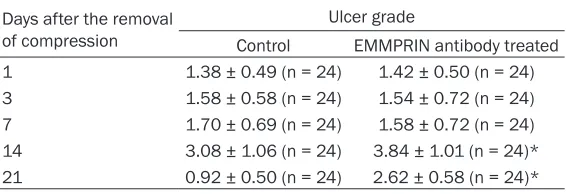
Control EMMPRIN antibody treated 1 1.38 ± 0.49 (n = 24) 1.42 ± 0.50 (n = 24) 3 1.58 ± 0.58 (n = 24) 1.54 ± 0.72 (n = 24) 7 1.70 ± 0.69 (n = 24) 1.58 ± 0.72 (n = 24) 14 3.08 ± 1.06 (n = 24) 3.84 ± 1.01 (n = 24)* 21 0.92 ± 0.50 (n = 24) 2.62 ± 0.58 (n = 24)* Data were presented as mean ± SE. *P < 0.05 compared with control.
tissue were hydrolyzed in 6 M HCl (50 mg tis-sue/1 ml 6 M HCl) at 100°C for 24 hours. The remaining HCl was neutralized with 2.5 M NaOH and was diluted 10-fold with deionized H2O. Chloramine T (0.05 M Sigma-Aldrich) solution was added to the neutralized/diluted solution
isometric (100 ml), and the mixture was incu-bated for 20 minutes at room temperature. Then, 100 ml of perchloric acid solution (3.15 M) was added and incubated for 5 minutes at room temperature. A volume of 100 mL of 20% P-dimethylaminobenzaldehyde (Sigma-Aldrich)
was added, and the final mixture was incubated
for 20 min at 60°C. Absorbance was measured at 560 nm using a microplate reader. The hydroxyproline content was determined accord-ing to a standard curve and was normalized to a milligram of wounded tissue homogenate for data analysis.
Gelatin zymogram analysis
The activity of MMP-2 and MMP-9 in the fluid
exudates and homogenized biopsies from pres-sure ulcers on day 14 after removing the com-pression were assessed using a gelatin zymo-gram, as described previously [9]. The protein concentration was determined using a BCA assay, and 50 µg of protein was loaded onto precast gelatin zymogram gel (7.5% Tris-glycine gels with 1 g/L gelatin). Following electrophore-sis, the gels were washed with renaturing buf-fer (2.5% Triton X-100, w/v) for 30 minutes at 370°C, and the gels were then incubated with developing buffer (50 mM Tris-HCl, 200 mM NaCl, 5 mM CaCl2, 0.2% Brij 35, w/v) on a rota-ry shaker at 370°C for 24 hours to allow the MMPs to digest. The gels were stained with 0.1% Coomassie blue R-250 and then destain- ed with 12.5% trichloroacetic acid. Then, the gel was visualized under a chemiluminescence
(Beyotime, China) and were transferred to PVDF membranes (Millipore). Membranes were probed with antibodies against EMMPRIN (1:200), VEGF (1:500), PDGF-A (1:200) and
β-actin (1:1000) overnight at 4°C, followed by 1
h incubation with HRP-conjugated secondary antibody, and were visualized with enhanced chemiluminescence following the manufactur-er’s instructions. Densitometric signals were
quantified by Quantity One software. Statistical analysis
The results are expressed as the mean ± SE. SPSS16.0 software was used for statistical analysis. The independent samples t test was used for quantitative continuous data, and the chi-square test was used for nominal data aris-ing from pressure ulcer grade. Values of P <
0.05 were considered statistically significant.
Results
Expression of EMMPRIN was elevated in pres-sure ulcers
In preparation for our studies investigating pressure ulcers, rats underwent magnet com-pression on the skin. After the model was established, we assessed EMMPRIN expres-sion in the pressure ulcer leexpres-sions. EMMPRIN was expressed in normal rat skin as detected by immunoblotting, and the EMMPRIN levels were increased substantially in the pressure ulcers of rat skin on day 7 after compression removal (Figure 1).
EMMPRIN antibody treatment prolonged ulcer healing
Table 2. Microvessel density of pressure ulcers after the removal of compression
Group 3 d 7 d 14 d 21 d
Control 21.36 ± 5.32 47.38 ± 6.12 62.48 ± 7.25 49.46 ± 6.39
EMMPRIN antibody treatment 19.24 ± 4.63 24.68 ± 5.38* 32.38 ± 6.84* 28.46 ± 8.37* Data were presented as mean ± SE. *P < 0.05 compared with control.
Table 1, the ulcer grades reached the maxi-mum at day 14 in both groups. Of the 48 ulcers,
there were only 8 (16.7%) superficial ulcers
(less than grade 2); however, only 3 (62.5%) of these were EMMPRIN antibody-treatment ulcers. In addition, the grades of ulcers in the
EMMPRIN antibody-treated group were signifi -cantly higher than in the control group at days 14 and 21 (P < 0.05). On day 21, 31.2% (15) of ulcers were completely closed, 26.7% (4) of which were EMMPRIN antibody treated.
EMMPRIN antibody treatment reduced angio-genesis in the ulcers
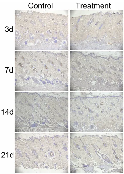
In view of angiogenesis playing an important role in the wound healing process, we evaluat-ed the microvessel density in ulcer tissues by immunohistochemistry using an antibody tar-geted to vascular endothelial cells marker CD 34 (Figure 2). EMMPRIN antibody-treated ulcers showed lower angiogenesis than that of the controls on days 7, 14 and 21 (Table 2). EMMPRIN antibody treatment did not affect the content of collagen in the pressure ulcers
Collagen biosynthesis in the pressure ulcers was assessed by detecting the hydroxyproline content. The results showed that there was no
significant difference of hydroxyproline content
in the EMMPRIN antibody treated group and the control group (9.26 ± 2.14 vs. 8.74 ± 1.68
μg/mg wounded tissue homogenate).
EMMPRIN antibody treatment did not affect the activity of gelatinase in pressure ulcers The results of the gelatinases spectrum analy-sis showed that the activity of MMP-2 and MMP-9 in the homogenates of wounds biop-sied from pressure ulcers were increased com-pared to the homogenates from normal tissue, and the activity reached the maximum on day 14 (Figure 3A). However, on day 14 after remov-al of compression, the activity of MMP-2 and MMP-9 of wounds biopsied from pressure ulcers in the EMMPRIN antibody treated group
was slightly lower than that of the control group, but the difference was not statistically
significant (Figure 3B). Accordingly, the activity
of MMP-9 in wound fluid exudates of the
EMMPRIN antibody treated group was slightly higher than that in the control group, but the
difference was not statistically significant. MMP-2 was not detected in the wound fluid
exudates (Figure 3C).
EMMPRIN antibody treatment inhibited the expression of angiogenic factors
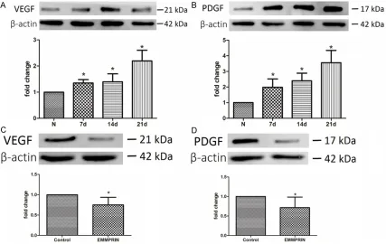
As mentioned above, EMMPEIN antibody treat-ed ulcers showtreat-ed lower angiogenesis, so we observed the level of angiogenic factors (VEGF and PDGF-A) in the pressure ulcers. The west-ern blot analysis showed that the expression of VEGF and PDGF-A gradually increased over time in the homogenates of wounds biopsied from pressure ulcers compared to the homog-enates from normal tissue (Figure 4). However, on day 14 after removal of compression, the expression of VEGF and PDGF-A in EMMPRIN
antibody ulcers was significantly lower com -pared to the control group.
Discussion
EMMPRIN has emerged as an important regu-lator for MMPs activity in cancer progression. Considering the correlation of MMPS with wound healing, we hypothesize that EMMPRIN is involved in pressure ulcer healing. The
pres-ent data showed the first evidence that 1)
EMMPRIN expression was up-regulated in pres-sure ulcers; 2) inhibiting EMMPRIN activity by
specific antibody prolongs the pressure ulcer
healing; 3) inhibiting EMMPRIN activity reduces the collagen content in pressure ulcers; and 4) inhibiting EMMPRIN activity also reduces the angiogenesis and the level of angiogenic fac-tors. Together, our data implicate an EMMPRIN correlative with pressure ulcer healing.
Figure 4. EMMPRIN antibody inhibited the expression of VEGF and PDGF-A. A, B: Western blot analysis showed that the expression of VEGF and PDGD-A in pressure was increased along with the pressure ulcer healing compare to normal tissue. N = 3, *P < 0.05 compared with normal tissue. C, D: On day 14 after the removal of compression, the expression levels of VEGF and PDGD-A were lower in the EMMPRIN treated group. N = 3, *P < 0.05 compared with control.
healing, evidence from in vivo studies is still lacking. In this study, we found that in a rat pressure ulcer model, the expression of EMMPRIN was up-regulated along with the pro-cess of ulcer healing. The results are consis-tent with previous investigative studies in
Because there is no EMMPRIN null animal
model, specific antibodies against EMMPRIN
[image:6.612.93.520.304.574.2]are widely used to block EMMPRIN function in many pathogeneses, such as cancer [11], asth-ma [12], and multiple sclerosis [13]. Thus, to investigate the role of EMMPRIN in regulating
body to inhibit the activity of this protein. We hypothesized that inhibiting EMMPRIN would promote the pressure ulcer healing by reducing the MMP activity. However, pressure ulcers receiving EMMPRIN antibody treatment exhib-ited poor healing quality. Furthermore, EMMPRIN antibody treatment did not affect the activity of MMP-2 and MMP-9. MMP expression can be induced in several cell types
(keratino-cytes, fibroblasts, endothelial cells and in inflammatory cells, such as monocytes, lym -phocytes and macrophages) in response to cytokines, hormones, oncogenes and cell con-tact with ECM or other cell types [14]. In the process of pressure ulcer healing, most of the
cell types mentioned above, such as fibro -blasts, monocytes, lymphocytes and macro-phages, were activated by soluble cytokines/ growth factors; thus, the main regulating mech-anism of MMPs in pressure ulcers may rely on the soluble factors rather than EMMPRIN, which is why EMMPRIN antibody treatment only minimally impacted MMPs activity.
Collagen content is a wisely used indicator for wound healing quality; it is primarily determined by the balance of synthesis and degradation. In this present study, we found that EMMPRIN antibody treatment reduced the content of col-lagen in pressure ulcers, independent of MMP activity, which was implicated in collagen deg-radation. Thus, EMMPRIN may play a role in regulating the collagen content by controlling the collagen synthesis process. However, more research is required to determine the exact relationship between EMMPRIN and collagen metabolism.
Angiogenesis is another indicator of wound healing. In the present study, blocking
EMMPRIN significantly reduced the microves -sel density in pressure ulcers; accordingly, expression of angiogenic growth factors (VEGF and PDGF-A) also decreased. Thus, the possi-ble mechanism by which EMMPRIN antibody treatment reduces angiogenesis is (at least partly) attributed to the impacted expression of these growth factors observed in the present study. Fibroblasts are the key original source of growth factors and collagen; we presume that
EMMPRIN may participate in regulating fibro
-blast function. This finding agrees with the data
of Huet et al. [10] that EMMPRIN promoted
fibroblast differentiation, but more research is
required to illuminate the relationship between EMMPRIN and these growth factors.
In conclusion, in the process of pressure heal-ing, the expression of EMMPRIN was up
regu-lated. Inhibiting EMMPRIN by a specific anti -body reduced the collagen contents and angio-genesis in pressure ulcers but only minimally impacted MMP activity, which led to the poor healing quality. More study is required to illumi-nate the intrinsic mechanism of EMMPRIN in regulating collagen metabolism and angiogen-esis, which has important implications for the control of pressure ulcer healing.
Acknowledgements
This study was supported in part by Fuling Science and Technology Commission project (FLKJ, 2012ABB1103).
Disclosure of conflict of interest
None.
Address correspondence to: Dr. Xi-Lan Zhao, Depart- ment of Nursing, Fuling Central Hospital, 2 Gaosuntang Road, Fuling District, Chongqing 40- 8099, China. E-mail: [email protected]
References
[1] Dini V, Bertone M and Romanelli M. Prevention and management of pressure ulcers. Dermatol Ther 2006; 19: 356-364.
[2] Hackam DJ and Ford HR. Cellular, biochemical, and clinical aspects of wound healing. Surg Infect (Larchmt) 2002; 3 Suppl 1: S23-35. [3] Norgauer J, Hildenbrand T, Idzko M, Panther E,
Bandemir E, Hartmann M, Vanscheidt W and Herouy Y. Elevated expression of extracellular matrix metalloproteinase inducer (CD147) and membrane-type matrix metalloproteinases in venous leg ulcers. Br J Dermatol 2002; 147: 1180-1186.
[4] Biswas C, Zhang Y, DeCastro R, Guo H, Nakamura T, Kataoka H and Nabeshima K. The human tumor cell-derived collagenase stimulatory factor (renamed EMMPRIN) is a member of the immunoglobulin superfamily. Cancer Res 1995; 55: 434-439.
[5] Iacono KT, Brown AL, Greene MI and Saouaf SJ. CD147 immunoglobulin superfamily recep-tor function and role in pathology. Exp Mol Pathol 2007; 83: 283-295.
noninvasive, clinically relevant model of pres-sure ulcers in the mouse. J Invest Surg 2004; 17: 221-227.
[7] Lardenoye JW, Thiefaine JA and Breslau PJ. Assessment of incidence, cause, and conse-quences of pressure ulcers to evaluate quality of provided care. Dermatol Surg 2009; 35: 1797-1803.
[8] Woessner JF Jr. The determination of hydroxy-proline in tissue and protein samples contain-ing small proportions of this imino acid. Arch Biochem Biophys 1961; 93: 440-447.
[9] Fini ME and Girard MT. The pattern of metallo-proteinase expression by corneal fibroblasts is altered by passage in cell culture. J Cell Sci 1990; 97 (Pt 2): 373-383.
[10] Huet E, Vallee B, Szul D, Verrecchia F, Mourah S, Jester JV, Hoang-Xuan T, Menashi S and Gabison EE. Extracellular matrix metallopro-teinase inducer/CD147 promotes myofibro -blast differentiation by inducing alpha-smooth muscle actin expression and collagen gel con-traction: implications in tissue remodeling. FASEB J 2008; 22: 1144-1154.
[11] Dean NR, Newman JR, Helman EE, Zhang W, Safavy S, Weeks DM, Cunningham M, Snyder LA, Tang Y, Yan L, McNally LR, Buchsbaum DJ and Rosenthal EL. Anti-EMMPRIN monoclonal antibody as a novel agent for therapy of head and neck cancer. Clin Cancer Res 2009; 15: 4058-4065.
[12] Gwinn WM, Damsker JM, Falahati R, Okwumabua I, Kelly-Welch A, Keegan AD, Vanpouille C, Lee JJ, Dent LA, Leitenberg D, Bukrinsky MI and Constant SL. Novel approach to inhibit asthma-mediated lung inflammation using anti-CD147 intervention. J Immunol 2006; 177: 4870-4879.
[13] Agrawal SM, Silva C, Tourtellotte WW and Yong VW. EMMPRIN: a novel regulator of leukocyte transmigration into the CNS in multiple sclero-sis and experimental autoimmune encephalo-myelitis. J Neurosci 2011; 31: 669-677. [14] Yan C and Boyd DD. Regulation of matrix