Original Article
Glutamine ameliorates intestinal ischemia-reperfusion
Injury in rats by activating the Nrf2/Are signaling
pathway
Ai-Li Wang1, Qiong Niu1, Ning Shi1, Jian Wang1, Xing-Fang Jia1, Hai-Feng Lian1, Zhiqiang Liu2, Cheng-Xia Liu1
1Department of Gastroenterology, Binzhou Medical College Affiliated Hospital, Binzhou, China; 2Center for Cancer
Immunology Research, Division of Cancer Medicine, The University of Texas MD Anderson Cancer Center, Houston, TX, USA
Received May 18, 2015; Accepted June 27, 2015; Epub July 1, 2015; Published July 15, 2015
Abstract: Ischemia-reperfusion (I/R)-mediated intestinal mucosal injury is usually induced by oxygen-derived toxic free radicals from the xanthine oxidase system after reperfusion, but the detailed molecular mechanisms underly-ing glutamine protection is still unclear. This study aims to elucidate whether glutamine prevents damage to the intestinal mucosa after I/R in rats and to investigate signaling by the Nrf2/ARE pathway induced by GLN in a rat model. Our results revealed that Glutamine pretreatment reduced jejunum injury and microvascular hyper-permea-bility induced by I/R. MDA level significantly increased while the SOD and GSH-Px levels decreased in the I/R group compared to the sham group and the GLN-I/R group. Both the mRNA and protein levels of the Nrf2 and HO-1 were significantly elevated by GLN pretreatment when compared to the I/R group. GLN treatment also elevated Bcl-2 levels, and accordingly suppressed apoptotic damage in the jejunum cells shown by decreased cleaved caspase-3 level. Mechanistic investigation revealed that GLN treatment augmented binding of Nrf2 onto Bcl2 gene promoter. These results indicate that glutamine has protective effects on I/R in vivo by activating the Nrf2/ARE signaling path-way to inhibit ROS production and reduce intestinal apoptosis.
Keywords: Glutamine, intestinal ischemia-reperfusion injury, Nrf2/ARE signaling pathway, Bcl2
Introduction
Intestinal ischemia/reperfusion (I/R) injury sec-ondary to splanchnic hypoperfusion is a com-mon event after a variety of clinical conditions [1, 2]. The intestine is vulnerable to hypoperfu-sion after injuries such as shock, neonatal nec-rotizing enterocolitis, intestinal transplant, and mesenteric ischemia [3]. Resuscitation allows
intestinal blood flow to recover, resulting in
ischemia/reperfusion. Previous studies sug-gest that the mechanism of I/R is complicated and that reactive oxygen species (ROS) attack,
inflammation infiltration, Ca2+ overload, energy metabolism obstacles, and apoptosis may be involved [4-6]. These mechanisms are intercon-nected, thus ultimately leading to apoptosis and necrosis. Recently, a better understanding of the mechanism(s) of oxidative stress in I/R injury has been achieved. ROS activation,
con-tributing to oxidative stress, is considered the key factor. When I/R occur, ROS activates
vari-ous signaling pathways and results in inflamma
-tory amplification, while other proteins may also
be activated to produce a protective effect. For example, the transcription factor NF-E2-related factor-2 (Nrf2) regulates a major environmental and oxidative stress response. It is held in the
cytoplasm by a cytoskeletal-associated specific
inhibitory protein, the Kelch-like ECH associat-ing protein 1 (Keap1) in normal quiescent cells [7-10]. Upon stimulation by oxidative stress, cysteine residues within the hinge region of
oxygen-ase 1 (HO-1), superoxide dismutoxygen-ase (SOD), GSH-Px, and NAD(P)H, which can antagonize oxidative stress induced by ROS.
Glutamine is the primary metabolic fuel of small intestinal enterocytes, it constitutes 50% of the free amino acid pool in the body, and is an essential metabolic component for the syn-thesis of glutathione, because its metabolism by the intestinal epithelium generates substan-tial quantities of glutamate, a precursor for glu-tathione synthesis [11]. Enterocytes rely domi-nantly on glutamine as an essential metabolic precursor in nucleotide, glucose and amino sugar, and protein synthesis [12]. Although
classified as “non-essential”, glutamine
appe-ars essential for the viability and growth of intestinal cells. Studies have established that glutamine reduced atrophy of the intestinal mucosa in rats on total parenteral nutrition (TPN). The potential mechanisms involved in I/R-mediated intestinal mucosal injury include oxygen-derived toxic free radicals, generated by the activation of the xanthine oxidase sys-tem after reperfusion [13]. Studiesshow that glutamine pre-supplementation improves sur-vival after I/R in mice models [14]. Other experi-mental studies [15] demonstrate that intralumi-nal injection of GLN protects the mucosa and diminishes the accumulation of neutrophils in the lamina propria of the small bowel during I/R.
With the discovery that the Nrf2/ARE signaling pathway can reduce the tissue damage induced by ROS, and increasing number of studies have shown that modulating Nrf2/ARE signaling plays a protective role in splanchnic ischemia reperfusion injuries, including heart, brain, and renal injuries. However, no reports addresses whether glutamine protects against intestinal ischemia reperfusion by activating the Nrf2/ ARE signaling pathway. The purpose of this study was to elucidate whether GLN prevents damage to the intestinal mucosa after II/R in rats and to investigate signaling by the Nrf2/ ARE pathway induced by GLN.
Materials and methods
Animals
Male Wister rats weighing 250-300 g were pro-vided by the Animal Research Center of Shandong University (Jinan, China). The rats
were maintained in a 12:12-hour day-night rhythm at a constant temperature of 23°C and a relative humidity of 40-60%. The experiments were performed in adherence with the National Institutes of Health Guidelines for the Use of Laboratory Animals and were approved by the Binzhou Medical University Committee on Animal Care. All efforts were made to minimize the number of animals used and their suf- fering.
Surgical protocols and histologic evaluation
The rats were randomly assigned into three groups (10 rats per group): the sham group served as a normal control, and the ischemia/ reperfusion group (I/R group) was the model control. These two groups received 1 g/kg of normal saline via metal tube gavage every day. The glutamine pretreatment group (GLN-I/R group) received 1 g/kg of glutamine every day for 7 days before surgery. The rats were anes-thetized with an intraperitoneal injection of 10% chloral hydrate (3 ml/kg body weight). Rats in the sham group were subjected to a laparotomy and the superior mesenteric artery (SMA) was exposed but not occluded; rats in the I/R and GLN-I/R groups were subjected to a laparotomy and the SMA was occluded with a microvascular clamp for 30 min. The laparoto-my incision was then closed, to be opened later for removal of the clamps after 30 min of
isch-emia [16]. After verification of blood supply to
the intestine, the abdominal incision was closed. After 24 h of reperfusion, the incision was opened again, and a 25-cm intestinal seg-ment was obtained from a point 10 cm distal to the ligament of Treiz for morphological and bio-chemical analysis; another 25-cm intestinal segment was collected and stored at -80°C for western blotting. Intestinal samples were
embedded in paraffin and stained with hema -toxylin and eosin (H&E staining). Three sections
were prepared from each fixed tissue sample,
and each slide was analyzed. A blinded observ-er pobserv-erformed the histologic analyses using the histologic scoring system described by Chiu et al [17]. All measurements were made in tripli-cate, and mean values were obtained.
Intestinal permeability
The permeability of the intestinal mucosa was assayed by measurement of D-lactate levels
sample (1800 μg/L) was diluted to 1200, 800, 400, 200, and 100 μg/L; then a standard curve
was plotted based on the absorbance (OD) of a standard sample at 450 nm. The OD of sam-ples was measured used to calculate the con-centration of the sample’s D-lactic acid in rats via the standard curve. Serum was separated from the blood sample by centrifugation and stored at -80°C until analysis. A chromomeric end-point tachypleus amebocyte lysate (CE TAL) test was used to measure serum endotox-in levels. Platelet-rich serum was prepared from the blood by centrifugation at 3000 rpm for 15 min. The serum samples were thawed at room temperature for ~30 min before the assay. LAL
(100 μl) was added to 100 μL of serum and
incubated at 37°C for 5 min. In the next step, 100 ml chromogenic substrate solution was added and incubated at 37°C for another 10 min. To terminate the reaction, 100 ml HCl was added the OD was read at 545 nm. A standard curve from 0.1 to 1.0 EU/mL was used to evalu-ate the concentration of endotoxin.
Spectrophotometric measurement of T-SOD, MDA, and GSH-Px in serum
The enzymatic activity of these factors in sam-ples was measured by colorimetric analyses using a spectrophotometer with relevant detec-tion kits according to the instrucdetec-tions of the manufacturer. All measurements of SOD activi-ty, MDA, and GSH-Px content in serum were performed in accordance with the technical manuals of the detection kits (Jianchen Biological Institute, China).
Detection of Nrf2, HO-1, caspase-3 and bcl-2 proteins by immunohistochemistry (IHC) analy-sis
IHC was performed using an SV
hypersensitivi-ty two-step kit (BOSTER, Wuhan, China). Briefly, slides were deparaffinized, and tissue sections
were hydrated through xylene and a graded alcohol series. Then, slides were incubated in 3% H2O2 for 30 min to block the endogenous peroxidase activity and treated with citrate buf-fer (pH 6.0) at 95°C for antigen retrieval. The slides were incubated overnight with primary antibody (Nrf2, HO-1, c-Caspase-3 or bcl-2; 1:100 dilution) from ImmunoWay (USA) at 4°C, washed three times with PBS, and incubated with HRP-labeled secondary antibody for 30
min at 37°C. The slides were again rinsed three times for 5 min with PBS, and the sections were incubated with DAB. Finally, the slides were counterstained with haemotoxylin, mounted with DPX, and visualized under the micro- scope.
RNA isolation and analysis of Nrf2, HO-1 mRNA by real-time fluorescence quantitative PCR (RT-PCR)
Total RNA was extracted, and cDNA was syn-thesized according to the instructions of the manufacturer (TaKaRa, Japan). qPCR analysis was performed using a real-time PCR System (Rotor Gene 3000) with the following primers: Nrf2, F 5-ACACGGTCCACAGCTCATC-3, R 5-TG- CCTCCAAGTATGTCAATA-3; HO-1, F 5-TCAGTCC- CAAACGTCGCGGT-3, R 5-GCTGTGCAGGTGTTG-
AGCC-3; β-actin, F
5-GAAGTGTGACGTTGACAT-CCG-3, R 5-TGCTGATCCACATCTGCTGGA-3. Rela- tive changes in expression were calculated using the 2−ΔΔCt method.
Immunoprecipitation (ChIP) assay
The ChIP assay was performed using a kit from
Cell Signal Technology (CST, CA, USA). Briefly,
intestinal tissues were lysed, and nuclei were
pelleted and sheared using a sonicator with five
20-s pulses. Sheared chromatin was immuno-precipitated with 2 µg of anti-Nrf2 or control IgG antibody. The cross-links were reversed overnight at 65°C, and the chromatin was deprotonated with 20 µg/ml proteinase K. PCR
amplification detected the Bcl-2 promoter
region containing Nrf2/AREr3 binding sites were performed using the following primers was used for PCR: Bcl2-BS1, F 5-GTTCTT- AAGCCCGATGTGGCAAC-3, R 5-GAGTAGTACCA- ATATGCTACCCTT-3; Bcl2-BS2, 5-ACCTTTCAGC- ATCACAGA-3, R 5-AATCACGCGGAACACTTG-3.
Statistical analyses
Statistical analyses were used to compare mean values from different groups by the homogeneity test. One-way analysis of variance (ANOVA) and Tukey’s multiple comparisons test
were used to evaluate the significance of differ -ences in other results by using Statistical Product and Service Solutions (SPSS) software package 17.0. P<0.05 was considered
Results
GLN reduces tissue injury in the jejunum after I/R
A sham operation was well-formed since
villos-ity cell did not show any lysis or inflammatory
process (Figure 1A). The I/R group displayed structural destruction of the villi (with only draft
of one of them formed by inflammatory cells
and necrotic material), hemorrhage and basal glandular ulceration (mainly for the intestinal
mucosa villus epithelium), la- mina propria disintegration,
and some fluff top bleeding gland was significantly
im-paired (Figure 1B). The GLN group displayed expansion of the intestinal mucosa villi subcutaneous space but not obvious villus edema, gener-ally normal glands, and lami-na propria edema (Figure 1C). The 24 h reperfusion injury degree of comparison was: Sham group, 0.313±0.063 points; I/R group, 4.120± 0.502 points; and GLN-I/R group: 2.461±0.372 points (Figure 1D). Comparing the I/R and the sham groups, the difference was statistically
significant (P<0.001), as was the difference between the GLN-I/R group and the I/R group (P<0.05).
GLN protects rats against I/R-induced microvascular hyper-permeability
Glutamine pretreatment att- enuated I/R-induced eleva-tion of plasma endotoxin and D-lactic acid levels. These lev-els in the I/R group were
sig-nificantly higher than in the
Sham and GLN-I/R groups (both P<0.001). There was
no significant difference
be-tween the sham group and the GLN-I/R groups (Figure 2), indicating glutamine suc-cessfully restore the micro-Figure 1. Histological characteristics of the of ileum segments in the I/R rat
[image:4.612.89.377.72.287.2]model. H&E staining showing normal histology of the ileum structure of rat in the sham group (A), showing the massive epithelial lifting, dilated capillar-ies, hemorrhage, and ulceration in the I/R group (B), and showing glutamine pretreatment results in significant structural improvement as evidenced by decreased edema and inflammatory cell infiltration in intestinal (C). Chiu’s score of comparison in these three groups (D). Magnification, 40×. **,P < 0.01 vs. I/R group.
Figure 2. GLN protects rats against I/R-induced microvascular hyper-perme-ability. Shown as the D-lactic acid levels (A) and endotoxin levels (B) of the sham group, the I/R group and the GLN-I/R group. **P < 0.01 vs. I/R group.
vascular hyper-permeability in ischemia/reper-fusion process.
Effects of GLN in oxidative stress
Oxidative stress plays a significant role in the I/R and can be reflected by MDA, GSH-Px, and
T-SOD levels. In this study, the serum
concen-trations of MDA and GSH-Px were significantly
[image:4.612.91.375.401.514.2]GLN-I/R group were obviously lower than that in the I/R group (P<0.05, Figure 3A, 3B), suggesting that treatment with glutamine generated a low risk of lipid peroxidation. The increase in serum T-SOD activities after 30 min of ischemia and 24 h of reperfusion in rats pretreated with
glu-tamine was significantly more pronounced than in I/R rats, thus confirming the anti-oxidative
properties of glutamine in rats (P<0.01, Figure
Effects of GLN on Nrf2 and HO-1 expressions
As shown in the Figure 4A, the expression of Nrf2 in the sham group showed light brown immunostaining in the cytoplasm and no
stain-ing in the nuclei. Significantly positive expres -sion of Nrf2 in the cytoplasm and nuclei were observed in the I/R group. Compared to the I/R group, the positive rates of Nrf2 expression
[image:5.612.93.521.72.188.2]sig-nificantly increased in the GLN-I/R group. The
Figure 3. Effects of glutamine on oxidative stress in I/R rats. Shown as comparison of serum (A) T-SOD activities, (B) GSH-Px, (C) MDA levels and in the sham group, the I/R group and the GLN-I/R group. **P < 0.01 vs. I/R group.
[image:5.612.92.521.237.545.2]expression of HO-1 in the sham group showed sparse brown immunostaining in the
cyto-plasm, but significant positive expression of
HO-1 was observed in the cytoplasm of the I/R group . Compared to the I/R group, the positive
rates of HO-1 expression increased significant -ly in the cytoplasm in the GLN-I/R group. The mRNA level and protein levels of the Nrf2 and Ho-1 were also validated by RT-PCR and west-ern blot respectively, and the results were con-sistent with the IHC staining assay (Figure 4B, 4C).
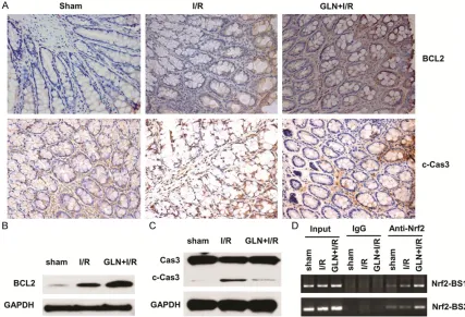
GLN promotes Bcl2 expression and inhibits intestinal apoptosis
Apoptosis is the major mechanism of cell death immediately following a short period of isch-emia with ensuing reperfusion. In this study, intestinal apoptosis was evaluated by expres-sions of Bcl-2 and the cleaved caspase-3. The expression of Bcl-2 was elevated in the GLN-I/R group compared with the I/R group (Figure 5A
upper panel, 5B). Compared with the I/R group,
the cleaved caspase-3 protein significantly
decreased in the GLN-I/R group (Figure 5A lower panel, 5C). These findings suggest that
pretreatment with GLN can reduce intestinal apoptosis. Since GLN activates Nrf2/ARE sig-naling, we also evaluated whether GLN-induced Bcl2 expression was regualated by Nrf2 tran-scriptional factor in I/R process. We used immunoprecipitation assay to examine the binding of Nrf2 protein on two sites on Bcl2 pro-moter, and found that GLN treatment augment-ed the binding of Nrf2 on both sites (Figure 5D), indicating that GLN promotes Bcl2 expression through Nrf2/ARE signaling pathway.
Discussion
[image:6.612.90.517.72.363.2]nism. To the best of our knowledge, our current
study is the first one to disclose the function of
glutamine against oxidative stress as well as the detailed molecular mechanism.
GLN is the most important and most available energy source in intestinal epithelial cells, lym-phocytes, and macrophages [18-20], as well as a precursor of nucleotide synthesis and gluta-thione in the important antioxidant defense system. Luo et al show that GLN supplementa-tion maintains gut glutathione levels during I/R [21]. Further, Wu and Ban’s studies demon-strate that GLN treatment has a protective effect during intestinal mucosal I/R injury, can reduce endotoxins elevation in I/R, and reduc-es bacterial translocation [22-24]. However, it is unclear whether GLN is protective for the intestinal mucosa against oxidative stress. Therefore, we established a rat I/R injury model to investigate the effects of GLN on I/R and its mechanism of action. Our results demonstrat-ed that the GLN pretreatdemonstrat-ed I/R rats had a great-er villous height and a lowgreat-er degree of intestinal permeability compared to the I/R rats. This suggests that GLN pretreatment plays an important role in the maintenance of the intes-tinal structure and barrier function in I/R-injured rats. Furthermore, we found that I/R leads to oxidative stress. Villi are rich in ROS-related enzymes, and I/R activates adenine-hypoxanthine metabolic pathways that produce abnormal increases in oxygen free radicals. Phospholipids are made of polyunsaturated fatty acids, which have a plurality of
unsaturat-ed bonds, and oxygen radicals have high affini -ty for these unsaturated bonds and can stimu-late excessive lipid peroxidation chain reac-tions. This results in cell membrane lipid peroxi-dation, inhibition of mitochondrial activity, and increased membrane permeability of the cell and lysosome, leading to cell swelling/rupture. Our results indicate that GLN pretreatment inhibited lipid peroxidation, improved the body’s ability to scavenge oxygen free radicals, and thus, inhibited membrane disintegration. The potential protective mechanisms may include upregulation of endogenous antioxidant factors (i.e.,SOD, Nrf2, and HO-1) and downregulation of MDA levels by the Nrf2/ARE signaling pathway.
In our I/R injury model, we also observed an interesting result in that GLN exerted potent apoptotic effects, as inferred by the
anti-(programmed cell death) is a rare event in healthy intestinal cells but is the early and pre-dominant form of cell death in intestinal necro-sis and has been linked to I/R [25]. The concen-trations of Bcl-2 have an important role in the protection or acceleration of intestinal apopto-sis after ischemia and/or reperfusion [26]. When the Bcl-2 levels in a cell decrease, the mPTP can be opened, resulting in a caspase cascade and ultimately leading to cell apopto-sis. In contrast, increased Bcl-2 can inhibit apoptosis [27]. A recent publications has revealed that Nrf2/AREr3 signaling regulates Bcl2 gene expression by directly binding to the promoter regions of Bcl2 gene and prevents human hepatoblastoma cell apoptosis [28]. In
our study, we also confirmed that GLN treat -ment increased Nrf2 level and promoted Nrf2/ AREr3 binding to the promoter of Bcl2 gene, therefore promotes Bcl2 gene expression in the I/R process.
In conclusion, our results demonstrate that GLN pretreatment protects the rat intestine from morphologic and functional mucosal inju-ry, as well as increases Nrf2 and HO-1 protein levels after I/R. The protective effect may be achieved by scavenging oxygen free radicals and inhibiting the ROS/pASK1/pJNK/Bcl-2/ Caspase/Apoptosis lipid peroxidation. These results suggest that GLN would be clinically useful in the treatment of I/R injury.
Acknowledgements
This work was supported by the Shandong Province Natural Science Foundation of China (Grants ZR2010hl051). The authors gratefully acknowledge Cui-ping Mao for her technical assistance during the course of the study and Yan-yan Wang for her help in the surgery. Disclosure of conflict of interest
None.
Address correspondence to: Dr. Cheng-Xia Liu, De- partment of Gastroenterology, Hospital Affiliated Binzhou Medical University, Binzhou, People’s Re- public of China. Tel: 86-138-5430-9199; Fax: 86-543-325-6764; E-mail: phdlcx@163.com
References
te heart failure. Int J Clin Exp Med 2008; 1: 171-180.
[2] Hassoun HT, Kone BC, Mercer DW, Moody FG, Weisbrodt NW and Moore FA. Post-injury mul-tiple organ failure: the role of the gut. Shock 2001; 15: 1-10.
[3] Young CM, Kingma SD and Neu J. Ischemia-reperfusion and neonatal intestinal injury. J Pediatr 2011; 158: e25-28.
[4] Fishman JE, Levy G, Alli V, Sheth S, Lu Q and Deitch EA. Oxidative modification of the intesti-nal mucus layer is a critical but unrecognized component of trauma hemorrhagic shock-in-duced gut barrier failure. Am J Physiol Gastro-intest Liver Physiol 2013; 304: G57-63. [5] Yang Y, Li W, Sun Y, Han F, Hu CA and Wu Z.
Amino acid deprivation disrupts barrier func-tion and induces protective autophagy in intes-tinal porcine epithelial cells. Amino Acids 2014; [Epub ahead of print].
[6] Bhattacharyya A, Chattopadhyay R, Mitra S and Crowe SE. Oxidative stress: an essential factor in the pathogenesis of gastrointestinal mucosal diseases. Physiol Rev 2014; 94: 329-354.
[7] Guo H, Li MJ, Liu QQ, Guo LL, Ma MM, Wang SX, Yu B and Hu LM. Danhong injection attenu-ates ischemia/reperfusion-induced brain dam-age which is associating with Nrf2 levels in vivo and in vitro. Neurochem Res 2014; 39: 1817-1824.
[8] Han J, Wang M, Jing X, Shi H, Ren M and Lou H. (-)-Epigallocatechin gallate protects against ce-rebral ischemia-induced oxidative stress via Nrf2/ARE signaling. Neurochem Res 2014; 39: 1292-1299.
[9] Li L, Dong H, Song E, Xu X, Liu L and Song Y. Nrf2/ARE pathway activation, HO-1 and NQO1 induction by polychlorinated biphenyl quinone is associated with reactive oxygen species and PI3K/AKT signaling. Chem Biol Interact 2014; 209: 56-67.
[10] Satoh T, McKercher SR and Lipton SA. Reprint of: Nrf2/ARE-mediated antioxidant actions of pro-electrophilic drugs. Free Radic Biol Med 2014; 66: 45-57.
[11] Alves WF, Aguiar EE, Guimaraes SB, da Silva Filho AR, Pinheiro PM, Soares Gdos S and de Vasconcelos PR. L-alanyl-glutamine preopera-tive infusion in patients with critical limb isch-emia subjected to distal revascularization re-duces tissue damage and protects from oxidative stress. Ann Vasc Surg 2010; 24: 461-467.
[12] Fukatsu K, Ueno C, Hashiguchi Y, Hara E, Kinoshita M, Mochizuki H and Hiraide H. Gluta-mine infusion during ischemia is detrimental in a murine gut ischemia/reperfusion model. JPEN J Parenter Enteral Nutr 2003; 27: 187-192; discussion 192.
[13] Zhang Y, Lv Y, Liu YJ, Yang C, Hu HJ, Meng XE, Li MX and Pan SY. Hyperbaric oxygen therapy in rats attenuates ischemia-reperfusion testic-ular injury through blockade of oxidative stress, suppression of inflammation, and re-duction of nitric oxide formation. Urology 2013; 82: 489, e489-489.e415.
[14] Ikeda S, Zarzaur BL, Johnson CD, Fukatsu K and Kudsk KA. Total parenteral nutrition sup-plementation with glutamine improves survival after gut ischemia/reperfusion. JPEN J Parent-er EntParent-eral Nutr 2002; 26: 169-173.
[15] Salomao AB, Aguilar-Nascimento JE, Percario S, Sano V, Marques NR and Dias CC. Intestinal intraluminal injection of glutamine increases trolox total equivalent antioxidant capacity (TEAC) in hepatic ischemia-reperfusion. Acta Cir Bras 2006; 21 Suppl 4: 69-73.
[16] Lindestrom LM and Ekblad E. Structural and neuronal changes in rat ileum after ischemia with reperfusion. Dig Dis Sci 2004; 49: 1212-1222.
[17] Chiu CJ, McArdle AH, Brown R, Scott HJ and Gurd FN. Intestinal mucosal lesion in low-flow states. I. A morphological, hemodynamic, and metabolic reappraisal. Arch Surg 1970; 101: 478-483.
[18] Zuhl MN, Lanphere KR, Kravitz L, Mermier CM, Schneider S, Dokladny K and Moseley PL. Ef-fects of oral glutamine supplementation on exercise-induced gastrointestinal permeability and tight junction protein expression. J Appl Physiol (1985) 2014; 116: 183-191.
[19] Han T, Li XL, Cai DL, Zhong Y and Geng SS. Ef-fects of glutamine-supplemented enteral or parenteral nutrition on apoptosis of intestinal mucosal cells in rats with severe acute pancre-atitis. Eur Rev Med Pharmacol Sci 2013; 17: 1529-1535.
[20] Kaufmann Y, Spring P and Klimberg VS. Oral glutamine prevents DMBA-induced mammary carcinogenesis via upregulation of glutathione production. Nutrition 2008; 24: 462-469. [21] Luo M, Bazargan N, Griffith DP, Estivariz CF,
Leader LM, Easley KA, Daignault NM, Hao L, Meddings JB, Galloway JR, Blumberg JB, Jones DP and Ziegler TR. Metabolic effects of enteral versus parenteral alanyl-glutamine dipeptide administration in critically ill patients receiving enteral feeding: a pilot study. Clin Nutr 2008; 27: 297-306.
[22] Wu GH, Wang H, Zhang YW, Wu ZH and Wu ZG. Glutamine supplemented parenteral nutrition prevents intestinal ischemia- reperfusion inju-ry in rats. World J Gastroenterol 2004; 10: 2592-2594.
novel mechanism. Am J Physiol Gastrointest Liver Physiol 2011; 301: G547-554.
[24] Ban K and Kozar RA. Enteral glutamine: a nov-el mediator of PPARgamma in the postisch-emic gut. J Leukoc Biol 2008; 84: 595-599. [25] Li JL, Wang QY, Luan HY, Kang ZC and Wang
CB. Effects of L-carnitine against oxidative stress in human hepatocytes: involvement of peroxisome proliferator-activated receptor al-pha. J Biomed Sci 2012; 19: 32.
[26] Ge M, Chi X, Zhang A, Luo G, Sun G, Xie H and Hei Z. Intestinal NF-E2-related factor-2 expres-sion and antioxidant activity changes in rats undergoing orthotopic liver autotransplanta-tion. Oncol Lett 2013; 6: 1307-1312.
[27] Ola MS, Nawaz M and Ahsan H. Role of Bcl-2 family proteins and caspases in the regulation of apoptosis. Mol Cell Biochem 2011; 351: 41-58.