MULTIPLE ROLES OF AFADIN IN EPITHELIAL MORPHOGENESIS
Kendall Jacob Lough
A dissertation submitted to the faculty at the University of North Carolina at Chapel Hill in
partial fulfillment of the requirements for the degree of Doctor of Philosophy in the Curriculum
of Genetics and Molecular Biology.
Chapel Hill
2020
Approved by
Scott Williams
Mark Peifer
Victoria Bautch
James Bear
Stephanie Gupton
© 2020 Kendall Jacob Lough ALL RIGHTS RESERVED
ABSTRACT
Kendall Jacob Lough; Multiple Roles of Afadin in Epithelial Morphogenesis (Under the direction of Dr. Scott Williams)
Afadin is an essential actin-binding protein which scaffolds between nectin- and cadherin-based cell-cell adhesions and the underlying cytoskeleton. Previous studies have shown that afadin isrequired for the establishment of polarity cues, maintaining epithelial integrity, bearing molecular contractile forces, and orienting cell divisions. Herein, I explore how afadin contributes to oriented cell division in embryonic epidermal progenitors in order to balance self-renewal and differentiation. Secondly, I detail how the afadin-nectin cell-cell adhesion system in craniofacial development.
During organogenesis, precise control of spindle orientation balances proliferation and
differentiation. In the developing murine epidermis, planar and perpendicular divisions yield symmetric and asymmetric fate outcomes, respectively. My research describes a novel mechanism which corrects erroneous anaphase orientations during telophase. The directionality of reorientation correlates with the maintenance or loss of basal contact by the apical daughter. The fidelity of telophase correction also relies on the adherens junction proteins vinculin, α-E-catenin, and afadin. Failure of this corrective mechanism impacts tissue architecture, as persistent oblique divisions induce precocious, sustained differentiation. The division orientation plasticity provided by telophase correction may enable progenitors to adapt to local tissue needs.
Familial genetic studies have shown that mutations in NECTIN1, an afadin binding partner, cause cleft lip/palate ectodermal dysplasia (CLPED1). Cleft palate (CP) is one of the most common congenital diseases and arises from failures in secondary palatogenesis during embryonic development. While the genetic link between NECTIN1 and CP is well established, mouse models have largely failed to
recapitulate these findings. Here, I provide the first evidence in an animal model that the nectin-afadin axis is essential for palatogenesis. We demonstrate that conditional loss of afadin (Afdn)—an obligate nectin-binding protein—induces a high penetrance of CP. In contrast, loss of Nectin1 or Nectin4 induces CP with low penetrance, while loss of both causes severe CP with a frequency similar to Afdn loss.
Finally, expression of the human disease mutant NECTIN1W185X causes CP with greater penetrance than
Nectin1 loss, suggesting this alteration may drive CP via a dominant interfering mechanism. These disparate projects share afadin as a common thread, weaving together two complex phenomena in embryonic epithelia morphogenesis.
ACKNOWLEDGMENTS
As co-first student to join the Williams lab, I often think of the lab’s growth and maturation as a metaphor for my own. In my eagerness to begin graduate school, I performed a summer rotation in Scott’s lab where I was Scott’s first employee—helping unpack boxes and set up equipment. After joining the lab at the same time as another student, Kevin and I spent those first few years somewhat naïvely throwing grandiose ideas and experiments at the wall—hoping anything would stick. Such was the optimism and invigorating positivity of our mentor, that we were unafraid to pursue nearly anything. At the same time, Scott’s unwavering commitment to his students (and a much-needed dose of scientific guidance) ensured we successfully landed on our feet. We both found wonderful projects, helped establish a positive, fun, and productive lab environment, and formed meaningful personal relationships. But this was only possible because Scott empowered us to take joint ownership of the lab—to contribute to something, rather than simply be a trainee. Scott always prioritized the people in the lab and in doing so, has set a wonderful example for how to be a good mentor—regardless of profession—for the numerous undergraduate and graduate students that have come through our doors. Scott has also been a fantastic scientific mentor, striking a balance between guiding and trusting that can be difficult to achieve. Through his mentorship, I’ve come to believe that I can have a career as an academic research scientist and—more importantly—that I can find real joy while doing so. For that, I am eternally grateful.
The Williams lab has always been a wonderful place to do research, in no small part thanks to the diverse, fun, and weird group of undergraduate and graduate students that have been a part of our team. I’ve had the great fortune to work with and mentor more than a dozen fantastic people, with many of whom I’ve formed lasting friendships. These people have taught me so much about being part of a team,
about being a rigorous scientist, and most importantly, how to be a supportive friend. Through burnt fingers (non-work related), grant and manuscript rejections, and personal and profession successes, the people of the Williams lab have kept me laughing and smiling through everything. Thank you all for your giddiness, your goofiness, and your encouragement.
I also want to thank my dissertation committee: Drs. Mark Peifer, Vicky Bautch, Jim Bear, and Stephanie Gupton, whose help and support have been invaluable. My committee meetings were always productive, insightful and extremely beneficial to both my work and my personal career. Their analysis and guidance significantly improved the rigor and impact of my thesis work and I always felt that they had my best interests at heart. Vicky’s expertise in vascular cell biology and collaborative inclination improved my thesis work and allowed me to explore the tractability of micropatterned substrates as a screening platform for regulators of oriented cell division in keratinocytes. Stephanie’s perspective as an expert cytoskeletal cell biologist and microscopist significantly benefited my studies of actin organization in cell-culture models and improved the quality of my fluorescent image analyses. Similarly, Jim’s expertise in cytoskeletal biology and willingness to share their abundance of genetic imaging tools allowed me to dream big and pursue exciting experiments in actin imaging. I especially want to thank Mark, who throughout my graduate career has served as a de facto—and with regards to my F31
fellowship, official—secondary mentor to me. Mark’s honest and blunt approach towards scientific rigor was extremely helpful, as was his expertise on Drosophila canoe and polarity systems. Mark has always been willing to give me his time and attention, whether to provide comments on a grant proposal, a manuscript, or just some confusing data, and his suggestions have always benefited the science.
I also feel it is important to acknowledge the support of Danelle Devenport, without whom the live imaging experiments in Chapter 3 would not have been possible. Danelle found me at the annual ASCB meeting in 2017 and freely offered to share their unpublished protocol to help me address a specific hypothesis. She similarly took the time to follow up and walk me through, step by step, the hurdles her lab had overcome to establish and optimize this protocol. Doing so did not help support any
ongoing projects in her lab, demonstrating an approach towards scientific collaboration worth aspiring too.
I’d like to thank the funding sources that have made my research possible. Scott Williams was supported by a Sidney Kimmel Scholar Award (SKF-15-165). I was supported for two years by an NIH Ruth L. Kirschstein Predoctoral National Research Service Award (F31 DE026956).
I’d also like to thank my family—both the Lough and Niederkorn sides—who have always been caring and supportive, and who demonstrate great patience and interest whenever I try to explain some science tidbit over dinner. Lastly, I’d like to thank Kendra, my wife and best friend, whose constant love, patience, and support has been my rock throughout my PhD.
PREFACE
This dissertation and the work presented herein attempts to describe the role of afadin in two distinct morphogenic processes: oriented cell division and secondary palatogenesis. Chapter 1 aims to summarize the discovery of afadin and its known binding partners (1.1), describe the known roles of afadin in cell-cell adhesion (1.2), introduce the process of asymmetric cell division (1.3) and highlight studies showcasing afadin’s role in embryonic development and disease (1.4).
Chapter 2 represents a review article I wrote with substantial support from my colleague, Dr. Kevin Byrd. While I primarily contributed the introduction (2.1) and literature summary of cell-cell adhesion molecules in palatogenesis (2.2 and 2.3), Dr. Byrd and our mentor, Dr. Scott Williams, co-wrote the portion summarizing the tools available for validating candidate genetic variants associated with cleft lip/palate (2.4). The review was published previous to writing this thesis with the following citation:
Lough, K.J., Byrd, K.M., Spitzer, D.C. & Williams, S.E. (2017) Closing the Gap: Mouse Models to Study Adhesion in Secondary Palatogenesis. J Dent Res 96, 1210-1220.
KJL, KMB, and SEW outlined the review topics and collaboratively wrote the manuscript with varying degrees of contribution. DCS provided extensive edits and intellectual input throughout manuscript preparation and revision. Permission to include the article in its entirety in a PhD dissertation was retained from SAGE (publisher of JDR), as explained at:
https://us.sagepub.com/en-us/nam/journal-author-archiving-policies-and-re-use
Chapter 3 is derived from a peer-reviewed publication on which I am the lead author. This work began as a simple investigation of a candidate gene (Afadin) and its role in oriented cell division in the embryonic epidermis. This project, however, took on a life of its own, eventually leading to the discovery
of a novel mechanism essential for establishing the bimodal distribution of division angles associated with the bipotency of basal progenitors. Excitingly, afadin, as well as additional actin binding proteins, proved to be essential for this particular mechanism. This article was published previous to the writing of this thesis with the following citation:
Lough, K.J., Byrd, K.M., Descovich, C.P., Spitzer, D.C., Bergman, A.J., Beaudoin III, G.M.J., Reichardt, L.F., Williams, S.E. (2019) Telophase correction refines division orientation in stratified epithelia. Elife 8 e49249. doi: 10.7554/eLife.49249.
KJL designed, performed, and analyzed all experiments and resultant data with significant intellectual input from KMB, CPD, and SEW. DCS and AJB provided extensive support in sample preparation and processing. GMJ and LFR created and provided an essential mouse strain. Permission to include the article in its entirety in a PhD dissertation was retained from eLife sciences, as explained at:
https://reviewer.elifesciences.org/author-guide/journal-policies
Chapter 4 represents unpublished work I have led with the support of several talented
undergraduate students including Danielle C. Spitzer, Abby J. Bergman, and Jessica J. Wu, investigating the role of the nectin-afadin complex in formation of the secondary or hard palate. This project is the result of astute observations by the Williams lab’s resident oral biologist, Dr. Kevin Byrd, who happened to notice that Afdn knockdown mutants displayed with cleft palate. I was equally fortunate to receive an NIH F31 predoctoral fellowship from the National Institute of Dental and Craniofacial Research (NIDCR; DE026956) to pursue this project. A first-author manuscript derived from this work is currently under peer-review at Development (submitted February 10, 2020):
Lough, K.J., Spitzer, D.C., Bergman, A.J., Wu, J.J., Byrd, K.M., Williams, S.E. (2020) Disruption of the nectin-afadin complex recapitulates features of the human cleft lip/palate syndrome CLPED1. Development. Under Revision.
KJL designed, performed, and analyzed all experiments and resultant data with significant intellectual input from KMB and SEW. DCS, AJB, and JJW provided extensive support in sample preparation, as well as significant edits and intellectual input during manuscript preparation.
TABLE OF CONTENTS
LIST OF FIGURES ... xv
LIST OF ABBREVIATIONS AND SYMBOLS ... xvii
Terminology ... xvii
Protein/Gene abbreviations ... xviii
Protein domains/epitopes ... xix
Symbols ... xix
CHAPTER 1 – INTRODUCTION. CELL-CELL ADHESION AND AFADIN
INFORM EPITHELIAL CELL IDENTITY AND MORPHOGENESIS ... 1
1.1 Identification and biochemical properties of afadin and its orthologs ... 1
1.2 Afadin is part of the adherens junction, a canonical cell-cell adhesion
complex ... 3
1.2.1 The afadin-nectin complex in cell-cell adhesion ... 5
1.3 Asymmetric cell division and cell fate specification ... 7
1.3.1 Asymmetric cell division in the Drosophila neuroblast ... 7
1.3.2 Coupling of division orientation and cell fate determination in the
mammalian epidermis ... 10
1.3.3 Biochemical architecture of the spindle orientation complex ... 13
1.3.4 Cell-cell adhesion and cell mechanics inform oriented cell division... 14
1.4 Role of afadin in tissue morphogenesis and disease ... 21
1.4.1 Roles of canoe in Drosophila morphogenesis ... 22
1.4.2 Roles of afadin and nectins in mammalian development and disease ... 23
CHAPTER 2 – INTRODUCTION. CLOSING THE GAP: MOUSE MODELS
TO STUDY CELL-CELL ADHESION IN MAMMALIAN SECONDARY
PALATOGENESIS ... 28
2.1 Introduction to secondary palatogenesis ... 28
2.2 Genes expressed in palatal epithelium underlie CL/P disorders ... 29
2.3 Cell-cell adhesion molecules in palatogenesis ... 30
2.3.1 Nectins and afadin ... 30
2.3.2 Cadherins and catenins ... 32
2.3.3 Other cell adhesion molecules ... 33
2.3.4 Connecting the dots between transcription factors and cell adhesion ... 34
2.4 Methods to investigate the genetics of CL/P ... 36
2.4.1 Ex vivo palate cultures ... 36
2.4.2 Animal models to investigate epithelial cell-cell adhesion in palate
closure ... 36
2.4.3 Other approaches to study epithelial contributions to secondary palate
closure ... 37
2.5 Conclusions ... 39
2.6 Figures ... 40
CHAPTER 3 – RESULTS. TELOPHASE CORRECTION REFINES DIVISION
ORIENTATION IN STRATIFIED EPITHELIA ... 45
3.1 Introduction ... 45
3.2 Results ... 47
3.2.1 Randomized division orientation persists into anaphase ... 47
3.2.2 Oblique anaphase divisions reorient during telophase ... 48
3.2.3 LGN mediates perpendicular telophase correction ... 50
3.2.4 Directionality of telophase correction is correlated with basement
membrane contact ... 51
3.2.5 The actin-binding protein, vinculin, regulates α-E-catenin
conformation. ... 52
3.2.6 Afadin is required for normal AJ morphology and is a novel regulator
of α-E-catenin conformation... 54
3.2.7 Ctnna1, Vcl and Afdn knockdown leads to randomized division
orientation ... 55
3.2.8 Tension-sensitive components of the AJ are essential for telophase
correction. ... 56
3.2.9 Telophase correction occurs independently of canonical polarity and
spindle-orienting cues. ... 57
3.2.10 Telophase correction and early mitotic spindle orientation function as
parallel pathways ... 60
3.2.11 Telophase correction also occurs during early stratification ... 60
3.2.12 Telophase correction impacts cell fate decisions ... 61
3.4 Discussion ... 63
3.4.1 A two-step mechanism for division axis determination ... 63
3.4.2 Corrective mechanisms in oriented cell divisions ... 64
3.4.3 Insights into epidermal cell fate specification... 65
3.5 Acknowledgements ... 66
3.6 Materials and Methods ... 67
3.7 Figures ... 76
3.8 Videos ... 106
CHAPTER 4 – RESULTS. DISRUPTION OF THE NECTIN-AFADIN
COMPLEX RECAPITULATES FEATURES OF THE HUMAN CLEFT
LIP/PALATE SYNDROME CLPED1 ... 113
4.1 Introduction ... 113
4.2 Results and Discussion ... 115
4.2.1 The obligate nectin binding partner, afadin, is required for secondary
palatogenesis ... 115
4.2.2 LUGGIGE presents advantages over epithelial-specific
Cre-recombinase approaches ... 117
4.2.3 Acute loss of Nectin1 or Nectin4 results in mild cleft palate with low
penetrance ... 117
4.2.4 Dual loss of Nectin1 and Nectin4 causes highly penetrant cleft palate ... 118
4.2.5 Causal CLPED1 mutations drive CP via dominant negative and/or
neomorphic activity ... 119
4.3 Acknowledgements ... 121
4.4 Materials and Methods ... 121
4.5 Figures ... 126
CHAPTER 5 – DISCUSSION ... 137
5.1 Updated model of OCD of epidermal progenitors ... 138
5.1.1 Roles of the Par/aPKC/Par6 apical polarity complex ... 139
5.1.2 Roles of the Crumbs apical polarity complex ... 139
5.1.3 Roles of the Scribble basolateral polarity complex ... 141
5.1.4 Roles for basal cell-matrix adhesions ... 142
5.2 Corrective mechanisms to ensure orientation of cell division ... 143
5.3 Speculative mechanisms promoting planar OCD in the epidermis ... 145
5.4 Insights into epidermal cell fate specification ... 146
5.4.1 Evidence supporting intrinsic fate determination ... 147
5.4.2 Evidence supporting extrinsic fate determination ... 148
5.5 Cell and Molecular mechanisms of CLPED1 ... 149
5.5 Exploring mutations in other cell-cell adhesion genes and their association
with CL/P ... 151
5.6 Figures ... 154
APPENDIX 2: GENETIC MOUSE MODELS OF CELL-CELL ADHESION
GENES ... 157
APPENDIX 3: K14-CRE LINES UTILIZED TO GENERATE GENETIC
MODELS OF CP ... 163
REFERENCES ... 165
LIST OF FIGURES
Figure 1.1 Afadin domain structure and notable binding partners ... 26
Figure 1.2 Involvement of afadin in the adherens junction ... 27
Figure 2.1 Secondary palatogenesis ... 40
Figure 2.2 Palatal gene expression and human orofacial clefting syndromes ... 42
Figure 2.3 Mutations in NECTIN1 and CDH1 associated with CL/P ... 43
Figure 2.4 LUGGIGE in oral epithelia ... 44
Figure 3.1 Telophase reorientation corrects oblique anaphase orientations ... 76
Figure 3.2 Telophase reorientation corrects oblique anaphase orientations ... 78
Figure 3.3 LGN mediates perpendicular but not planar telophase correction ... 80
Figure 3.4 Maintenance of basal contact correlates with planar-directed telophase
correction ... 82
Figure 3.5 A basal endfoot mediates planar telophase correction ... 84
Figure 3.6 Vinculin and afadin regulate α-E-catenin conformation and AJ linkage
to the actin cytoskeleton... 86
Figure 3.7 The α-E-catenin/vinculin/afadin complex demonstrates reciprocal
regulation to form mature adherens junctions in vitro ... 88
Figure 3.8 The α-E-catenin/vinculin/afadin pathway is required for normal
division orientation ... 90
Figure 3.9 AJ loss-of-function mutants display errors in division orientation ... 92
Figure 3.10 AJ mutants fail at both planar and perpendicular telophase correction. ... 93
Figure 3.11 AJ loss-of-function mutants display errors in division orientation ... 95
Figure 3.12 AJ mutants alter division orientation via LGN-independent
mechanisms ... 96
Figure 3.13 Afdn loss-of-function does not affect functional apicobasal polarity or
downstream components of spindle orientation ... 98
Figure 3.14 AJ components alter division orientation in an LGN-independent
manner... 99
Figure 3.15 Planar telophase correction limits precocious and sustained
differentiation and biases clones towards symmetric cell divisions ... 101
Figure 3.16 Failed telophase correction induces precious, sustained
hyperstratification without impacting proliferation or delamination ... 103
Figure 3.17 Two-step model of division orientation ... 105
Video 3.1.1 Planar anaphase orientation is fixed ... 106
Video 3.1.2 Perpendicular anaphase orientation is fixed ... 107
Video 3.1.3 Oblique anaphase orientations undergo planar telophase reorientation ... 108
Video 3.1.4 Oblique anaphase divisions display perpendicular correction ... 109
Video 3.10.1 Persistent oblique division in Ctnna1
912knockdown mitotic basal
cell ... 110
Video 3.10.2 Persistent oblique division in Vcl
3466knockdown mitotic basal cell ... 111
Video 3.10.3 Persistent oblique division in Afdn
2711knockdown mitotic basal cell ... 112
Figure 4.1 Afadin is essential for secondary palatogenesis ... 126
Figure 4.2 Validation of transgenic models highlights improved efficacy of
LUGGIGE ... 128
Figure 4.3 Loss of Nectin1 or Nectin4 does not cause highly penetrant CP ... 130
Figure 4.4 In vitro validation of Nectin1/4 shRNA efficacy using calcium-shift
assays ... 131
Figure 4.5 Nectin1 and Nectin4 cooperatively prevent highly penetrant CP ... 132
Figure 4.6 Dual loss of Nectin1 and Nectin4 in a novel, double shRNA construct
delays PS elevation and can result in residual MES ... 134
Figure 4.7 CLPED1 variant W185X acts in a dominant negative fashion to cause
CP ... 136
LIST OF ABBREVIATIONS AND SYMBOLS Terminology
ACD Asymmetric cell division; type of division in which daughter cells adopt distinct fates AJ Adherens junction; a classic cell-cell adhesion complex composed of cadherins/catenins CLPED1 Cleft lip/palate ectodermal dysplasia syndrome
CL/P Cleft lip/palate
CP Cleft palate
E Embryonic day; e.g. E14 =14-days into gestation
ED Ectodermal dysplasia
F-actin Filamentous actin
GMC Ganglion mother cell
LUGGIGE Lentiviral ultrasound-guided gene inactivation and gene expression MDCK Madin-Darby Canine Kidney; commonly utilized epithelial cell line.
MEE Medial edge epithelia
MES Medial epithelial seam
MOI Multiplicity of infection
MT Microtubule
OCD Oriented cell division
OFC Orofacial clefting
PS Palatal shelf/shelves
RNAi RNA-interference
SB Suprabasal; non-basal cells within the epidermis
shRNA Short-hairpin RNA
TJ Tight junction; classic, impermeable cell-cell adhesion.
Protein/Gene abbreviations
α-cat α-catenin/Ctnna1; component of the adherens junction
Afdn Afadin; Mus musculus ortholog of Canoe
aPKC Atypical protein kinase C; component of the apical polarity Par complex
Baz Bazooka; component of the Drosophila apical polarity Par complex
Brat Brain Tumor; fate determinant in Drosophila neuroblast ACD
Cno Canoe; Drosophila protein
Crb Crumbs; Drosophila protein; component of the apical Crumbs polarity complex
Cre Cre-recombinase
Dlg Discs Large; component of basolateral Scribble polarity complex
Insc Inscuteable; component of the Drosophila spindle orientation complex K5/14 Cytokeratin-5/14; basal cell specific keratins
K10 Cytokeratin-10; suprabasal cell specific keratin
Lgl Lethal Giant Larvae; component of basolateral Scribble polarity complex LGN Leucine/Glycine/Asparagine repeats; Mus musculus protein and Pins ortholog mInsc Inscuteable; Mus musculus protein and Insc ortholog
Mira Miranda; fate determinant in Drosophila neuroblast ACD
Mud Mushroom Body Defect; component of the Drosophila spindle orientation complex
NuMA Nuclear Mitotic Apparatus; Mus musculus protein and Mud ortholog
Par3 Partitioning defective-3; ortholog of Baz/component of the apical polarity Par complex Par6 Partitioning defective-6; component of the apical polarity Par complex
pHH3 phospho-histone H3; a mitotic biomarker
Pins Partner of Inscuteable; component of the Drosophila spindle orientation complex
Pon Partner of Numb; fate determinant in Drosophila neuroblast ACD
Pros Prospero; fate determinant in Drosophila neuroblast ACD
Stau Staufen; fate determinant in Drosophila neuroblast ACD
Std Stardust; Drosophila protein; component of the apical Crumbs polarity complex
Vcl Vinculin; binding partner of α-catenin
Protein domains/epitopes
a18 a18 epitope of a-catenin; cryptic epitope exposed by “active” conformation of a-catenin EC Extracellular cadherin domain; protein interaction domain in cadherins
CBD Catenin-binding domain; protein interaction domain in cadherins
GFP Green fluorescent protein
JMD Juxtamembrane domain; protein interaction domain in cadherins
NICD Notch Intracellular Domain
PDZ PSD-95/Dlg/ZO-1 binding domain; a common protein-protein interaction domain
RA Ras associated; afadin protein binding domain
RFP Red fluorescent protein
TPR Tetratricopeptide repeat; protein structural motif present on the N-terminus of LGN/Pins
YFP Yellow fluorescent protein
Symbols
φ Orientation of cell division at anaphase onset (t=0 min)
θ Orientation of cell division in telophase (t=+60 min)
CHAPTER 1 – INTRODUCTION. CELL-CELL ADHESION AND AFADIN INFORM EPITHELIAL CELL IDENTITY AND MORPHOGENESIS
1.1 Identification and biochemical properties of afadin and its orthologs
Mammalian afadin (gene name AFDN; also known as MLLT4 or AF6) was independently
discovered by two separate lines of investigation, each bestowing it with separate gene names. It was first identified from patient samples with acute mixed-lineage leukemia, who present with a series of
chromosomal translocations1, one of which resulted in a fusion protein joining both the AFDN and ALL-1
genes (the name AF-6 is derived from ALL-1 fusion partner located on chromosome 6)2. Several years
later, while searching for actin binding proteins, a separate group isolated afadin via actin
co-sedimentation assays from rat brain samples3. The same study localized the afadin protein at sites of
cell-cell contact, where it colocalized with classic adherens junction markers such as E-cadherin, suggesting a role in cell-cell adhesion. Numerous studies since have demonstrated that afadin primarily functions as a scaffold, binding numerous protein partners with pleiotropic functions in cell-cell adhesion and cell signaling.
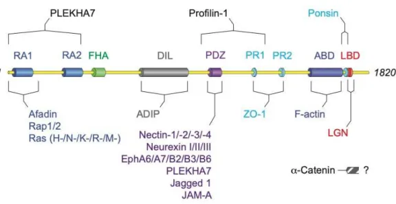
Afadin is a multidomain protein, with two N-terminal Ras-associated (RA) domains, a forkhead-associated (FHA) domain, a DIL domain, a PDZ domain, three proline-rich (PR) domains, and a C-terminal F-actin binding domain (Fig. 1.1). Myosin-competition experiments suggest afadin binds along the sides of actin filaments3. Afadin also scaffolds with an α-actinin binding protein, ADIP, through its
DIL domain4, and to the actin binding protein profilin-15. Afadin’s RA domains primarily bind to the
small GTPase Rap1, but can also bind other Ras family members6, and also mediate homodimerization7.
Through its PDZ domain, afadin primarily interacts with the four nectin proteins8, 9, transmembrane
domain also contributes to interactions involved in neuronal synapsing, binding to members of the neurexin10 and ephrin family11. Furthermore, afadin harbors two bindings sites—one including the PDZ
domain and another including the RA domains—for PLEKHA7, a p120-catenin binding protein12, 13.
Interestingly, PLEKHA7 and afadin play a significant role in clustering ADAM10 and its trafficking partner TspanC8s to cell-cell junctions, which is required for Staphylococcus aureus-mediated cell death14. Afadin also interacts the tight junction proteins JAM-A15 and the scaffold ZO-116 through its PDZ
and PR1/2 domains, respectively. The third PR domain binds to the vinculin-binding protein ponsin, which afadin recruits to site of cell-cell adhesion17. Afadin can also bind to the Notch ligand Jagged 118,
which has tissue specific functions in cellular differentiation. Importantly, afadin also interacts with the actin binding protein α-catenin19, though the interaction has not been mapped to a specific region of
afadin. More recently, studies have characterized an interaction between afadin and the spindle
orientation protein LGN, which binds to a region of afadin proximal to the F-actin binding domain20, 21.
In 1984, the famous Drosophila melanogaster forward genetic screen by Jurgens, Wieschaus, Nusslein, Volhard and Kluding22-24 characterized more than one-hundred novel mutants, including one
aptly named canoe (cno). cno mutants present with large gaps in the dorsal cuticle, granting the embryos a unique ‘canoe-like’ appearance22. Cno was later identified as an afadin ortholog, sharing significant
sequence homology and domain structure6. As with afadin, Cno has a pair of N-terminal RA domains
which can interact with both Ras6, 25, Ran26 and Rap127. The internal PDZ domain of Cno binds to the
nectin ortholog, echinoid28, but also binds to DE-cadherin29, a classic transmembrane cell-cell adhesion
protein, and to the ZO-1 ortholog, polychaetoid30. Cno also binds directly to F-actin29 and the LGN
ortholog Pins26 via distal C-terminal domains. The scaffolding activity of Cno and afadin—binding
numerous proteins involved in adhesion and cell signaling—grants the molecules varied function in cell-cell adhesion (1.2), cell-cell fate determination (1.3), and tissue morphogenesis (1.4).
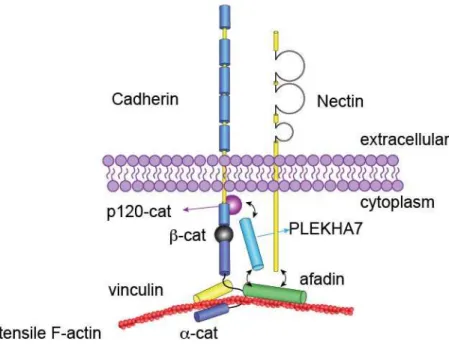
1.2 Afadin is part of the adherens junction, a canonical cell-cell adhesion complex
The adherens junction (AJ) is a cell-cell adhesion complex, classically composed of both cadherins and catenins (Fig. 1.2). Transmembrane cadherins form calcium-dependent homodimers in trans via the distalmost five extracellular cadherin (EC) domains31. There are four classical type I
cadherins, specified by the cells or tissues that express them (e.g. E-cadherin is the primary cadherin expressed by epithelia)32-37. In the cytoplasm, cadherins interact with α -, β-, and p120-catenin38-40.
p120-catenin stabilizes cadherins at the plasma membrane41, 42 while β-catenin—in addition to its
extensively studied role in Wnt signaling43-45—enables recruitment of α-catenin into the AJ46. There are
three α-catenin homologs; α-N-catenin, α-T-catenin and α-E-catenin, the latter of which is the primary a-catenin expressed in most epithelia and will be simply referred to as α-catenin for the remainder of this dissertation. α-catenin links the actin cytoskeleton to the plasma membrane via a C-terminal actin binding domain. However, scaffolding between AJs and the actin cytoskeleton has proven to be a biomechanically complicated process. While α-catenin alone can bind F-actin in solution, the α-catenin/β-catenin/E-cadherin ternary complex has only weak interactions with actin in solution47, suggesting that proper
AJ/actin scaffolding is allosterically regulated.
Ultimately, it was discovered that the minimal cadherin/catenin complex binds actin in a tension dependent manner48, 49. In the presence of non-tensile F-actin, α-catenin’s internal M-domain exists in an
autoinhibited closed conformation50, 51. This behavior is unsurprising given that α-catenin is a homolog
of vinculin, another autoinhibited actin scaffold52-56. When α-catenin’s actin binding domain engages
with tensile actin, the M domain opens, exposing cryptic binding sites for two additional actin binding proteins: the aforementioned vinculin, and afadin19, 51, 55, 57. α-catenin’s M domain is split into three
distinct subdomains (M1, M2, M3), with the vinculin binding domain embedded within M1. In
coimmunoprecipitation experiments, significantly more afadin bound to a truncated fragment of α-catenin compromising M2 and M3 (residues 385-651) than to full-length α-catenin19. Similarly, an
cardiomyocyte junctions58. These studies conclude that both afadin and vinculin require F-actin mediated
tension to bind the open conformation of α-catenin, where they occupy exclusive regions within the tension-sensitive M-domain.
These findings suggest that afadin’s primary role in AJs may be to increase the actin scaffolding potential of the cadherin-catenin complex, thereby allowing it to bear higher tensile forces. Such a role has been already described for vinculin, which is recruited to AJs in a tension-dependent manner and is capable of stabilizing the open conformation of α-catenin49, 59. Similarly, using a ZO-1/2 double
knockdown model60 in Madin-Darby Canine Kidney (MDCK) epithelial cells, Choi et al. demonstrated
that afadin accumulates at hypercontractile adhesions, particularly tricellular junctions where numerous actin bundles are anchored end-on61. In this context, afadin was required to maintain normal cell shape
and epithelial barrier function. In other studies, mosaic knockdown of afadin was sufficient to reduce E-cadherin (E-cad) junctional accumulation in MDCK cells, without affecting nectin-1 or α-, β-, and p120- catenin62, 63. Strangely, despite these changes in E-cad junctional accumulation, afadin knockdown didn’t
reduce E-cad cell surface accumulation, nor its association with the catenins63. Using a different cell line
(EpH4 murine mammary epithelial cells), similar experiments demonstrated that the interaction of afadin with PLEKHA7 promotes the junctional accumulation of p120-catenin and E-cadherin13. However, these
findings are complicated by results from Choi et al., where afadin knockdown had no effect on E-cad localization or accumulation61. One key difference between these studies lies in the fact that those
observing reduced E-cad junctional accumulation used mosaic cultures, while Choi et al. used monocultures, suggesting that non-cell autonomous effects such as anisotropic adhesion/mechanical tension may contextualize afadin’s role. For example, afadin may be specifically essential for balancing anisotropic cortical tension to maintain AJ molecular structure and epithelial integrity under tensile stress. Regardless, these findings suggest that cooperation between numerous actin-binding scaffolds is required to maintain normal cell-cell adhesion and epithelial integrity. However, whether afadin regulates
α-catenin function or conformation—or recruitment of vinculin into the AJ—remains unknown. This knowledge gap is addressed in Chapter 3.
1.2.1 The afadin-nectin complex in cell-cell adhesion
Afadin also interacts with the nectins, a separate family of transmembrane cell-cell adhesion molecules8. In contrast to cadherins, nectins interact in a calcium-independent manner, and are capable of
forming both homodimers in cis and heterotetramers in trans8, 64. Nectins, of which there are four distinct
family members in mammals (nectin-1, -2, -3, and -4) are part of the immunoglobulin-like superfamily, each having three extracellular Ig-like loops9, 65. The first Ig-like loop is essential for trans interactions,
while the second Ig-like loop is required to form cis homodimers66, 67. Evidence suggests the 1st Ig-like
loop of each nectin is capable of interacting with all four family members, albeit with variable affinity. For example, nectin-1 can interact in trans with either nectin-2 or nectin-1, but preferentially binds nectin-3 or nectin-49. The cytoplasmic tail of each nectin forms an obligate interaction with afadin’s PDZ
domain using an EAXYV motif. While nectin-4 lacks this specific motif, it nonetheless binds afadin with comparable affinity9. Subsequently, afadin is responsible for recruiting nectins to cadherin-based AJs via
its interaction with α-catenin8, 65, 68. However, this interaction is not essential for nectin cis dimerization
or trans interactions, and nectins can still be observed at the cell membrane, albeit with diminished accumulation, when they lack the afadin-binding motif8, 65. While nectins frequently colocalize with AJs,
as observed by both fluorescent and electron microscopy, their overlap is not universal, complicating their inclusion as canonical AJ molecules8.
While the role of nectins in cell-cell adhesion is still being uncovered, some studies suggest they may serve as pioneer adhesion factors, preceding cadherins at sites of nascent adhesion assembly69. While
the requirement of endogenous nectin for cadherin-based AJ assembly has not been shown, overexpression of nectin-1 in cultured cells accelerates assembly of E-cad based AJs70. Similar
observations have been made for nectin-2 and nectin-3, which can promote cell-cell adhesion in a nonadherent cell line expressing low levels of E-cad71. Interestingly, overexpression of nectin-3 can
override the dominant-negative function of a truncated N-cadherin lacking the EC domains to induce adhesion formation 72. Nectins also bind to apical polarity proteins, including the canonical polarity
pioneer factor Par373, as well as Mupp1 (Mpdz) and PATJ74, two PDZ-scaffolds of the Crumbs complex
(discussed in Chapter 5). Similarly, junctional recruitment of zyxin, an actin regulator and α-actinin binding partner, can be modulated through an interaction with nectin-2. Taken together, these findings suggest that while the nectin-afadin complex may not be required for nascent AJ assembly, it has the potential to augment AJ dynamics and recruit additional polarity and cytoskeletal modulators.
The role of nectins as modulators of cell-cell adhesion is reinforced by their mutant phenotypes in mice. Germline knockouts of Nectin175, 76, Nectin277, and Nectin375 are all viable, though dual loss of both
Nectin1/Nectin3 results in early embryonic lethality78. Both Nectin2 and Nectin3 knockouts display
diminished male fertility, which has been attributed to defects in cell-cell adhesion between Sertoli cells and haploid spermatids during spermatogenesis77, 79, 80. Loss of nectin-1 or nectin-3 similarly causes
separation between the pigmented and non-pigmented epithelial layers in the ocular ciliary body, resulting in microphthalmia75. Additionally, the fact that nectins display greater affinity for heterotypic
trans-interactions affords them unique functionality in cell sorting. The inner-ear cochlear epithelium is composed of hair cells and supporting cells, which form a checkerboard pattern where no two hair cells share a cell boundary. This pattern is maintained by the trans-interactions of nectin-1 and nectin-3, which are specifically expressed by hair cells and supporting cells, respectively81, 82. Similarly, in the olfactory
epithelium, nectin-2 is expressed by olfactory cells (OC) while nectin-3 is expressed in support cells (SC). Nectin-2/3 trans-interactions cooperate with cadherins to increase the adhesive strength of the OC-SC cell contact at the expense of OC-OC contacts, ensuring normal isolation of OCs83. Nectin heterotypic
trans-interactions also play a significant role in the deposition of dental enamel of the murine incisors. While nectin-1 is expressed by enamel-secreting ameleoblasts, nectin-3 is expressed by juxtaposed stratum intermedium and their trans-interaction is required to prevent separation of these two layers76, 78. The role
of nectins in the oral cavity is particularly interesting, as mutations in the human NECTIN1 gene cause cleft lip/palate ectodermal dysplasia syndrome (CLPED1). However, homozygous null nectin-1 mice do
not present with cleft palate, nor do combination Nectin1/Nectin3 mutants with loss of three alleles78. This
discrepancy is the subject of my work in Chapter 4, which is preceded by an introduction to palatogenesis and the role of cell-cell adhesion proteins in affecting this developmental program (Chapter 2).
Interestingly, germline Nectin1 knockouts display nonlethal neonatal defects in epidermal development. Loss of nectin-1 causes reduced expression of loricrin, a marker of suprabasal
differentiation84. Differentiation and stratification of the embryonic epidermis is partially regulated by
asymmetric cell divisions (ACD) of basal epidermal progenitors85. Additionally, the epidermis of
newborn Nectin1 null mice is red and shiny, and this phenotype has previously been correlated with perturbation of ACD86. Furthermore, loss of the obligate nectin binding partner, afadin, causes ACD
defects in murine cortical progenitors87 and Drosophila neuroblasts88. However, whether afadin plays a
significant role in ACD in murine epidermal progenitors is unknown. This knowledge gap is further outlined below (1.3) and experimentally addressed in Chapter 3.
1.3 Asymmetric cell division and cell fate specification
Classically, mitosis allows for one mother cell to duplicate its genetic code and generate two identical daughter cells. While a majority of cell divisions are symmetric—producing progeny of identical size with identical fates—stem and progenitor cells are uniquely capable of undergoing asymmetric cell divisions (ACDs), generating two daughter cells with distinct fates. While these asymmetries may refer to any aspect of cellular identity (size, shape, transcript inheritance, protein inheritance, etc.), for the
purposes of this dissertation, I’ll be referring to asymmetry in cellular identity as determined through biomarkers of differentiation.
1.3.1 Asymmetric cell division in the Drosophila neuroblast
One classic model for studying ACDs is the Drosophila neuroblast, an embryonic neuronal precursor. At mitotic onset, neuroblasts invaginate from the neuroepithelial monolayer and then orient
their metaphase spindle orthogonal to the plane of the tissue. During metaphase, numerous fate
determinants—including Numb, Prospero (Pros)89, Miranda (Mira)90, 91, Partner of Numb (Pon)92, 93 and
Brain Tumor (Brat)94, 95—are polarized towards the basal cell cortex. Numb inhibits Notch signaling96,
which normally promotes proliferation and maintenance of the neuroblast identity97, 98. Pros is a
transcription factor which both suppresses transcription of hundreds of neuroblast genes, and enhances expression of differentiation genes, suggesting that it is capable of acting as both a transcriptional inhibitor and activator to promote neuronal differentiation99. Mira and Pon are adapter proteins which
promote the basal polarization of Pros90, 91 and Numb92, 93, respectively. Similarly, Mira encourages the
basal segregation of Brat, which acts cooperatively with the transcription factors Pros, Deadpan, and Zelda to suppress proliferation and promote differentiation94, 95, 100. As cytokinesis completes, these
proteins are asymmetrically inherited by the basal daughter and together promote differentiation into a ganglion mother cell (GMC) fate. The absence of these factors in the apical daughter allows it to maintain its progenitor identity and undergo additional mitoses.
During the identification of these fate determinants, questions arose regarding how their asymmetric inheritance was established. This led Knoblich, Jan and Jan to speculate that additional positional cues acted to orient the mitotic spindle: “… localization of Numb and Pros is microtubule independent, but recognizes positional information that is provided for the placement of the centrosome and the mitotic spindle.89” This line of investigation led to the discovery of the spindle orientation
effector, Inscuteable (Insc), loss of which altered both the orientation of the mitotic spindle and the localization of fate determinants101. Insc localizes to the apical cell cortex, opposite the fate determinants,
and its polarization is dependent on an interaction with the canonical polarity protein Bazooka (Baz, the
Drosophila ortholog of vertebrate Par3)102. Searching for additional Insc binding partners, Yu, et al.
identified an additional regulators of mitotic spindle orientation, Partner of Inscuteable (Pins)103, which
was later shown to form a tripartite complex with Insc and the heterotrimeric G-protein Gαi/o subunit104.
As with insc mutants, pins mutants fail to orient their metaphase spindle along the apico-basal axis and fail to properly segregate their fate determinants104. Despite their clear effects on spindle orientation, the
mechanism of action of the Insc/Pins/Gαi complex remained elusive; none of the complex members bind
to microtubules, and thus the complex lacked a spindle effector. One potential downstream component was identified in HeLa cells as the microtubule binding protein NuMA, which was recruited to the cell cortex through its interactions with the Pins ortholog, LGN105, 106. A fly relative of NuMA, Mushroom
Body Defect (Mud)107, binds to both dynactin and Lis1108, a regulator of the dynein microtubule motor.
Each of these mutants not only fail to properly align their metaphase spindle, but live imaging of larval neuroblasts show they are incapable of normal spindle rotation, suggesting the complex functions as not just a spindle anchor, but can also physically rotate the spindle until it aligns with a particular polarity axis.
While proper coordination of the spindle orientation machinery promotes the asymmetric segregation of fate determinants, there are also several mechanisms which directly link the spindle orientation complex to the basal polarization of the fate determinants. Bazooka (Baz) binds to additional polarity proteins including Par6 and aPKC, both of which reinforce apical polarization of Baz and are required to properly localize the spindle orientation machinery and ensure basal segregation of fate determinants109. aPKC directly phosphorylates Mira within its cortical localization domain, which
displaces it from the apical cell cortex110. Lgl, a non-polarized cytoskeletal adapter protein, cooperates
with the cortical proteins Dlg and Scrib to competitively inhibit aPKC activity, restricting aPKC to the apical cortex to enable basal-specific cortical polarization of Mira110-114. Additionally, Insc binds directly
to and colocalizes with the RNA-binding protein Staufen (Stau)115. During interphase, Stau is apically
polarized in an Insc-dependent manner and binds pros mRNA in the 3’ UTR. As the neuroblast enters mitosis, Stau then binds to Mira, allowing pros RNA to be basally segregated, where it promotes the GMC identity116-118. These pathways highlight the interconnectivity of apical polarity complexes, spindle
orientation machinery, and fate determinants themselves.
Together, these series of studies spanning multiple decades culminated in the identification of a spindle orientation complex which captures astral microtubules emanating from the centrosome in order to rotate the metaphase spindle through the Dynein motor. The apical polarization of the
Baz/Insc/Gαi/Pins complex promotes spindle alignment along the apico-basal axis, which encourages the
asymmetric inheritance of basally-polarized fate determinants. Subsequent studies have relied on this pathway as a blueprint for elucidating mechanisms of fate determination and morphogenesis in numerous tissue systems. Since their discovery, these proteins have been implicated in division orientation of mammalian cortical neural progenitors119 and epithelia of the lung120, intestine121, 122, mammary gland 123-125, and skin85, 86, 126, 127, among others. Studies in these vertebrate systems and mammalian cell culture
models like MDCK and HeLa cells, as well as invertebrate systems of the C. elegans embryo128 and D.
melanogaster sensory organ precursors129, have furthered our understanding of the machinery required for
both oriented and asymmetric cell divisions. However, it is important to retain distinct terminology for referring to oriented cell divisions (OCD), in which the spindle is positioned in response to polarity cues, and asymmetric cell divisions (ACD), which result in mitotic progeny with distinct cell fates through asymmetric inheritance of intrinsic or extrinsic fate determinants.
1.3.2 Coupling of division orientation and cell fate determination in the mammalian epidermis The mammalian epidermis is a stratified epithelium consisting of numerous cell layers which are both morphologically and functionally distinct. During embryogenesis, the epidermis originates as a simple, monolayered epithelium which, during the 14th through 17th day of murine gestation (E14-17),
undergoes rapid stratification and differentiation to produce a fully functional barrier prior to birth. The mechanisms governing epidermal development have been studied for many decades, but the molecular underpinnings have been only more recently elucidated. Early insights from culture models suggested delamination—the separation and upward migration of basal progenitors from the underlying
extracellular matrix—correlated with stratification and cellular differentiation130, 131. This suggested a
model wherein cycling progenitors, restricted to the basal layer of the epidermis, would divide symmetrically within the plane of the epithelium and stratification would be explicitly driven by delamination132. However, this model neglected earlier observations suggesting that basal progenitors
displayed divisions with a variety of orientations, including both within the tissue plane (i.e. planar divisions) or orthogonal to it (i.e. perpendicular divisions)126.
More recent studies have revealed that basal progenitors display bipotent OCDs, with planar divisions promoting progenitor expansion while perpendicular divisions serve as potent drivers of stratification85. The canonical spindle orientation machinery proved essential for this behavior, as the
mammalian orthologs of Pins (LGN/Gpsm2), Insc (mInsc), Gαi (Gαi3), Baz (Par3), Mud (NuMA) and
p150glued (dynactin) are all specifically required for perpendicular divisions86, 127. In this model, interphase
apical Par3 and Gai3 recruit the mitotic-specific mInsc. In turn, the mInsc/Par3 and Gαi3 polarity proteins
cooperatively recruit LGN to the apical cell cortex, which then scaffolds with NuMA and the
dynein/dynactin complex on astral microtubules to align the mitotic spindle along the apico-basal axis. This orientation drives the apical daughter into the superficial layers of the tissue—promoting
differentiation—while the basal daughter retains contact with the basement membrane and can undergo additional mitoses127.
Importantly, LGN is only recruited to the apical cortex in ~50% of mitoses of (pro)metaphase cells. In later stages of mitosis (ana/telophase), LGN is present in the majority of perpendicular divisions, but noticeably absent from planar divisions, suggesting the presence/absence of LGN specifically serves as an instructive cue for perpendicular divisions86. Fittingly, loss of LGN (Gpsm2) or NuMA (Numa1)
results in nearly all divisions becoming planar/symmetric, rather than randomized as in the neuroblast. Given the bimodal distribution of division behaviors, it is not surprising that multiple groups have found evidence suggesting the metaphase spindle rotates before determining a final axis of division. Live imaging of cultured keratinocytes shows that the metaphase spindle rotates within the epithelial plane prior to cytokinesis (i.e. planar rotation)133. Similarly, while division orientation measurements made at
telophase in vivo display the bimodality of perpendicular/planar division angles, measurements made at metaphase result in a near-random distribution of spindle orientations, suggesting the spindle has not yet committed to a final division axis86. Given that the spindle effector machinery
(LGN/NuMA/dynein/dynactin) is assumed to operate on astral microtubules emanating from the metaphase spindle, division orientation (and thereby cell fate) should become fixed at anaphase onset. While anaphase division orientation of epidermal progenitors has never been specifically reported, our findings in Chapter 3 contradict this assumption, demonstrating that anaphase orientation is not significantly different from metaphase. Through live imaging of embryonic explants, we discover that epidermal progenitors frequently initiate anaphase at oblique orientations and these divisions undergo “correction” during telophase to generate the bimodal distribution of divisions associated with progenitor self-renewal and differentiation. These findings significantly improve our understanding of OCD in embryonic epidermal progenitors and are further detailed in Chapter 3 and their implications regarding the current model of OCD discussed in Chapter 5.
In the Drosophila neuroblast, Numb operates as an intrinsic fate determinant whose asymmetric inheritance by the GMC daughter suppresses Notch signaling in order to promote GMC differentiation. While no such fate determinants have been reported in embryonic epidermal ACDs86, numerous studies
still implicate Notch signaling as a potent regulator of proliferation and cell fate134, 135. The core Notch
signaling pathway in vertebrates is composed of four transmembrane Notch receptors and a variety of ligands: Delta, Delta-like, Serrate, and Jagged136. Following ligand binding in trans, the Notch receptor is
cleaved, releasing the Notch intracellular domain (NICD). NICD then enters the nucleus in complex with RBPJ and the coactivator MAML (of which there are three homologs in vertebrates). This transcriptional complex activates Notch effector genes such as Hes1. Overall, Notch activity appears to be suppressed in basal epidermal progenitors and most active in the suprabasal differentiated progeny, as determined by fluorescent RBPJ or Hes1 reporters86, 137, 138. While the Notch repressor Numb is not asymmetrically
segregated during ACD in embryonic epidermal progenitors86, it can be asymmetrically segregated in
planar divisions in the adult139. Interestingly, knockdown of LGN resulted in a cell-autonomous decrease
in Notch-reporter activity in suprabasal cells86. Furthermore, stratification defects observed in LGN
mutants can be rescued by overexpression of active Notch86, suggesting some asymmetry of Notch
inform cell fate through competition for niche occupancy, as occurs in other systems121, 140-142. Early
studies of epidermal stem cells using colony formation assays identified high levels of β1-integrin expression—which may promote strong basement membrane adhesion—to be strongly correlated with colony formation, a surrogate for “stemness.”132, 143 It is thus enticing to speculate that basement
membrane contact may be sufficient for—or at the very least contribute to—defining basal progenitor identity.
1.3.3 Biochemical architecture of the spindle orientation complex
The essential scaffold LGN functions as a linchpin within the spindle orientation complex, linking the upstream polarity components Par3/mInsc/Gαi3 to the downstream spindle effectors
NuMA/dynactin/dynein. Through its four C-terminal GoLoco motifs, LGN interacts with four copies of the GDP-bound heterotrimeric Gαi/o subunit144. LGN also interacts with mInsc (mammalian Insc) via its
eight N-terminal TPR repeats, which bind to the Insc N-terminal LGN-binding domain (LBD) and structural studies indicate that this interaction is highly conserved between fly and mammalian orthologs145. In vitro studies using purified proteins confirm that mInsc is required to
coimmunoprecipitate LGN with Par3, suggesting that mInsc is required for LGN to respond to apical polarity established by the Par3/Par6/aPKC complex. Furthermore, Pins and Insc form a 2:2 symmetric tetramer, dependent on dimerization of both the C-terminal Armadillo repeats of Insc and the
complementation between TPR3-4-5 of Pins125. Similarly, size-exclusion chromatography of a
reconstituted Pins/Insc/Baz/Gαi ternary complex support a 2:2:2:8 stoichiometry, which allows for a
Pins/Insc tetramer existing within the higher order spindle orientation complex125.
One complicating factor, however, is that mInsc and NuMA cannot bind LGN simultaneously, and in fact compete for LGN binding sites along the TPR repeats. In addition, the affinity of LGN for Insc is orders of magnitude higher than for NuMA, suggesting that NuMA is incapable of displacing mInsc from the mInsc/LGN complex146. If NuMA cannot outcompete mInsc from the complex, how does
polarized LGN engage NuMA to capture astral MTs and orient the mitotic spindle? Studies have shown that LGN can adopt an autoinhibited conformation via intramolecular interactions between its GoLoco and TPR repeats147. Interestingly, this conformation allows for interactions with Gai, which releases the
LGN autoinhibitory conformation and enables NuMA binding148. While early experiments utilizing a
small NuMA fragment (residues 1900-1928) suggested a 1:1 binary interaction with the LGN TPR domains, recent evidence using a longer fragment (1821-2001) suggests they form a higher order hetero-hexameric complex with 3:3 stoichiometry149. This oligomerization is required for planar division
orientation of HeLa cells and normal Caco-2 cell cyst formation, but dispensable for recruitment of the spindle motor, dynein/dynactin. The same study suggests that this oligomer promotes the formation of multimeric protein aggregates, which in turn are required to properly coordinate spindle motor function to align the mitotic spindle149. However, these findings do not explain how diverse populations of
mInsc/LGN tetramers and NuMA/LGN hexamers may coexist or cooperate to orient the spindle. While it is possible these complexes form from distinct pools of Gai-bound LGN and serve unique functions in the segregation of fate determinants (mInsc/LGN) and spindle orientation (NuMA/LGN), as recently
suggested125, the fact that mInsc is required for LGN polarization complicates this hypothetical model.
Alternatively, it remains possible that the LGN/mInsc interaction is allosterically regulated or post-translationally modified by an unknown effector which promotes the exchange of TPR-bound mInsc for NuMA. Characterization of multiple mutants which impair mInsc- or NuMA-LGN interactions and oligomerization in vivo would further our understanding regarding the role each interaction plays in modulating division orientation and symmetry.
1.3.4 Cell-cell adhesion and cell mechanics inform oriented cell division
Since Hertwig’s observations more than 100 years ago that sea urchin oocytes align their mitotic spindle with the interphase long axis150, it has been understood that cell shape and mechanics can provide
instructive cues for division orientation. For example, as tissues develop, the epithelia lining them must respond to growth-induced stretch by orienting division to align with these tissue-scale forces and
maintain a contiguous barrier. This phenomenon has been observed in cultured keratinocytes, where uniaxial stretch results in a bias in spindle orientation alignment with the stretched axis151. The
mechanisms that allow cells to sense and adapt to these external forces varies but several key cellular factors have been described which translate mechanical forces into cell polarity and orientated cell division: acto-myosin contractility152 (i), cell-matrix adhesion153 (ii), and cell-cell adhesion154 (iii).
Additionally, as cells enter and progress through mitosis, they undergo a series of drastic shape changes, which depend on proper coordination of the underlying cytoskeleton and remodeling of their cell-cell and cell-matrix adhesions. More recently, it has become clear that these structural components of the cell machinery are also essential for coordinating mitotic spindle positioning with intrinsic polarity cues. Taken together, there is an undeniable body of evidence suggesting that the acto-myosin cytoskeleton, in cooperation with cell-matrix and cell-cell adhesions, is an essential component of OCD.
i) Actomyosin contractility in OCD. Numerous studies have demonstrated that the orientation of cell division requires an intact actin cytoskeleton, most frequently using actin depolymerization agents to perturb OCD89, 101, 142, 155. Early experiments in D. melanogaster neuroblasts confirmed that localization
of the apical polarity complex, including the spindle effector Insc, was compromised by actin depolymerization by cytochalasin D treatment101. More recently, actomyosin contractility during cell
rounding and mitotic progression was shown to play a significant role in regulating the size asymmetry generating during neuroblast ACD. In Drosophila neuroblasts, during anaphase onset, myosin flows from the apical cortex towards a basal-biased lateral domain to position the cleavage furrow156. This flow of
contractile components induces internal anisotropic hydrostatic pressure which encourages swelling of the apical domain, supporting the resultant size asymmetry between the apical neuroblast and basal GMC daughters156, 157. The basal bias of myosin is dependent on the intrinsic spindle polarity protein Pins, loss
of which reduces size asymmetry158.
Actomyosin contractility can also function as a force generator to position the mitotic spindle in scenarios where the Pins/LGN spindle orientation complex is not required. For example, in the two-cell
not159. Instead, physical contact is sufficient to dictate the orientation of AB cell division orthogonal to the
site of contact, and coordinated cortical myosin flows are required for normal positioning of the spindle and cleavage furrow160. Similarly, during D. melanogaster segmentation, cells along the parasegment
boundary break Hertwig’s rule and instead consistently orient their division orthogonally to this boundary in a Pins- and Mud-independent manner161. Each boundary is demarcated by a contiguous actomyosin
cable running orthogonal to the ventral midline. Reduced contractility achieved by either myosin inhibition or laser ablation of the cable causes boundary-adjacent cells to fail to orient their division orthogonally, suggesting that actomyosin contractility is both essential and sufficient to dictate the orientation of cell division. These studies highlight the adaptability of actomyosin tension, which can serve as either a spindle positioning force or a sufficient polarity cue to dictate OCD.
In contrast, the mammalian embryonic epidermis more closely mirrors early studies from neuroblasts. Actin depolymerization via latrunculin A treatment diminishes apicobasal polarity, causing errors in apical LGN recruitment and division orientation155. Similarly, epidermal loss of the transcription
factor Srf—a key regulator of many cytoskeletal and contractility genes—causes errors in mitotic cell rounding and oriented cell division, which have been attributed to defects in myosin accumulation at mitotic onset155. Epidermal knockout of the Arp2/3 actin branching complex causes defects in
differentiation of the suprabasal layers, possibly through hyperactivation of the YAP/TAZ growth pathway; though no effect on OCD was reported162. More recently, epidermal loss of the actin bundling
protein T-plastin resulted in defects in basement membrane deposition and cell polarity, with related alterations to OCD of basal progenitors163. Taken together, these studies highlight that the actomyosin
contractile cytoskeleton is an essential component of OCD and ACD in a variety of tissue systems, either in cooperation with, or independent of, the canonical spindle orientation machinery.
ii) Cell-matrix adhesion in OCD. Fifteen years ago, Thery, et al. performed exciting experiments using micropatterned fibronectin substrates to demonstrate that HeLa cells orient their division in response to the shape and force applied by their cell-matrix adhesions164. This behavior was
rounding165, 166. The spindle was oriented via cooperation with NuMA and subcortical “actin clouds” that
align with retraction fibers and orient the spindle in a myosin-10 dependent manner167. However, the
relevance of this mechanism to an in vivo, multicellular context remained untested.
Early experiments in the D. melanogaster follicular epithelium demonstrated that perturbation of cell-matrix adhesions alters OCD, without impairing apicobasal polarity168. Additional experiments in
other models continued to demonstrate that cell-matrix adhesions may be essential for OCD in a variety of contexts. Loss of β1-integrin—a key component in cell-matrix hemidesmosomal adhesions—in the murine mammary epithelium results in abnormal out-of-plane divisions with consequences for mammary ductal morphogenesis169. Integrin-linked kinase (ILK)—a β1-integrin binding partner—can form a direct
interaction with dynactin and regulates its cortical recruitment to coordinate division orientation in HeLa cells122. Additionally, loss of ILK in the murine intestine increases the rate of inappropriate divisions
oriented towards the luminal surface122. Cell-matrix adhesions operate upstream of apicobasal polarity in
the Drosophila intestine and are required for normal OCD of intestinal stem cells170. Similar observations
have been made in the embryonic epidermis, where loss of β1-integrin impairs apicobasal polarity, LGN cortical localization and division orientation85. However, due to the fact that perturbation of the ECM or
cell-matrix adhesion complexes consistently alters apicobasal polarity in the epidermis, it is hard to determine whether cell-matrix adhesions regulate epidermal OCD in a direct or indirect manner. Moreover, although loss of β1-integrin—a component of both basal-localized focal adhesions and desmosomal junctions at lateral cell-cell junctions—impacts division orientation, loss of β4-integrin—a component of hemidesmosomes at cell-matrix adhesions—does not85. Nonetheless, given the
aforementioned association between β1-integrin expression levels and epidermal progenitor capacity132, 143, it is intriguing to speculate that these adhesions may play important roles in regulating cell fate
through OCD. Fittingly, our experiments in Chapter 3 suggest that the maintenance or loss of basement membrane contact directs obliquely oriented epidermal progenitors towards a basal or suprabasal fate, respectively. How integrin based basal adhesions may impact this process is discussed in Chapter 5.