A thesis submitted in fulfilment of the requirements for the degree of Doctor of Philosophy
Madiha Tariq
MS Mechatronics Engineering, Air University, Islamabad, Pakistan B.Eng. Mechatronics Engineering, Air University, Islamabad, Pakistan
School of Engineering
College of Science, Engineering and Health RMIT University
ii I certify that except where dueDFNQRZOHGJPHQW has been made, the work is that of the author alone;the work has not been submitted previously, in whole or in part, to qualify for any other academicaward; the content of the thesis is the result of work which has been carried out since the officialcommencement date of the approved researchprogram; any editorial work, paid or unpaid, carriedout by a third party is
acknowledged; and, ethics procedures and guidelines have been followed.
,DFNQRZOHGJHWKHVXSSRUW,KDYHUHFHLYHGIRUP\UHVHDUFKWKURXJKWKHSURYLVLRQRI $XVWUDOLDQ*RYHUQPHQW5HVHDUFK7UDLQLQJ3URJUDP6FKRODUVKLS
Madiha Tariq Dated:-1-2019
iii existing.
v To
my most precious Ami Abu, dear husband, and my world, Maheen & Zeenya.
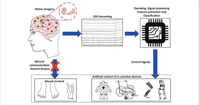
vi Over recent years significant advancements in the field of assistive technologies have been observed. One of the paramountneeds for the development and advancement that urged researchers to contribute in the field other than congenital or diagnosed chronic disorders, isthe rising number of affectees from accidents, natural calamity (due to climate change),or warfare,worldwide resulting in spinal cord injuries (SCI), neural disorder, or amputation (interception) of limbs,that impede a humanto live a normal life. In addition to this, morethan ten million people in the worldareliving with VRPH form of handicap due to the central nervous system (CNS) disorder,which is precarious. Biomedicaldevices for rehabilitation DUH the center of UHVHDUFKfocusIRU many years. For people with lost motor control or amputation, but unscathed sensory control, instigation of control signals from the source, i.e. electrophysiological signals, is vital for seamless control of assistive biomedical GHYLFHs. &ontrol signals, i.e. motion intentions arouse in the sensorimotor cortex of the brain that can be detected using invasive or non-invasive modality. With non-invasive modality, the electroencephalography (EEG) is used to record these motion intentions encoded in electricalactivityof the cortex, and are deciphered to recognize user intent for locomotion. They are furthertransferred to the actuatoror end effector of the assistive devicefor control purposes. This can be executed via the brain-computer interface (BCI) technology.
BCI is an emerging research field that establishes a real-time bidirectional connection between the human brain and a computer/output device. Amongst its diverse applications, neurorehabilitation to deliver sensory feedback and brain controlled biomedical devices for rehabilitation are most popular. While substantial literature on control of upper-limb assistive technologies controlled via BCI is there, less is known about the lower-limb (LL)control of biomedical devices for navigation or gait assistance via BCI.The types of EEG signals compatible with an independent BCI are the oscillatory/sensorimotor rhythms (SMR) and event-related potential (ERP). These signals have successfully been used in BCIs for navigation control of assistive devices. However, ERP paradigm accounts for a voluminoussetup forstimulus presentation to the user during operation of BCIassistive device. Contrary to this, the SMRdoes not requirelarge setup for activation ofcortical activity; it instead dependson the motor imagery(MI) that isproduced
vii termed kinaesthetic motor imagery (KMI) and elicitsclearly after rigorous training trials, in form of event-related desynchronization (ERD) or synchronization (ERS), depending on imagery activity or resting period.It usually comprises oflimb movementtasks, but isnot limited to itin a BCI paradigm.In order to produce detectable features that correlate to the user’s intent, selection of cognitive task is an importantaspectto improve the performance of a BCI.MI used in BCI predominantly remainsassociated with the upper-limbs, particularly hands, due to the somatotopic organization of the motor cortex. The hand representation area is substantially large,in contrast to the anatomical location of the LL representation areas in the human sensorimotor cortex. The LL area is located within the interhemispheric fissure, i.e. between the mesial wallsof both hemispheres of the cortex. This makes it arduous to detect EEG features prompted upon imagination of LL.Detailed investigation of the ERD/ERS in the mu and betaoscillatory rhythms during left and right LL KMI tasks is required, as the user’s intent to walk is of paramount importance associated to everyday activity. This is an important area of research, followed by the improvisation of the already existing rehabilitation system that serves the LL affectees. Though challenging, solution to these issues is also imperativefor the development of robust controllers that follow the asynchronous BCI paradigms to operate LL assistive devices seamlessly.
This thesis focusses on the investigation of cortical lateralization of ERD/ERS in the SMR, based on foot dorsiflexion KMI and knee extensionKMI separately. This research infers the possibility to deploy these features in real-time BCI by finding maximum possible classification accuracy from the machine learning (ML) models. EEG signal is non-stationary, as it is characterized by individual-to-individual and trial-to-trial variability, and a low signal-to-noise ratio(SNR), which is challenging. Theyare high in dimension with relatively low number ofsamples available for fitting ML models to the data. These factors account forML methods that ZHUHdeveloped into the tool of choice toanalyse single-trial EEG data.Hence the selection of appropriate ML model for true detection of class label with no tradeoff of overfitting is crucial. The feature extraction part of the thesisconstituted of testing the band-power (BP) and the common spatial pattern (CSP) methods individually. The study focused on the synchronous BCI paradigm.This was to ensure the exhibition ofSMRfor the possibility of a practically viable control system in a BCI. For the left vs.right foot KMI, theobjective was to
BCI for controlling/navigating arobotic/prosthetic LL for rehabilitation. Similar was the approach for left-right knee KMI. The research was based on four main experimental studies. In addition to the four studies, the research is also inclusive of the comparison of intra-cognitive tasks within the same limb, i.e. left foot vs. left knee and right foot vs. right knee tasks, respectively(Chapter 4).This added to another novel contribution towards the findings based on comparison ofdifferent tasks within the same LL. It provides basis toincrease thedimensionality of control signals within one BCI paradigm, such as a BCI-controlled LL assistive device with multiple degrees of freedom (DOF)for restoration of locomotion function. This study was based on analysis of statistically significant muERD feature using BP feature extraction method.
The first stage of this research comprised ofthe left vs. right foot KMI tasks, wherein the ERD/ERS that elicited in the mu-betarhythms were analysed using BPfeature extraction method(Chapter 5).Three individual features, i.e. muERD, beta ERD, and beta ERS were investigated on EEG topography and time-frequency (TF) maps, and average time course of power percentage, using the common average reference and bipolar reference methods. A comparative study was drawn for both references to infer the optimal method. This was followed by ML, i.e. classification of the three feature vectors (muERD, beta
ERD, and betaERS), using linear discriminant analysis (LDA), support vector machine (SVM), and k-nearest neighbour (KNN) algorithms, separately. Finally the multiple correction statistical tests weredone, in order to predictmaximum possible classification accuracy amongst all paradigms for the most significant feature.All classifier models were supported with the statisticaltechniques of k-fold cross validation and evaluation of area under receiver-operator characteristic curves (AUC-ROC) for prediction of the true class label. The highest classification accuracy of 83.4% ± 6.72 was obtained with KNN model for betaERS feature. The next study was based on enhancing the classification accuracy obtained from previous study. It was based on using similar cognitive tasksas study in Chapter 5, however deploying different methodology for feature extraction and classification procedure. In the second study, ERD/ERS from mu and betarhythms were extracted using CSPand filter bank common spatial pattern (FBCSP) algorithms, to optimize the individual spatial patterns(Chapter 6). This was followed by ML process, for which the supervised logistic regression (Logreg) and LDA were deployed separately. Maximum classification accuracy resulted in 77.5% ± 4.23with FBCSP feature vector viii
ix of agreement between the two classes. The left vs. right foot discrimination results were nearly same, however the BP feature vector performed better than CSP.
The third stagewas based on the deployment of novel cognitive taskof left vs. right knee extension KMI. Analysis of the ERD/ERS in the mu-beta rhythms was done for verification of cortical lateralizationvia BP feature vector (Chapter 7).Similar to Chapter 5, in this studythe analysis of ERD/ERS features was done on the EEG topography and TF maps, followed by the determination of average time course and peak latency of feature occurrence.However, for this study, only muERD and betaERS features were taken into consideration and the EEG recording method only comprised of common average reference. This wasdue to the established results from the foot study earlier, in Chapter 5, where beta ERD features showedless average amplitude. TheLDA and KNN classification algorithms were employed. Unexpectedly, the left vs. right knee KMI reflected the highest accuracy of 81.04% ± 7.5and an AUC-ROC = 0.84,strong enough to be used in a real-time BCI as two independent control features.This was using KNN model for betaERS feature. The final study of this research followed the same paradigm asusedin Chapter 6, but for left vs. right kneeKMI cognitive task (Chapter 8).Primarily this study aimed at enhancing the resulting accuracy from Chapter 7, using CSP and FBCSP methods with Logreg and LDA models respectively. 5esults were in accordance with those of the already established foot KMI study, i.e. BP feature vector performed better than the CSP. Highest classification accuracy of 70.00% ± 2.85 with kappa score of 0.40 was obtained with Logreg using FBCSP feature vector. 5esults stipulated theutilization ofERD/ERS inmuand betabands,as independent control features for discrimination of bilateral foot or the novel bilateral knee KMI tasks. Resulting classification accuraciesimplicatethat any 2-class BCI employing unilateral foot or knee KMI issuitable for real-timeimplementation.
In conclusion, this thesis demonstrates the possible EEG pre-processing, feature extraction and classification methodsto instigate a real-time BCI from the conducted studies. Following this, the critical aspects of latency in information transfer rate, SNR, and tradeoff between dimensionality and overfitting needs to be taken careof, during design of real-time BCI controller. It also highlights that there is a need for consensus over the development of standardizedmethods of cognitive tasks forMI based BCI.
x contribute to lay the foundations of the development of independent asynchronous BCI based on SMR.
xii First and foremost my heartiest gratitude goes to Almighty, who gave me the strength, confidence to work on this research idea, and good health to complete this thesis within the allocated timeframe. Further, I would like to acknowledge the assistance and support provided by the following people in the completion of this thesis.
Professor Pavel Trivailo (Principal Supervisor) for his continuous support, guidance, constructive feedbacks and valuable insights on this thesis. I acknowledge his perseverance for conducting high-level research and patience in reading my research articles and this thesis. Despite his busy schedule and lot of responsibilities, he has always been available for discussing different research ideas and has always encouraged me to transform those ideas into constructive work. I practically learnt from him the meaning of ‘never give up’. I would confidently say that Professor Trivailo is one of the best leading supervisors at RMIT University and I feel honoured to be under his research supervision.
Dr. Milan Simic (Associate-Supervisor) for his technical discussions, assistance and support throughout my candidature. He is a great source of motivation and has always encouraged me to bring some innovative ideas in my research. With a sincere commitment towards his profession he was always readily available for discussions on research ideas. I am truly privileged to have worked closely with him that helped me to improve my practical skills in conducting research work.
Dr. Yutaka Shoji from RMIT (Biomedical Engineering) for technical discussions and suggestions on different software that embedded the time stamps on recorded EEG signals involved in this work, and installation of unstable versions of EEGLAB and BCILAB. I am really grateful to my PhD research sponsors RMIT University for awarding me the RMIT PhD International Scholarship (RPIS). I also wish to thank my master student, Lena Uhlenberg for her assistance in establishing the experimental protocol for synchronous BCI in this project.
xiii Bundoora east campus forshowing trust in me andgiving me after-hours access tothe Mechatronics Lab (253.01.028). I would also like to thank MrSebastian Naselli for providing me with storage space, toolsand access in the MechatronicsLab.
Iam thankful toall my family members: My father and mother for their endless physical and moral support, countless prayers,and ever encouragingwords,I always look up to them for they have been the torchbearers in my life,my sisters(Jaweria Tariqand Bushra Komal Tariq)for never letting me lose hope and never letting the cheerful side of mine go away, my dearhusband(Dr.Khurram S. Munir)without whom it was impossible to pull this degree off,he is my confidant, inspiration and support system,my preciousand prettiest daughters (Maheen Khurram and Zeenya Khurram) who mean the world to me, they make me a proud mother, Ihope one day they will read this thesis. Finally,I thank my in-laws for their unconditionalencouragement, loveand supportthat instilled in me a beliefto complete this research,and make my and our family’sdream come true.I can proudly say that IZLllbe the first woman from my generation in my immediate family (the Qureshi clan),after my grandfather’s brother Dr.Imtinan Elahi Qureshi,to confer onthe title of Doctor of Philosophy.
xv The candidate would like to acknowledge the help of all-co-authors who assisted and contributed to the papers included in this thesis. All co-authors have given approval for the relevant papers to be included as part of this dissertation. As reflected by the candidate’s position as first author on all included papers, the candidate confirms that she has made the major contribution to all papers forming this dissertation. All papers were primarily written by the candidate. The author indication forms are provided in the appendices.
Declaration ... ii
Abstract ... vi
Acknowledgement... xii
Contribution to jointly authored papers ... xv
Table of contents ... 1
List of figures ... 4
List of tables ... 7
List of author’s published papers incorporated into this thesis ... 10
List of author’s published papers incorporated into appendices ... 11
List of author’s published papers not incorporated into this thesis ... 12
List of conference papers and conference abstracts ... 13
List of technical and research presentations... 14
List of abbreviations ... 15
1. Introduction ... 19
1.1. Overview of brain-computer interface controlled actuators/output devices ... 20
1.2. Research aim and objectives ... 22
1.3. Thesis structure ... 25
1.4. References ... 27
2. Literature Review ... 29
2.1.Introduction ... 31
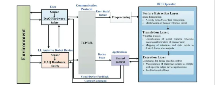
2.2.General control framework for BCI wearable lower-limb and Assistive-robot devices ... 32
2.3.User adaptability and EEG signal Acquisition work Processing ... 34
2.3.1 User Adaptability ... 34
2.3.2 EEG signal acquisition ... 34
2.4.Communication protocol ... 35
2.5.BCI operator ... 35
2.3.3 Preprocessing ... 36
2.3.4 Feature Extraction Layer ... 36
2.3.5 Translational Layer ... 37
2.3.6 Execution Layer ... 38
2.6. Shared Control ... 38
2.7.Lower-limb assistive-robot applications in different Environments ... 3
2
2.7.2. BCI Orthosis ... 39
2.7.3. BCI Wheelchairs, Humanoids, and Mobile Robots ... 39
2.8. Practical challenges ... 43
2.9. Conclusions ... 44
2.10. Acknowledgements ... 45
2.11. References ... 45
3. Materials and Methods ... 50
3.1. Introduction... 51
3.2.Experimental paradigm and data acquisition ... 51
3.2.1. Subjects and experimental design ... 51
3.2.2. Measuring brain activity ... 55
3.2.3. 10-20 System used in EEG ... 55
3.2.4. Data acquisition... 56
3.3.Pre-processing techniques to reduce noise and artifacts from EEG ... 57
3.3.1. Spatial filtering (ICA and CSP) ... 58
3.4.Feature extraction techniques ... 59
3.4.1. Time-frequency analysis: the wavelet transform ... 59
3.4.2. Band power (percentage power change in ERD/ERS) features transform . ... 62
3.4.3. Common spatial pattern (CSP) features ... 63
3.5.Classification techniques ... 64
3.5.1. Linear discriminant analysis (LDA)... 64
3.5.2. Support vector machine (SVM) ... 65
3.5.3. k nearest neighbour (KNN) ... 66
3.5.4. Logistic regression (Logreg) ... 67
3.6.Evaluation criteria for BCI performance ... 67
3.6.1. Bootstrap statistic ... 67
3.6.2. Cross-validation ... 68
3.6.3. Misclassification rate ... 68
3.6.4. Kappa coefficient ... 69
3.6.5. Receiver operator characteristic (ROC) curve and area under the ROC curve ... 70
3.6.6. Family-wise error rate /Bonferroni correction for multiple comparisons ... 71
3.6.7. False discovery rate correction ... 72
3
3.8.References ... 73
4. Comparison of event-related changes in oscillatory activity during different cognitive imaginary movements within same lower-limb ... 76
4.1.Introduction ... 78
4.2.Materials and methods ... 79
4.3.Results ... 83
4.4.Discussion ... 89
4.5.Conclusions and future work ... 90
4.6.Acknowledgements ... 90
4.7.References ... 90
5. Mu-beta event-related (de)synchronization and EEG classification of left-right foot dorsiflexion kinaesthetic motor imagery for BCI ... 93
5.1.Introduction ... 95
5.2.Materials and methods ... 97
5.3.Results ... 106
5.4.Discussion ... 113
5.5.Conclusions ... 116
5.6.Acknowledgements ... 117
5.7.References ... 117
6. Classification of left and right foot kinaesthetic motor imagery using common spatial pattern ... 120
6.1.Introduction ... 122
6.2.Materials and methods ... 123
6.3.Results ... 130
6.4.Discussion ... 134
6.5.Conclusions ... 137
6.6.Acknowledgements ... 138
6.7.References ... 138
7. Analysis and classification of EEG event-related (de)synchronization induced by left-right knee motor imagery for BCI applications ... 1
7.1.Introduction ... 14 7.2.Methods ... 14 7.3.5HVXOWV... 14 7.4.'LVFXVVLRQ... 15 7.5.Conclusion... 15 7.6.Acknowledgements ... 15 7.7.References ... 15
4
spatial pattern for BCI applications ... 15
8.1.Introduction... 15
8.2.Methodology ... 1
8.3.Results ... 1
8.4.Discussion and conclusion ... 16
8.5.References... 16
9. Conclusions and future work 9.1.Conclusions ... 16
9.2. Suggestions for future studies ... 1
Appendices ... 17
Appendix A. Other published articles and articles in press ... 17
AppendixB. Ethics approval...20
List of figures
Figures in Chapter 2 Figure 2-1 Generic concept/function diagram of BCI controlled assistive LL devices based on motor imagery. ... 32Figure 2-2Generalized framework in BCI controlled wearable LL and assistive devices for rehabilitation. ... 33
Figure 2-3Electrophysiological signals used in BCI controlled wearable LL and assistive-robot devices. ... 34
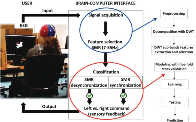
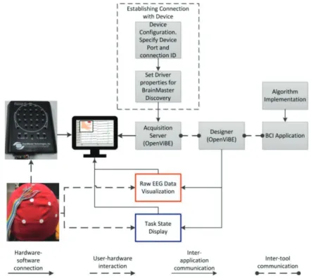
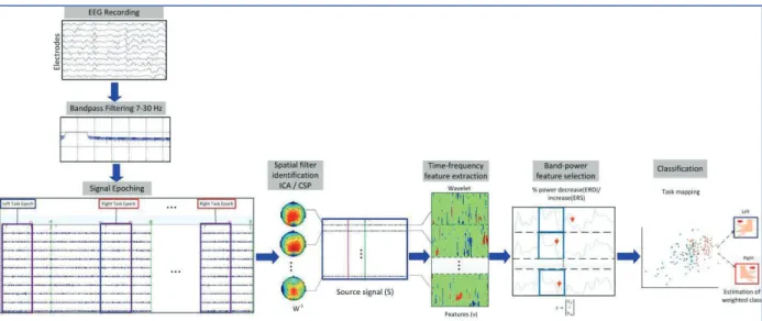
Figures in Chapter 3 Figure 3-1 Systematic overview of the established experimental setup for ERD/ERS band-power feature extraction and classification using machine learning... 53
Figure 3-2Overview of experimental setup for feature extraction using common spatial pattern (CSP) and filter-bank CSP (FBCSP), and classification using machine learning ... 53
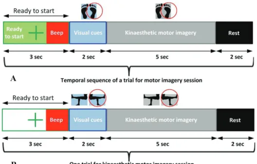
Figure 3-3Experimental Protocol for each trial reflecting timing of visual cues, with audio beep for first trial only, for (A) foot KMI and (B) knee KMI ... 54
Figure 3-4Experimental plan reflecting details of each run (session) ... 54
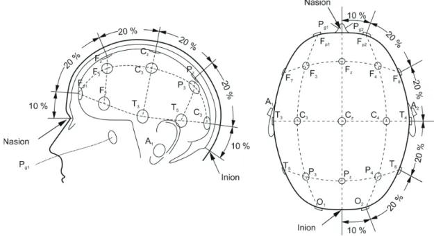
Figure 3-5 The standard 10-20 system for electrode placement over scalp used in EEG cap [5]... 56
5 Bundoora east campus RMIT... 56 Figure 3-7 As part of experimental setup, the established hardware-software connection between Discovery 24E amplifier and OpenViBE acquisition software. ... 57 Figure 3-8 Band-power/Common spatial pattern feature decoder and classifier training in one fold of the cross-validation ... 59 Figure 3-9Layout of study carried out for band-power features ... 63 Figure 3-10 Two projections of population from the same class onto different vectors ɘෝ. The left projection makes the classification of data simple, the right projection makes the data inseparable... 65 Figure 3-11Two possible linear decision boundaries, the left decision boundary with a larger margin is preferred over the small margin (on the right) by the SVM ... 66 Figures in Chapter 4
Figure 4-1Experimental protocol timing in seconds (left) and 10-20 electrode channel locations (right)... 80 Figure 4-2 Visual cues in the experimental protocol, for (a) left - right foot dorsiflexion, and (b) left- right knee extension... 81 Figure 4-3 Established connection for real-time EEG data acquisition and incorporation of event time-stamps in the data stream ... 82 Figure 4-4Topographical scalp maps of each subject during left-right foot and left-right knee imagery respectively between frequencies of 8-12 Hz ... 87 Figure 4-5 ERD and ERS time course for mu rhythm (8-12 Hz) of subject 3 at electrode position C3 for right foot and right knee imagery alongside their average, C4 for left foot and left knee imagery alongside their average, and Cz for left and right foot and knee imagery respectively alongside their average. The green window indicates visual cue presentation from 0 and 2 seconds... 88 Figure 4-6Average amplitude of mu ERD from all subjects based on common average reference derivation at central electrode positions. The red and blue bars indicate left foot and left knee motor imageries, respectively, and pale red and pale blue bars indicate right foot and right knee motor imageries, respectively. Error bars represent standard deviations ... 89 Figures in Chapter 5
Figure 5-1 (A) EEG electrode/channel locations. (B) Experimental protocol for foot kinaesthetic motor imagery tasks, with cue timings expressed in seconds, during one trial ... 98 Figure 5-2Band-power feature decoder with classifier training and testing in one fold of the cross-validation... Figure 5-3 Time-frequency maps (participant 1, 2, and 4). Common average reference channel Cz, and two bipolar channels, C3-Cz and Cz-C4, are shown. The left columns show left foot dorsiflexion kinaesthetic motor imagery (KMI), and the right columns show right foot dorsiflexion KMI. Significant (P < 0.05)
6 line indicates thebeginning of KMI...10 Figure 5-4Average amplitude of significant mu ERD, beta ERD and beta ERS from all nine participants (N=9). The blue bars show average amplitude of each feature after left foot task whereas red bars represent right foot task. The error bars depict standard deviations. The significant values for corrected p < 0.017 are plotted...10 Figure 5-5 Average time course (participant 2) for ERD and ERS of common average reference channel Cz and two bipolar channels C3-Cz and Cz-C4 are shown. The left column reflects power changes in mu rhythm, mid column for low beta and right column for high beta...1 Figure 5-6 Average EEG topographies of ERD/ERS during foot KMI of all participants. Mu ERD is shown in the left column for left foot and right foot respectively, following this, beta ERD is in the mid column, and beta ERS is in the right column with distinguished ERS pattern. The top row illustrates ERD/ERS patterns for common average reference and the bottom for bipolar reference...11 Figure 5-7 Classifiers performance accuracy in percentage, using (A) common average reference, and (B) bipolar reference. The error bars represent standard deviations...11 Figures in Chapter 6
Figure 6-1The temporal sequence of a trial for foot kinaesthetic motor imagery session followed in the experiment... 124 Figure 6-2 Experimental setup reflecting the methodology of common spatial pattern (CSP) and filter bank CSP (FBCSP) algorithms for training, testing and prediction. ... 126 Figure 6-3 A set of common spatial patterns (CSPs) filters of participant P01, where CSPs are optimized for the discrimination of right and left foot kinaesthetic motor imageries with respect to reference period... 131 Figure 6-4 Receiver operator characteristics curves reflecting area under the curves for all participants... 132 Figure 6-5 Average classification accuracies (in percentage) for each algorithm across participants, red dotted line shows average on and above chance level (p <0.01). The error bars represent standard deviationsǤ... 134
Figure 6-6 Resulting misclassification rate (in percentage) of LDA, CSP-Logreg, FBCSP-LDA, and FBCSP-Logreg algorithms for individual participant (N=9). The error bars represent standard deviations... 134 Figures in Chapter 7
Figure 7-1 Time-frequency maps reflecting ERD/ERS of all participants. Left column shows left knee extension kinaesthetic motor imagery (KMI), and right column shows right knee extension KMI. Significant (P < 0.05) band-power changes are shown during the trial period of -3 to 7 s. The pink dotted line indicates the beginning of the visual cue...14 Figure 7-2Average amplitude of beta ERS and mu ERD from five participants (N=5). Blue bars show average amplitude of respective feature after left knee task,
7 ...14 Figure 7-3 Average EEG topographies of ERD/ERS during knee KMI of all participants. Beta ERS is shown in the top row for left and right knee respectively, and mu ERD is in the bottom row...14 Figure 7-4Average classifier models performance in percentage, using common average reference. The error bars represent standard deviations...14 Figure 7-5Individual participant prediction accuracies in percentage for N=5.. ...1 Figure 7-6AUC-ROC plots with maximum AUC values for beta ERS using KNN model and mu ERD using SVM model, during classification of left vs. right knee KMI...1 Figures in Chapter 8
Figure 8-1Temporal sequence of one trial of knee kinaesthetic motor imagery followed in the experiment...1 Figure 8-2 Paradigm of common spatial pattern (CSP) and filter bank CSP (FBCSP) algorithms for training, testing and prediction phases, adapted from [13] ...1 Figure 8-3 (a) Classification accuracies in percentage across participants where blue line shows average on and above chance level (p<0.01). (b) Individual misclassification rate in percentage (for N=5) of CSP-LDA, CSP-Logreg, FBCSP-LDA, and FBCSP-Logreg algorithms...1 Figure 8-4A set of common spatial patterns (CSPs) filters of participant P01. The CSPs are optimized for the discrimination of right and left knee kinaesthetic motor imageries from the reference period...1 Figure 8-5 Receiver operator characteristics curves depicting area under the curves for all participants ...16
List of tables
Tables in Chapter 2
Table 2-1 Key features of EEG-based activity mode recognition exoskeletons, orthosis, wheelchairs and assistive robots for rehabilitation... 40 Tables in Chapter 3
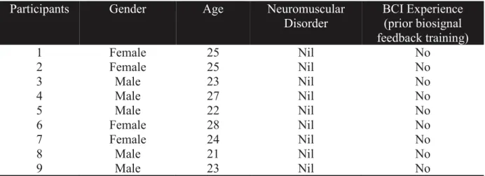
Table 3-1Characteristics of participants volunteering in BCI experiments ... 51 Table 3-2Sample confusion matrix for a binary classification problem. It displays probabilities for each occurrence; P11 and P22 representing correctly classified samples... 69
8 Table 3-4Errors in multiple testing of N hypotheses [37]... 72 Tables in Chapter 4
Table 4-1Unsupervised feature extraction-based approach ... 84 Table 4-2Task combinations within the same lower-limb... 85 Tables in Chapter 5
Table 5-1 Resulting test-statistic values of significant features with 95% confidence interval, by comparing left foot KMI vs. right foot KMI, using common average and bipolar references... 103 Table 5-2Individual peak latencies from cue-onset for significant mu ERD, beta ERD, and beta ERS, using common average reference (CAR) and bipolar reference (BIP), across participants ... 104 Table 5-3Errors in multiple testing of N hypotheses ... 106 Table 5-4 The 5-fold cross-validation classification accuracy of left-right foot KMI using mu ERD, beta ERD, and beta ERS for common average reference.. ... 112 Table 5-5 The 5-fold cross-validation classification accuracy of left-right foot KMI using mu ERD, beta ERD, and beta ERS for Bipolar reference ... 112 Table 5-6 False discovery rate (FDR) corrections for LDA, SVM and KNN classifiers... 116 Tables in Chapter 6
Table 6-1 The 10-fold cross-validation performance of misclassification rate using CSP and FBCSP with linear discriminant analysis (LDA) and logistic regression (Logreg) classifiers ... 132 Table 6-2The 10-fold cross-validation performance in terms of maximum kappa value and the Area under ROC Curve (AUC) using CSP and FBCSP with Linear discriminant analysis (LDA) and logistic regression (Logreg) models. 133 Tables in Chapter 7
Table 7-1 Individual peak latencies from cue-onset for significant mu ERD and beta ERS...14 Table 7-2The 5-fold cross-validation classification accuracy and AUC-ROC values of left-right knee KMI using beta ERS and mu ERD...14 Table 7-3Mann-Whitney U test for SVM and KNN machine learning models... ...15 Tables in Chapter 8
Table 8-1Misclassification rate using CSP and FBCSP with linear discriminant analysis (LDA) and logistic regression (Logreg) classifiers with 5x5-fold of cross-validation...16 Table 8-2 Area under (ROC) curve (AUC) and kappa scores using CSP and FBCSP with linear discriminant analysis (LDA) and logistic regression (Logreg) classifiers...16
10 M. Tariq, P. M. Trivailo, and M. Simic.EEG-Based BCI Control Schemes for Lower-Limb Assistive-Robots. Frontiers in Human Neuroscience, 12, 312, 2018
M. Tariq, P. M. Trivailo, Yutaka Shoji, and M. Simic. Comparison of event-related changes in oscillatory activity during different cognitive imaginary movements within same lower-limb. Acta Polytechnica Hungarica, 16 (2) 77-92, 2019.
M. Tariq, P. M. Trivailo, M. Simic. Mu-beta event-related (de)synchronization and EEG classification of left-right foot dorsiflexion kinaesthetic motor imagery for BCI. PLOS One (Under Review).
M. Tariq, P. M. Trivailo, and M. Simic. Classification of left and right foot kinaesthetic motor imagery using common spatial pattern. Biomedical Physics and Engineering Express (Accepted).
M. Tariq, P. M. Trivailo, M. Simic. Classification of left and right knee extension motor imagery using common spatial pattern for BCI applications. International Journal of Knowledge-Based and Intelligent Engineering Systems: Procedia Computer Science, 159, 2598-2606, 2019.
11
0 7DULT, Pavel M. Trivailo, and MilanSimic.Event-related changes detection in
sensorimotor rhythm.International Robotics & Automation Journal, 4(2) 119-120,2018. M. Tariq, P.M. Trivailo, and M. Simic.Motor imagery based EEG features visualization for BCI applications. International Journal of Knowledge-Based and Intelligent Engineering Systems:Procedia Computer Science126,1936-1944, 2018.
M. Tariq, L. Uhlenberg, P.M. Trivailo, K.S. Munir, and M. Simic.Mu-Beta Rhythm ERD/ERS Quantification for Foot Motor Execution and Imagery Tasks in BCI Applications. 8th IEEE International Conference on Cognitive Infocommunications (CogInfoCom), 2017, pp. 091-096. IEEE, 2017,Debrecen, Hungary.
M. Tariq, P.M. Trivailo, and M. Simic.Detection of Knee Motor Imagery by Mu ERD/ERS Quantification for BCI Based Neurorehabilitation Applications. 11thAsian Control Conference (ASCC), 2017,pp. 2215-2219. IEEE, 2017,Gold Coast, Australia.
12 M. Tariq, Z.U. Koreshi, and P.M. Trivailo. Optimal Control of an Active Prosthetic Ankle. 3rd International Conference on Mechatronics and Robotics Engineering (ICMRE), 2017, pp.113-118. ACM, 2017, Paris, France.
13 M. Tariq, L. Uhlenberg, P.M. Trivailo, K.S. Munir, and M. Simic. Mu-Beta Rhythm ERD/ERS Quantification for Foot Motor Execution and Imagery Tasks in BCI Applications. 8th IEEE International Conference on Cognitive Infocommunications (CogInfoCom), 2017, pp. 091-096. IEEE, 2017, Debrecen, Hungary.
M. Tariq, P.M. Trivailo, and M. Simic. Detection of Knee Motor Imagery by Mu ERD/ERS Quantification for BCI Based Neurorehabilitation Applications. 11th Asian Control Conference (ASCC), 2017, pp. 2215-2219. IEEE, 2017, Gold Coast, Australia. M. Tariq, Z.U. Koreshi, and P.M. Trivailo. Optimal Control of an Active Prosthetic Ankle. 3rd International Conference on Mechatronics and Robotics Engineering (ICMRE), 2017, pp.113-118. ACM, 2017, Paris, France.
14 M. Tariq, P.M. Trivailo, and M. Simic. Brain actuated bionic foot. Pitch for US-AUS Robotics & Autonomy Workshop, organized by Defence Science Institute, Adelaide Australia, 2017.
15
ACC Accuracy
ALS Amyotrophic lateral sclerosis
ANN Artificial neural network
ANOVA Analysis of variance
AUC Area under curve
AUX1 Auxiliary channel-1
AUX2 Auxiliary channel-2
BCI Brain-computer interface
BP Band-power
BSS Blind source separation
CAN Controller area network
CHEAN College human ethics advisory network
CMC Corticomuscular coherence
CNN Convolutional neural network
CNS Central nervous system
CPCA Class-wise principal component analysis
CPG Central-pattern-generators
CSP Common spatial pattern
CVA Canonical variate analysis
DFT Discrete Fourier Transform
DOF Degrees of freedom
16
EMG Electromyography
ERD Event-related desynchronization
ERDS Event-related desynchronization-synchronization
ERP Event-related potential
ERS Event-related synchronization
ERSP Event-related spectral perturbation FBCSP Filter bank common spatial pattern
FDR False discovery rate
FES Functional electrical stimulation
FIR Finite impulse response
FP False positive
FPR False positive rate
GMM Gaussian mixture model
ICA Independent component analysis
ICT Information and communication technology
IP Internet protocol
ITC Inter-trial coherence
ITV Inter-trial variance
KMI Kinaesthetic motor imagery
KNN K-nearest neighbors
LDA Linear discrimination analysis
17
LL Lower-limb
Logreg Logistic regression
LSL Lab streaming layer
MAFO Motorized ankle-foot orthoses
mcr misclassification rate
ME Motor execution
MI Motor imagery
ML Machine learning
MLP Multi-layer perceptron
MRCP Movement-related cortical potential
PCA Principal Component analysis
PSD Power spectral density
RF Right foot
RK Right knee
ROC Receiver-operator characteristic curve
RoGO Robotic gait orthosis
SCI Spinal cord injury
SCP Slow cortical potential
SGRM Sparse group representation model
SMA Supplementary motor area
SMR Sensorimotor rhythm
18
SVM Support vector machine
TCP Transmission control protocol
TF Time-frequency
TMS Transcranial magnetic stimulation
TP True positive
TPR True positive rate
TSGSP Temporally constrained sparse group spatial pattern
VEP Visual evoked potential
VRE Virtual reality environment
VRPN Virtual reality peripheral network
19
Chapter 1
Introduction
1.1. Overview of brain-computer interface controlled actuators/output devices
1.2. Research aim and objectives
1.3. Thesis structure
1.4. References
Chapter Overview
The objective of this chapter is to highlight the significance of various factors involved in the development of brain-computer interface (BCI) controlled actuators/output devices. In this framework, the chapter discusses key issues and challenges involved in the feature selection and classification of sensorimotor-based EEG signals. The motivation and significance of the present study are highlighted, followed by a detailed discussion of the scope of this particular study. In the last section, the structure of the thesis is presented, including a brief summary of each chapter.
20
1.1 Overview of brain-computer interface controlled actuators/output devices
From a recent survey it is observed that due to central nervous system (CNS) disorder, more than 10 million people in the world live with various forms [1] of disability. Existing assistive lower-limb (LL) devices based on sensors and smart control algorithms are known to be suitable for rehabilitation of lost mobility compared to passive devices that account for high metabolic cost of transport [2]. In order to account for such critical aspects, research has been carried out to develop algorithms matching user’s motion intention that could generate correct walking trajectories with wearable robots. However, the control features offered by these devices solely rely on actuation of the system derived from artificial sensors along finite state controller that attempts to implicate biomechanical gait mechanism [3, 4], resulting in a restricted seamless control. This lead to the development of controllers based on actuation signals directly driven by cortical activity in correlation with the user intent for volitional movements [5]. The state of the art brain-computer interface (BCI) provides an augmentative communication source by creating a muscle-free channel between the brain and the output device. It accentuates real-time bidirectional connection between the brain and actuator/output device. With these superior control properties, BCI is considered to be a novel engineering tool for neurorobotics, neuroprosthesis and assistive rehabilitation device applications for patients with neural disorders, spinal cord injury (SCI), or amputation. Successful design with seamless control and development of brain actuated assistive devices with improved classification accuracy, information transfer rate, and signal-to-noise-ratio, remains a significant research area through which it would be possible to minimize occurrence of non-volitional control and enhance the reliability of these devices under severe operating conditions of users.
Since the first successful experiment on creating a direct link between a patient’s motor cortex and the external device (a projector), by Grey Walter in 1963 and later by NIH laboratory, to control artificial actuators via cortical neuron recordings [6], significant research has been carried out to monitor and decipher cortical neuron activity using cortical implants. However, the modality employed was invasive. It has significant properties as high spatial resolution (tenths of millimeters), broader bandwidth (0 to 500 Hz), high characteristic amplitude (50 to 100 V), less vulnerability to artifacts and less user-BCI system adaptability (training); followed by higher cost with an inflated risk of scar tissue formation [7]. The
non-21
invasive modalities offer better solutions. Electroencephalography (EEG) is one such viable tool that offers effective specifications as, lower in cost, ease of use, safest method to record brain activity and high time resolution (millisecond scale temporal resolution)[8]; however there is a tradeoff between performance and features as, lower spatial resolution (centimeters), lower bandwidth (0 to 50 Hz), lower characteristic amplitude (10 to 20 V), high susceptibility to artifacts, and several hours of training for user-BCI adaptability [7]. Nevertheless, EEG account for the safest technology therefore remains popular choice for BCI modality.
While BCIs may not require any voluntary muscle control, they are certainly dependent on normal brain function to some degree therefore the choice of BCI type depends on user's condition [9]. Research on EEG based BCIs for assistive device applications have been carried out since 2000 [9]. Majority of the electrophysiological input signals employed by researchers included event-related potentials (ERPs), steady-state visually evoked potentials (SSVEPs), slow cortical potentials (SCPs) and oscillatory/sensorimotor rhythms (mu and beta oscillatory activity also termed SMRs). The most challenging signals discerned, arose in the motor cortex against the execution or imagery of a motor task, i.e. SMRs. This is because of the proprioceptive feedback involved during execution of the task and the varying level of user concentration. Active research contributions were from Graz BCI and Wadsworth BCI research centers that focused on ERPs and SMRs [10, 11]. Their applications addressed amyotrophic lateral sclerosis (ALS)/totally locked-in patients to restore basic communication needs including, 1D-2D cursor control on a computer, answering spoken Yes/No commands, basic word processing speller, first point development of prototype systems integrating submenu for everyday use in people’s homes, and control of orthotic device for opening and closing paralyzed limb (hand grasp) [12]. However, less emphasis reflected LL movement restoration for patients with spinal cord injury (SCI), disarticulated leg muscles, inactive residual LL or amputees, until recently. Concept was made that the central-pattern-generators with less supraspinal control are involved in the control of bipedal locomotion [5, 13]. EEG-based activity mode recognition for assistive portable devices has been deployed recently such as wheelchairs, assistive/guiding robots, orthosis and exoskeletons, [5]. However, challenges still remain in the field.
For BCI systems that employ SMR-based EEG as input signals, the long training process of users to adapt to the BCI system is challenging [14]. Same cortical areas should activate during the actual performance of a limb movement and imagination of the same movement [15]. Although supervised classification methods are employed to learn how to recognize
22
specific patterns of EEG activities, i.e. to learn the mapping between the EEG data and classes corresponding to mental tasks, such as movement of the left or right hand. However, the learning task is challenging as various governing factors impact the output result, such as the varying physical and mental state (degree of attention and concentration), eye blinks and muscle artifacts that contaminate the EEG signal.
Based on the somatotopic arrangement of sensory and motor cortices, the upper limbs particularly hand representation is on the mantle of the human cortex. It is lateralized, the reason why left and right hand movement’s ERD patterns can be spatially distinguished. In contrast, the LL e.g. the foot and knee’s motor area representation on the homunculus is located deep inside the interhemispheric fissure with low spatial resolution, which makes it very difficult for the detection of these patterns through the EEG signal [16], hence more exploration needs to be done in this area. There is a need to investigate the probability of using band-power and common spatial pattern features as input control signals to a BCI system.
Before 2009, BCIs to control prosthetic devices were limited to upper limb prosthetics e.g. the DARPA modular prosthetic limb [17]. This was attributable to lack of analysis tools for analyzing cortical dynamics with EEG due to excessive proprioceptive feedback during walking. Until recently, the concept was made that the central-pattern-generators with less supraspinal control is involved in the control of bipedal locomotion [5]. Though scientific contribution has been made in the field of rehabilitative robotics controlled via BCI, yet no contribution has been made to the direct user intent control of active prosthetic LL device via BCI employing SMR-based EEG only.
1.2 Research aims and objectives
The rationale for this research project is to prove the possibility to deploy mu and beta sensorimotor rhythms, elicited upon LL kinesthetic motor imageries, as input control signals for development of an augmentative communication channel in order to restore lost motor control in subjects with LL amputation, SCI, disarticulated leg muscles, or inactive residual LL. In a BCI paradigm, output device is controlled via input commands extracted from cortical activity but these commands surpass the brain's usual output pathways of peripheral muscles and nerves, and are encoded in an electroencephalographic activity (in case of non-invasive EEG). Henceforth, providing an alternate source of basic communication and control paradigm to the completely or partially paralyzed subjects, in order to express their needs to caregivers, or independently operate program and control neuroprostheses
23
seamlessly in real-time. Present-day BCIs that determine the intent of the user employ SMR electrophysiological signals. SMR-based BCI are either synchronous or asynchronous. Unlike the P300 (ERP) and SSVEP-based BCIs that require minimum/no training to adapt BCI system and vice versa, SMR-based BCIs typically require much longer training periods to attain high levels of performance. The training process is deployed, both to familiarize the user to system, and to provide calibration data for the system’s classifier(s).
Despite advancements in BCI expansion in the recent decade, less literature is available on the employment of SMR based on LL tasks, in particular there is no evidence on the knee kinesthetic motor imagery. Therefore challenges still exist in the development of non-invasive SMR based-BCIs in regards to assistive (wearable) LL devices with a minimal probability of non-volitional output commands. The feature extraction and classification of feature vector for reduced error rate, effective information transfer rate and improved signal-to-noise ratio (SNR) remain an open research problem in BCI systems.
Gaps in the Research Field
1. Less known facts and investigations are observed on the LL kinaesthetic motor imagery (KMI) tasks based band-power and common spatial pattern features, that are deployed as control signals, in any BCI protocol.
2. Selection of optimal frequency-band that consists of the maximum useful features and reliable feature vectors to be used as input control signals to a BCI system is still an open research problem.
3. Cognitive states used in BCI system i.e. the types of mental states/motor actions for motor imagery is still an area open for research [9]. For instance, there is no comprehensive approach to the detection of signals associated to knee imagery.
4. Similarly most efficient algorithms for translating LL SMR signals into device commands are not conclusively defined.
5. No explicit development of ankle-foot prosthesis actuated by LL KMI is available.
The important research questions addressed in this project include:
What is the interaction platform between human brain signals and output device or outer world?
How we can quantitatively predict which precise cortical activity is associated to specific tasks?
24
How can a BCI system prove to be reliable?
How BCI can effectively contribute to the use of prosthetic or assistive robotic devices for rehabilitation?
The ultimate objective is to analyze and classify the sensorimotor rhythms i.e. mu and beta band-power (BP) and common spatial pattern (CSP) features elicited upon user’s intent of locomotion, i.e. LL KMI tasks including foot dorsiflexion and knee extension, for BCI applications.
The research objectives include:
i. Development of a clear understanding on human brain anatomy and ankle-foot biomechanics followed by the control mechanism between neurons, LL (motor tasks) and central pattern generators for walking gait. Analysis of the non-invasive modalities to detect and monitor brain activity. Review of conventional and existing BCI systems (based on EEG modality) that have been incorporated in different LL wearable robotic applications.
ii. Establish the experimental set up from scratch and satisfy criteria for synchronous BCI protocol (cue-paced) streaming data and timestamps/event markers to describe the time course of the experiment.
iii. Recruitment of participants in the experiments, train them in LL motor execution and kinesthetic imagery tasks and ensure progressive output in performance.
iv. Observe significant changes in oscillatory activities, in relation to an internally, or externally paced events that are time-locked, but not phase-locked (induced) associated to event-related desynchronization (ERD) or event-related synchronization (ERS). Following this, test statistics for evaluation of significant feature vectors. Consequently, to analyze the ERD-ERS and significant BP changes of most reactive muandbetacomponents and CSP for LL tasks, to comply with the already built notion and results from literature referring to the cortical lateralization of ERD/ERS during left-right foot and knee tasks in sensory motor cortex. Establish correlation between the motor execution and motor imagery tasks for the same limb.
v. Employ classification techniques for the 2-class BCI i.e. discrimination between left and right tasks, by comparing results from linear discriminant analysis (LDA), linear support vector machine (SVM), k-nearest neighbors (KNN), and logistic regression (Logreg) (algorithm) models. This includes
25
data standardization, and training of the classifier to improve the classification accuracy, signal-to-noise ratio, reduce error rate and prove its statistical significance. Consequently conducting the test statistics i.e. multiple comparison corrections for each classifier model outcome.
1.3 Thesis structure
This thesis is assembled as a combination of publications and submitted manuscripts resulting in 9 chapters as follows:
Chapter 1gives an introduction to the field of research employed and an overall structure of the thesis.
Chapter 2presents an overview of the related literature on control schemes employed by LL motor imagery based BCIs for controlling LL assistive robots. Particular emphasis is put on the output of different methodologies adopted for the EEG signal pre-processing, feature extraction and training of the classifiers. The assistive LL robotic systems that employ sensorimotor rhythms and event-related potentials as input signals in a BCI for rehabilitation, such as BCI wheelchair, BCI controlled humanoid and guidance robots, BCI orthotic and exoskeleton devices are reviewed here. The role of the shared control paradigms for wearable assistive devices is highlighted. A general framework for BCI controlled LL portable and assistive-robot devices for rehabilitation is represented in the novel form of a three-level hierarchical operational structure coupled to the shared controller and together connected to the portable output device and its surrounding environment. Latest developments in field of EEG-based BCIs are included. The findings of this work were published in the journal of Frontiers in Human Neuroscience.
Chapter 3provides a methodological description of the materials and methods for the EEG data acquisition. The pre-processing methods employed to de-noise and filter the EEG signal are demonstrated. Techniques employed for ERD/ERS percentage power change BP features and CSP feature extraction is shown. Following this, in order to characterize the features belonging to the two classes i.e. left vs. right motor imagery, the classification models are presented. In order to evaluate the performance of each classifier, test statistical analysis methods are discussed and the multiple comparison correction procedures adopted in each chapter.
26
InChapter 4, the study aimed at highlighting any observed differences in the muoscillatory rhythm derived BP changes during different LL KMI tasks i.e. left vs. right foot dorsiflexion and left vs. right knee extension tasks for the same limb. Despite a small LL sensorimotor area representation in the homunculus, the foot and knee movement imagery elicited ERD patterns. An increase in the mid-central ERD was observed overall with all the participants. The kinaesthetic knee imagery triggered mu ERD, mainly in the cortical foot area representation. No contralateral dominance of cortical areas was present in the case of left-right knee imagery tasks, unlike with foot tasks. Results indicate the possibility of discriminating different movements within the same LL. This could increase the dimensionality of control signals in a BCI system. The findings of this work are published in the journal of Acta Polytechnica Hungarica.
Chapter 5presents the experimental outcomes from analysis and classification of ERD/ERS that elicit in the band-power feature vector, for mu and beta rhythms. This was based on cognitive tasks of left vs. right foot KMI. The analysis was carried out for two EEG montages, common average reference and bipolar reference to draw a comparison of resulting features that are significant enough to confirm the cortical lateralization. Analysis of mu and beta features was done using time-frequency (TF) maps, scalp topographies, and average time course for ERD/ERS. Consequently machine learning (ML) models were deployed for classifying left vs. right foot KMI. The study comprised of three different models to conclude the best one with maximum classification accuracy and AUC. All test statistics including multiple comparisons correction was included for evaluation of statistically significant model outcomes. The cortical lateralization was confirmed, which proved that mu ERD, beta ERD, and beta ERS can be deployed as independent control features in a BCI. The findings from this study are under review in the journal of PLOS One.
Chapter 6 reflects the findings from analysis of ERD/ERS patterns exhibited in the oscillatory rhythm from CSP and filter bank CSP (FBCSP) feature vectors respectively. Experimental protocol was based on left vs. right foot KMI for synchronous BCI. This was followed by deployment of ML models including linear LDA and Logreg. The study was carried out to improve the classification accuracy earlier established by literature. Results proved a successful improvement in the accuracy outcomes from the suggested FBCSP-LDA model. All test statistics including multiple comparisons correction was conducted for statistical evaluation of models. Classification results contribute to the possibility of
27
exploiting muandbetaERD/ERS features as control commands in a BCI. The findings from this work are accepted in the journal of Biomedical Physics and Engineering Express.
Chapter 7 highlights the classification accuracies of mu ERD and beta ERS features based on left and right knee extension KMI. This cognitive task involved full knee extension while in sitting posture. The spatial proximity of left and right knee in the mesial wall of the sensorimotor cortex hinders the discrimination between the left and right tasks. Consequently ERD/ERS patterns were only reflected in the foot area of somatosensory cortex. However, this research’s results established the possibility to deploy knee KMI as cognitive input signals and use mu and beta as independent features for operating a 2 degrees of freedom BCI-controlled prosthetic or robotic knee.
Chapter 8 reflects the results from classification of oscillatory mu andbeta ERD/ERS from CSP and FBCSP feature vectors respectively, following the synchronous BCI protocol of left vs. right knee extension KMI. In order to confirm the cortical lateralization of ERD/ERS, ML models, LDA and Logreg were used for an enhancement in the classification accuracy established from the previous chapter. FBCSP-Logreg model showed maximum classification accuracy amongst other models. Classification results provide the basis for the possibility of exploiting mu andbeta ERD/ERS features as control commands in a BCI. The results from this research are published in the International Journal of Knowledge-Based and Intelligent Engineering System (Procedia Computer Science).
In the end, Chapter 9 outlines all the significant findings of this thesis and suggests some recommendations for further studies.
1.4 References
1. Chéron, G., et al., From spinal central pattern generators to cortical network: integrated BCI for walking rehabilitation. Neural plasticity, 2012. 2012.
2. Au, S.K., J. Weber, and H. Herr, Powered Ankle--Foot Prosthesis Improves Walking Metabolic
Economy. IEEE Transactions on Robotics, 2009. 25(1): p. 51-66.
3. Au, S.K., et al. Powered ankle-foot prosthesis for the improvement of amputee ambulation. in Engineering in Medicine and Biology Society, 2007. EMBS 2007. 29th Annual International Conference of the IEEE. 2007. IEEE.
4. Jimenez-Fabian, R. and O. Verlinden, Review of control algorithms for robotic ankle systems in lower-limb orthoses, prostheses, and exoskeletons. Medical engineering & physics, 2012. 34(4): p. 397-408.
5. Tucker, M.R., et al., Control strategies for active lower extremity prosthetics and orthotics: a review. Journal of neuroengineering and rehabilitation, 2015. 12(1): p. 1.
28 6. Lebedev, M.A. and M.A. Nicolelis, Brain-Machine Interfaces: From Basic Science to
Neuroprostheses and Neurorehabilitation. Physiological Reviews, 2017. 97(2): p. 767-837. 7. Schalk, G. and J. Mellinger, A practical guide to brain–computer interfacing with BCI2000: General-purpose software for brain-computer interface research, data acquisition, stimulus presentation, and brain monitoring. 2010: Springer Science & Business Media.
8. Repovs, G. Dealing with noise in EEG recording and data analysis. in Informatica Medica Slovenica. 2010.
9. Wolpaw, J.R., et al., Brain–computer interfaces for communication and control. Clinical neurophysiology, 2002. 113(6): p. 767-791.
10. Pfurtscheller, G., et al., 15 years of BCI research at Graz University of Technology: current projects. IEEE Transactions on Neural Systems and Rehabilitation Engineering, 2006. 14(2): p. 205-210.
11. Vaughan, T.M., et al., The wadsworth BCI research and development program: at home with
BCI. IEEE transactions on neural systems and rehabilitation engineering, 2006. 14(2): p. 229-233.
12. Daly, J.J. and J.R. Wolpaw, Brain–computer interfaces in neurological rehabilitation. The Lancet Neurology, 2008. 7(11): p. 1032-1043.
13. Presacco, A., et al., Neural decoding of treadmill walking from noninvasive
electroencephalographic signals. Journal of neurophysiology, 2011. 106(4): p. 1875-1887. 14. He, B., et al., Noninvasive brain-computer interfaces based on sensorimotor rhythms.
Proceedings of the IEEE, 2015. 103(6): p. 907-925.
15. Pfurtscheller, G., et al., Graz-BCI: state of the art and clinical applications. IEEE Transactions on neural systems and rehabilitation engineering, 2003. 11(2): p. 1-4.
16. Hashimoto, Y. and J. Ushiba, EEG-based classification of imaginary left and right foot movements using beta rebound. Clinical neurophysiology, 2013. 124(11): p. 2153-2160. 17. Presacco, A., L. Forrester, and J.L. Contreras-Vidal. Towards a non-invasive brain-machine
interface system to restore gait function in humans. in Engineering in Medicine and Biology Society, EMBC, 2011 Annual International Conference of the IEEE. 2011. IEEE.
29
Literature Review
2.1. Introduction
2.2. General control framework for BCI wearable lower-limb and assistive-robot
devices
2.3. User adaptability and EEG signal acquisition
2.4. Communication protocol
2.5. BCI operator
2.6. Shared control
2.7. Lower-limb assistive-robot applications in different environments 2.8. Practical challenges
2.9. Conclusions
2.10. Acknowledgements
2.11. References
Chapter Overview
A brief introduction of BCI systems for controllingLL assistive robots, their potentials,
challenges, and the objectives of tKH UHVHDUFK were drawn in Chapter 1. This
chapter reviews the existing literature oncontrol schemes, i.e. preprocessing, feature
extraction techniques and classification algorithmsdeployed by EEG-BCIsLL assistive
robotsfor rehabilitation. BCIs employing SMR (mu and beta rhythms) and ERP
as control commands have critically been reviewed. A general control framework
is novelly presented for BCI controlled LL portable and assistive-robot devices.
Practical challenges associated to thefield havealso been highlighted.
This workLV publishedinFrontiers in Human Neuroscience.
M. Tariq, P.M. Trivailo, and M.Simic.EEG-Based BCI Control Schemes for Lower-Limb Assistive-Robots.Frontiers in Human Neuroscience, 12, 312, 2018.
Frontiers in Human Neuroscience | www.frontiersin.org 1 August 2018 | Volume 12 | Article 312
Edited by:
Mikhail Lebedev, Duke University, United States
Reviewed by:
Vera Talis, Institute for Information Transmission Problems (RAS), Russia Yuri Levik, Institute for Information Transmission Problems (RAS), Russia
*Correspondence: Milan Simic [email protected] Received:03 May 2018 Accepted:16 July 2018 Published:06 August 2018 Citation:
Tariq M, Trivailo PM and Simic M (2018) EEG-Based BCI Control Schemes for Lower-Limb Assistive-Robots. Front. Hum. Neurosci. 12:312. doi: 10.3389/fnhum.2018.00312
EEG-Based BCI Control Schemes for
Lower-Limb Assistive-Robots
Madiha Tariq, Pavel M. Trivailo and Milan Simic* School of Engineering, RMIT University Melbourne, Melbourne, VIC, Australia
Over recent years, brain-computer interface (BCI) has emerged as an alternative communication system between the human brain and an output device. Deciphered intents, after detecting electrical signals from the human scalp, are translated into control commands used to operate external devices, computer displays and virtual objects in the real-time. BCI provides an augmentative communication by creating a muscle-free channel between the brain and the output devices, primarily for subjects having neuromotor disorders, or trauma to nervous system, notably spinal cord injuries (SCI), and subjects with unaffected sensorimotor functions but disarticulated or amputated residual limbs. This review identifies the potentials of electroencephalography (EEG) based BCI applications for locomotion and mobility rehabilitation. Patients could benefit from its advancements such as wearable lower-limb (LL) exoskeletons, orthosis, prosthesis, wheelchairs, and assistive-robot devices. The EEG communication signals employed by the aforementioned applications that also provide feasibility for future development in the field are sensorimotor rhythms (SMR), event-related potentials (ERP) and visual evoked potentials (VEP). The review is an effort to progress the development of user’s mental task related to LL for BCI reliability and confidence measures. As a novel contribution, the reviewed BCI control paradigms for wearable LL and assistive-robots are presented by a general control framework fitting in hierarchical layers. It reflects informatic interactions, between the user, the BCI operator, the shared controller, the robotic device and the environment. Each sub layer of the BCI operator is discussed in detail, highlighting the feature extraction, classification and execution methods employed by the various systems. All applications’ key features and their interaction with the environment are reviewed for the EEG-based activity mode recognition, and presented in form of a table. It is suggested to structure EEG-BCI controlled LL assistive devices within the presented framework, for future generation of intent-based multifunctional controllers. Despite the development of controllers, for BCI-based wearable or assistive devices that can seamlessly integrate user intent, practical challenges associated with such systems exist and have been discerned, which can be constructive for future developments in the field.
Keywords: brain-computer interface (BCI), electroencephalography (EEG), spinal cord injury (SCI), exoskeletons, orthosis, assistive-robot devices
INTRODUCTION
The field of assistive technologies, for mobility rehabilitation, is ameliorating by the introduction of electrophysiological signals to control these devices. The system runs independent of physical, or muscular interventions, using brain signals that reflect user’s intent to control devices/limbs (Millán et al., 2010; Lebedev and Nicolelis, 2017), called brain-computer interface (BCI). Commonly used non-invasive modality to record brain signals is electroencephalography (EEG). EEG signals are deciphered to control commands in order to restore communication between the brain and the output device when the natural communication channel i.e., neuronal activity is disrupted. Recent reviews on EEG-BCI for communication and rehabilitation of lower-limbs (LL) could be found in (Cervera et al., 2018; Deng et al., 2018; He et al., 2018a; Lazarou et al., 2018; Semprini et al., 2018; Slutzky, 2018).
About five decades ago, EEG-BCIs used computer cursor movements to communicate user intents for patient-assistance in various applications (Vidal, 1973; Wolpaw et al., 2002; Lebedev and Nicolelis, 2017). The applications are now widespread, as machine learning has become one essential component of BCI, functional in different fields of neurorobotics and neuroprosthesis. For lower extremity, applications include human locomotion assistance, gait rehabilitation, and enhancement of physical abilities of able-bodied humans (Deng et al., 2018). Devices for locomotion, or mobility assistance, vary from wearable to (non-wearable) assistive-robot devices. Wearable devices such as exoskeletons, orthosis, prosthesis, and assistive-robot devices including wheelchairs, guiding humanoids, telepresence and mobile robots for navigation are the focus of our investigation.
Control schemes, offered by these systems, rely on the inputs derived from electrophysiological signals, electromechanical sensors from the device, and the deployment of finite state controller that attempts to implicate user’s motion intention, to generate correct walking trajectories with wearable robots (Duvinage et al., 2012; Jimenez-Fabian and Verlinden, 2012; Herr et al., 2013; Contreras-Vidal et al., 2016). Input signals are typically extracted from the residual limb/muscles i.e., amputated or disarticulated lower-limbs (LL), via electromyography (EMG), from users with no cortical lesion or intact cognitive functions. Such solutions consequently preclude patient groups whose injuries necessitate direct cortical input to the BCI controller, for instance users with neuromotor disorders such as spinal cord injury (SCI) and stroke, or inactive efferent nerves/synergistic muscle groups. In this case direct cortical inputs from EEG could be the central-pattern-generators (CPG) that generate basic motor patterns at the supraspinal or cortical level (premotor and motor cortex); or the LL kinesthetic motor imagery (KMI) signals (Malouin and Richards, 2010). The realization of BCI controllers solely driven by EEG signals, for controlling LL wearable/assistive devices, is therefore possible (Lee et al., 2017). Several investigations reinstate that CPG with less supraspinal control is involved in the control of bipedal locomotion (Dimitrijevic et al., 1998; Beloozerova et al., 2003; Tucker et al., 2015). This provides the basis for the development of controllers, directly driven from
cortical activity in correlation to the user intent for volitional movements (Nicolas-Alonso and Gomez-Gil, 2012; Angeli et al., 2014; Tucker et al., 2015; Lebedev and Nicolelis, 2017) instead of EMG signals. Consequently, controllers with EEG-based activity mode recognition for portable assistive devices, have become an alternative to get seamless results (Presacco et al., 2011b). However, when employing EEG signals as input to the BCI controller, there necessitates a validation about the notion that EEG signals from the cortex can be useful for the locomotion control.
Though cortical sites encode movement intents, the kinetic and kinematic changes necessary to execute the intended movement, are essential factors to be considered. Studies indicate that the selective recruitment of embedded “muscle synergies” provide an efficient means of intent-driven, selective movement, i.e., these synergies, stored as CPGs, specify spatial organization of muscle activation and characterize different biomechanical subtasks (Chvatal et al., 2011; Chvatal and Ting, 2013). According toMaguire et al. (2018), during human walking, Chvatal and Ting (2012)identified different muscle synergies for the control of muscle activity and coordination. According toPetersen et al. (2012), the swing-phase was more influenced by the central cortical control, i.e., dorsiflexion in early stance at heel strike, and during pre-swing and swing phases for energy transfer from trunk to leg. They also emphasized the importance of cortical activity during steady unperturbed gait for the support of CPG activity. Descending cortical signals communicate with spinal networks to ensure that accurate changes