Pre-clinical
in vivo Imaging
P R O D U C T N O T E
Key Benefits:
• High performance, ultra-sensitive PET technology
designed for pre-clinical imaging
• 3D PET and X-ray whole body images
• Purpose built bench-top footprint
• Fast and automatic image reconstruction
• Real-time animal monitoring
• Simple data acquisition and advanced analysis
Small animal PET is an essential tool for
translational research. The G4 is an uncompromising high performance solution that offers flexibility for the researcher in a compact footprint. PerkinElmer believes it should be convenient, cost-effective, and deliver high performance in an easy-to-use compact footprint.
PerkinElmer’s Solution
High performance and quantitative benchtop PET imaging with intuitive workflows for seamless integration into any laboratory. G4 PET/X-ray introduces a unique, highly sensitive detection technology that enables physiologically translatable dosages in research models that are relevant to the clinic while reducing exposure.
Next Generation Small
Animal PET Technology
PerkinElmer is the exclusive, global provider of Sofie Pre-clinical PET platforms.
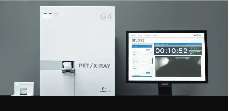
G4 PET/X-ray
Compact Size
Generate 3D quantitative PET and X-ray data in record time
At only 18 inches wide and 24 inches tall, G4 is the smallest PET imaging system in the industry. G4 can be installed in any laboratory setting with minimal site planning and training to quickly get your existing staff up and running.
Technology
New generation of PET imaging
G4 breaks away from a conventional ring-based detection system by surrounding the animal with panel detectors resulting in one of the most sensitive PET systems on the market.
This sensitivity allows imaging of trace amounts of probes, which translates to 10x less dose to the animal, while maintaining beautiful image quality.
Figure 2. Dimensions of G4 PET/X-ray imaging system.
18"
24"
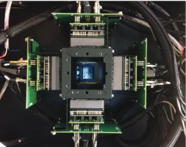
Figure 3. G4 PET/X-ray is a benchtop PET scanner dedicated to high
sensitivity and high resolution imaging of small rodents. New panel based architecture breaks away from the ring of detectors. Gu Z. et al. Phys Med Biol. 2013 Jun 7;58(11):3791-814.
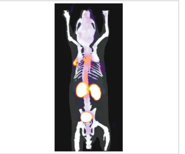
Figure 4. Integrated multi-modality imaging systems provide important information on the biological process of disease combined with an anatomical reference. Bone
metabolism can be imaged using [18F]NaF through the rapid incorporation of NaF into bone. Increased bone metabolism is an indicator of oncologic processes in the bone, both metastatic disease and de novo cancer. NaF PET Scan (left), X-ray, (middle), PET + X-ray (right).
G4 Workflow
Get more done with less effort
Make research studies successful day after day. Merging simplicity with performance is THE focal point of this system.
G4 is designed to maximize workflow efficiency while keeping experimental accuracy in mind. Queue up an assembly line of multiple animals in imaging chambers and docking stations to increase throughput and decrease set-up time.
Queue up animals
• Automatic anesthesia and heating • Efficient animal prep
• Simplified workflow • High-throughput capability
Figure 5. Seamless subject handling.
GLYCOLSYS
Figure 6. Simplified G4 workflow allows for a single person to image groups
of animals with multiple tracers. Images from Lazari et al, Fully Automated Production of Diverse [18F]-Labeled PET Tracers on the ELIXYS
Multireactor Radiosynthesizer Without Hardware Modification, JNMT, 2014.
DNA SYNTHESIS BONE METABOLISM
Imaging Chamber and Docking Station
Regulation of temperature and oxygen are critical to a well-executed study. These key factors can affect the physiology of your animal, and therefore your data. Simply glide the Imaging Chamber into the Docking Station to automatically deliver heat and anesthesia.
• Nose cone anesthesia and constant heating (37°C) • Reproducible positioning of the mouse for imaging • Pathogen barrier for environmental protection • Provisions
- Catheter line
- Blood sampling lines for kinetic studies - Slot to hold syringe for dynamic studies • Multi-modality connectivity for MRI, CT, SPECT
Simple Operation, Fast Results
With only three clicks away from whole body scanning, registration, and organ analysis, G4 may be small in size, but big on performance. With its high sensitivity, spatial resolution, ease-of-use, and heavy duty computing power, beautiful PET images will be in your hands quickly with minimal effort.
Ultrafast Automatic Image Reconstruction
G4 takes advantage of an array of CPUs between gantry and workstation, enabling fast and automatic histogramming, image processing and 3D image reconstruction – all completed in only a few minutes post acquisition.
Live Link to Animal
Real-time respiratory and video monitoring
The built-in video camera provides a live-link to the animal allowing monitoring of its physiologic condition in real-time, taking the guess-work out of anesthesia management for an optimal, safe, and stable imaging environment.
Rat and Mouse Compatible
A smaller size doesn’t mean a reduction in capacity. Use G4 for both your mouse and small rat studies.
G4 Software
Experience the G4 acquisition engine
Whether an experienced user or just beginning with PET imaging, G4 acquisition engine helps to generate 3D images in just a few clicks.
Built for speed, G4 acquisition engine combines the latest in computer processing power, advanced tomographic image reconstruction, and networking to quickly acquire, process, and manage image data. G4 software:
• Encourages optimized workflow through an intuitive user interface increasing study throughput.
• Optimized workflow enables simultaneous study creation, acquisition, and reconstruction.
• Reduces tedious bookkeeping and user input error through automation in software.
• Data is available in minutes.
• Robust administration tools for logging user and study activity for billing and study parameter cross checking.
• DICOM standard for output data.
Figure 8. Never before have you been able to setup a PET/x-ray system on a benchtop. What typically sits on the floor, occupies the majority of a room, and requires
Mouse Registration System
Mouse registration system is a proprietary algorithm that brings together PET images, X-ray projections, and a database of CT images from a variety of mice to produce a 3D integrated result specific to this mouse. This registration system gives the user an automatic, robust, and repeatable organ level quantification. As a use case, there are over 3,000 PET imaging probes in development that assay a wide variety of biological questions. The mouse registration system allows for those developing PET probes to produce a quick output of biodistribution of the compound, organ by organ.
G4 Visualization and Analysis
VivoQuant™ image viewer and analysis software
VivoQuant image analysis software processes and analyzes data from multiple pre-clinical imaging modalities such as PET, MR, CT, Optical and SPECT.
• 3D Region of Interest (ROI) tool enables easy ROI segmentation of volume data sets.
• Supports multimodal and dynamic data.
• Quantification tools enables easy analysis of activity concentration and SUV calculations.
• Easy exportation of results in various image and movie format. • Time activity curves which show a rate of change of activity in
plasma and tissues over time.
• InviCRO’s iPACS data management tool featuring
comprehensive image storage, version control, study planning, batch image processing, and report generations is an option available to support your data management needs.
Figure 9. G4 PET/X-ray data with mouse registration systems applied to a
Johns Hopkins Novel PET Test Article.
Figure 11. Visualization and analysis of G4 PET/X-ray with mouse registration
Applications
Oncology[18F]FDG PET is a common assay of glycolysis which represents the
vast majority of clinical PET studies.
CNS
Project evaluating the regulation of dopamine D2/D3 receptors in rat brain using [18F]Fallypride PET.
Cardiology
[18F]FDG PET can provide important insights into the physiology
and biology of the heart enabling researchers to explore the effects of new therapeutics
CORONAL
Figure 12. Image above shows a subcutaneous patient-derived
glioblastoma xenograft.
SAGITTAL
Figure 13. Balb/C mouse injected with 20 uCi [18F]Fallypride, one hr post injection. Acquisition time 10 minutes (Left). Sprague-Dawley rat injected with 50 uCi [18F]Fallypride, one hr post injection. Acquisition time 30 minutes (Right).
CORONAL
Figure 14. Non-gated [18F]FDG PET study of 26 g mouse. Without cardiac monitoring, still able to resolve the elusive right ventricle (top), coronal (lower left), and sagittal (lower right).
Biodistribution
G4 is capable of performing dynamic studies to measure the kinetics of transport and metabolism. In the end, it’s not just about images, but about quantitative measurements of kinetics.
ImmunoPET
Antibodies are becoming central in the development of targeted therapeutics and provides a powerful class of molecular imaging probes for interrogating cell surfaces in vivo. Antibody imaging is a sensitive, non-invasive means for molecular characterization which can guide diagnosis, prognosis, therapy selection, and
monitoring treatment in cancer. These radiolabeled antibodies, di-bodies and mini-bodies are commonly tagged with [64Cu], [89Zr],
and [124I]. Figure 15. Selected frames from the 1 hour dynamic scan of a mouse following
a tail vein injection of 43 uCi [18F]FDG. Different coronal images are shown for each frame to better reveal the activity distribution for different time periods.
Figure 16. Time activity curves in major organs of a mouse during 1 hour dynamic
[18F]FDG. (a) Organ uptake for the 1 hour period. (b) A detailed graph showing the first 26 s following injection.
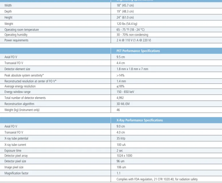
Engineering Specifications
Width 18” (45.7 cm)
Depth 19” (48.3 cm)
Height 24” (61.0 cm)
Weight 120 lbs (54.4 kg)
Operating room temperature 65 - 75 °F (18 - 24 °C)
Operating humidity 30 - 70% non-condensing
Power requirements 2 A @ 110 V (1 A @ 220 V)
PET Performance Specifications
Axial FO V 9.5 cm
Transaxial FO V 4.4 cm
Detector element size 1.8 mm x 1.8 mm x 7 mm
Peak absolute system sensitivity* >14%
Reconstructed resolution at center of FO V* 1.4 mm
Average energy resolution ≤18%
Energy window range 150 - 650 keV
Total number of detector elements 4,992
Reconstruction algorithm 3D ML-EM
Weight (kg) (instrument only) 46
X-Ray Performance Specifications
Axial FO V 9.0 cm
Transaxial FO V 4.0 cm
X-ray tube potential 35 kVp
X-ray tube current 100 uA
Exposure time 2 sec
Detector pixel array 1024 x 1000
Detector pixel size 96 um
Image pixel size 106 um
Magnification factor 1.1
Complies with FDA regulation, 21 CFR 1020.40, for radiation safety
For a complete listing of our global offices, visit www.perkinelmer.com/ContactUs
Copyright ©2014, PerkinElmer, Inc. All rights reserved. PerkinElmer® is a registered trademark of PerkinElmer, Inc. All other trademarks are the property of their respective owners.
011818_01 PerkinElmer, Inc. 940 Winter Street Waltham, MA 02451 USA P: (800) 762-4000 or (+1) 203-925-4602 www.perkinelmer.com
‘For laboratory use only. This product is intended for research purposes only and not for use in humans.’

![Figure 14. Non-gated [ 18 F]FDG PET study of 26 g mouse. Without cardiac monitoring, still able to resolve the elusive right ventricle (top), coronal (lower left), and sagittal (lower right).](https://thumb-us.123doks.com/thumbv2/123dok_us/460777.2553926/6.891.80.449.605.867/figure-cardiac-monitoring-resolve-elusive-ventricle-coronal-sagittal.webp)
![Figure 16. Time activity curves in major organs of a mouse during 1 hour dynamic [ 18 F]FDG](https://thumb-us.123doks.com/thumbv2/123dok_us/460777.2553926/7.891.54.423.193.554/figure-time-activity-curves-major-organs-mouse-dynamic.webp)