From Medical Epidemiology and Biostatistics
Karolinska Institutet, Stockholm, Sweden
CONTRALATERAL BREAST CANCER
- Risk and Prognosis
Maria E.C. Sandberg
All previously published papers were reproduced with permission from the publisher.
Published by Karolinska Institutet. Printed by Larserics Digital Print AB © Maria E.C. Sandberg, 2012
Illustrations
The front page picture is an illustration of contralateral breast cancer, conceived and created by the author. The photograph of dual channels lightings is taken by Steve Horsburgh and used with his permission. The female silhouette illustration is copyrighted by Can Stock Photo Inc./Eraxion, and used with permission. The photograph of a breast cancer survivor’s parade (Foreword) is taken by Robert Thivierge in Calgery, Canada 2009 and used with his permission. The anatomical illustration of the female breast (p.2) is produced by Patrick J. Lynch; illustrator and C. Carl Jaffe; MD; cardiologist, at Yale University Center for Advanced Instructional Media and made available by Wikimedia Commons. The mammograms (p.22) are examples of the actual mammograms used in Study IV.
Och dock, om du i tvivel sjunkit ner
och dröjer dystert grubblande vid vägen,
du griper åter ditt baner
och bär det genom öknen oförvägen.
Vad mer, om spejarblicken ser,
hur bort från fästet tusen solar fejas?
Vad mer, om stjärneskördar mejas
som gyllne säd av tidens lie ner?
Vad rätt du tänkt, vad du i kärlek vill,
vad skönt du drömt, kan ej av tiden härjas,
det är en skörd, som undan honom bärgas,
ty den hör evighetens rike till.
Gå fram, du mänsklighet! var glad, var tröst,
ty du bär evigheten i ditt bröst.
FOREWORD
Half of science is putting forth the right questions.
Sir Francis Bacon (1561-1626)
Today, many millions of women worldvide are ‘breast cancer survivors’, most of these women will luckily have long lives after their diagnosis, both due to lower breast cancer mortality and to longer life expectancy in general. Women surviving breast cancer are, however, at increased risk of several diseases, partly because they are a selected subpopulation; a diagnosis of breast cancer indicates
proneness for cancer, thus increased risk of other cancers. In addition; adjuvant cancer therapy increases the risk of secondary cancers, as well as e.g. heart disease and potentially also serious infections. Part of the increased risk of different diseases in this population could probably also be accounted for by the more intense surveillance of breast cancer patients as compared to healthy women. One of the most common and serious diseases after breast cancer is
contralateral breast cancer (CBC); a new primary cancer in the opposite breast.
With this cancer added to the first, these women have significantly worse prognosis compared to women with only one breast cancer. The incidence of breast cancer is very high and as the mortality after breast cancer keeps improving, CBC will be of increasing importance.
My rationale to study CBC is twofold; it is a potentially deadly but woefully understudied disease that will affect an
increasing number of women and is thus worth to study in its own right, to potentially improve the reality in the clinic for these patients. Also, in the rich part of the world, breast cancer patients are followed up after their diagnosis and treatment to a substantial cost, both measured in inconvenience and anxiety for the patients and in resources for the society, therefore, CBC-risk stratification should also be seen as an important goal. Further, I believe that by studying CBC we might, in the long run, also have a possibility to draw conclusions about breast cancer in general. For example, to study how endocrine therapy affects the risk, development and characteristics of CBC will most likely give an indication of how endocrine chemoprevention will affect unilateral breast cancer, which might otherwise be difficult to study as the population at risk is cancer-free women.
The lightning actually does strike twice in the same place… Let’s learn something from it.
ABSTRACT
“Therefore, since brevity is the soul of wit, and tediousness the limbs and outward flourishes, I will be brief”
William Shakespeare (1564-1616)
The objective of this thesis was to investigate different aspects of contralateral breast cancer. This disease is of increasing importance as the mortality of breast
cancer is decreasing while the incidence remains very high, consequently the population at risk for non-simultaneous (metachronous) contralateral breast cancer is increasing. Further, both simultaneously occurring (synchronous) contralateral breast cancer and non-simultaneous (metachronous) contralateral breast cancer have a worse prognosis than breast cancer in general. The
population-based cohort used in all four studies includes all patients diagnosed with contralateral breast cancer in the Stockholm region during 1976-2005 (N=1422).
In the first paper, which investigates the timing of diagnosis of contralateral breast cancer, we conclude that the diagnostic work-up has not improved over the last 25 years; the second cancers are neither found earlier, nor at smaller tumor size. In our second study we investigate the effect of adjuvant radiotherapy for the first breast cancer and conclude that radiotherapy seems to worsen tumor
characteristics (TNM-stage and differentiation grade) and prognosis after contralateral breast cancer. In analyzing estrogen receptors of the two breast tumors we, in our third study, show how estrogen receptors of both tumors taken together have an important prognostic value. We also find several indications of endocrine therapy resistance in patients with two metachronous estrogen receptor positive tumors. The fourth and final study investigates mammographic density as a risk factor for contralateral breast cancer, studying both
mammographic density at diagnosis of the first cancer and changes in density following the first cancer. We find no effect on the risk of contralateral breast cancer by mammographic density at the time of diagnosis of the first cancer; however, there is a significant risk decrease for patients who experience a decrease in breast density during follow-up after the first cancer.
LIST OF PUBLICATIONS
I. Diagnostic work-up of contralateral breast cancers has not improved over calendar period
Maria EC. Sandberg MSc, Mikael Hartman PhD, Gustaf Edgren PhD, Sandra Eloranta MSc, Alexander Ploner PhD, Per Hall PhD, Kamila Czene PhD
Breast Cancer Res Treat. 2010 Aug;122(3):889-95
II. Aggressiveness of contralateral breast cancer is influenced by radiotherapy for the first tumor
Maria EC. Sandberg MSc, Sara Alkner MD, Mikael Hartman PhD, Sandra Eloranta MSc, Lisa Rydén PhD, Alexander Ploner PhD, Hans-Olov Adami PhD, Per Hall PhD, Kamila Czene PhD
Manuscript submitted
III. Prognostic implications of estrogen receptor pattern of both tumors in contralateral breast cancer
Maria EC. Sandberg MSc, Mikael Hartman PhD, Daniel Klevebring PhD, Sandra Eloranta MSc, Alexander Ploner PhD, Per Hall PhD, Kamila Czene PhD
Breast Cancer Res Treat. 2012 Jul;134(2):793-800
IV. Change of mammographic density predicts the risk of contralateral breast cancer
Maria EC. Sandberg MSc, Jingmei Li PhD, Per Hall PhD, Mikael Hartman PhD, Isabel dos-Santos-Silva PhD, Keith Humphreys PhD, Kamila Czene PhD
CONTENTS
1 Introduction ... 1 1.1 Epidemiology ... 1 1.2 Breast ... 2 1.3 Cancer ... 3 2 Background ... 5 2.1 Breast cancer ... 5 2.1.1 Occurrence ... 5 2.1.2 Risk factors ... 52.1.3 Diagnosis and treatment ... 7
2.1.4 Prognosis ... 9
2.2 Contralateral breast cancer ... 10
2.2.1 Occurrence ... 11
2.2.2 Risk factors ... 14
2.2.3 Diagnosis and treatment ... 15
2.2.4 Prognosis ... 15
3 Aims of this thesis ... 17
4 Materials ... 18
4.1 Registers ... 18
4.1.1 Swedish cancer register ... 18
4.1.2 Stockholm breast cancer register (SBCR) ... 18
4.2 CBC-cohort ... 19
4.2.1 Inclusions ... 19
4.2.2 Exclusions ... 19
4.2.3 Populations for study I-IV ... 20
4.2.4 Medical records ... 21
4.2.5 Mammograms ... 22
5 Methods ... 24
5.1 Design and Statistical analysis ... 24
5.1.1 Cohort design ... 24 5.1.2 Case-control design ... 25 5.2 Analytical approch ... 27 5.2.1 Study I ... 27 5.2.2 Study II ... 28 5.2.3 Study III ... 28 5.2.4 Study IV ... 29
6 Results and implications ... 31
6.1 Study I: Diagnostic work-up of CBC ... 31
6.1.1 Results ... 31
6.1.2 Implications ... 32
6.2 Study II: Aggressiveness of CBC after radiotherapy ... 33
6.2.1 Results ... 33
6.2.2 Implications ... 34
6.3 Study III: Estrogen receptor pattern in CBC ... 35
6.3.1 Results ... 35
6.4 Study IV: Mammographic density affects risk of CBC ... 38 6.4.1 Results ... 38 6.4.2 Implications ... 39 7 Discussion ... 41 7.1 Methodological considerations ... 41 7.1.1 Validity ... 41 7.1.2 Precision ... 44 7.1.3 CBC-specific challenges ... 45
7.2 Findings and interpretation ... 48
7.2.1 Why does short latency time infer bad prognosis? ... 48
7.2.2 What can estrogen receptor status tell us? ... 49
7.2.3 Is synchronous/metachronous CBC different diseases? 49 7.2.4 How should metachronous CBC be handled? ... 50
7.2.5 Did CBC teach lessons about general breast cancer? ... 51
8 Conclusions ... 53 9 Future perspective ... 54 10 Afterword ... 56 11 Svensk sammanfattning ... 57 12 Acknowledgements ... 58 13 References ... 60
LIST OF ABBREVIATIONS
CBC ER TNM CPM SBCR ICD MRI MLO CC HRT IRR OR RR CI SEContralateral breast cancer Estrogen receptor
Tumor size, nodal status, metastatic status Contralateral prophylactic mastectomy Stockholm breast cancer register International classification of disease Magnetic resonance imaging
Media-lateral-oblique Cranio-caudal
Hormone replacement therapy Incidence rate ratio
Odds ratio Risk ratio
Confidence intervals Standard error
1
INTRODUCTION
1.1 EPIDEMIOLOGYIt could be said for epidemiology, with respect to disease etiology and prevention, what is frequently said about democracy as a system of government: they both have many problems and weaknesses, but they still represent the best available approach for the achievement of their respective objectives.
Prof. Dimitrios Trichopoulos (1938-), Harvard School of Public Health
Epidemiology can be broadly defined as the knowledge how and why diseases occur.
Naturally, such short definition needs clarification; how refers to questions of
descriptive epidemiology; in which areas are the disease more common, in which age groups, in which periods, etc. Why refers to questions of association, which
almost always aim towards a causal interpretation; to assess what factor/s cause the disease. Further; not all epidemiology study diseases, not even when we define it
broadly, e.g. a disease being ‘metastasis after breast cancer’. Oftentimes epidemiologists are interested in, if not disease, then at least health-related outcomes, e.g. ‘attitudes towards cancer screening’ but sometimes the outcome studied is not related to health, like ‘occurrence of left handedness’ or even to physical traits, e.g. studies of emigration.
I believe most epidemiologists would agree with the above quoted Professor
Trichopoulos; there is no perfect epidemiological study, they all have some defect. As an epidemiologist, but also as a consumer of epidemiological research, it is of
uttermost importance to be able to spot the weaknesses of a study and make up your mind about whether the weakness render the findings irrelevant, or whether credibility can still be given to the findings, despite the weakness of the study. Every time a researcher makes a too generous judgment of his/her own study and publish the findings, the creditability that is lost in the eye of the public potentially harms the entire field of epidemiology. On the other hand, to quote another man of great formulations; Sir Winston Churchill have said “’Nothing prevails but perfection’ can be spelled shorter; paralysis”. To that we can add that in order to make progress new
and innovative ideas should be tested, and tested hard, preferentially by different researchers, in different settings by different methods. In order to make this possible also imperfect studies have to be published so that the findings can be brought up for discussion and testing, but the key is to not over interpret, nor under interpret, the findings.
As in most research, there is somewhat of a difference between epidemiological studies aiming primarily towards understanding the underlying etiology/biology of the outcome and studies aiming towards results that can immediately improve the real-world handling of the outcome. Preferentially there should be elements of both these types of questions in all studies; an epidemiological finding without any type of plausible underlying mechanism should be regarded with caution, on the other hand; if the study findings have no potential connection to a clinical reality perhaps time and resources could be better spent.
1.2 BREAST
“Your breasts are like two fawns, twin fawns of a gazelle grazing among the lilies.” Songs of Salomon 4;5
The English word breast derives from the Proto-Indo-European base bhreus (to
swell, to sprout). In this thesis ‘breast’ refers to the human mammary glands. These are made up mainly of lobular cells, which constitute the alveoli, where milk
production takes place, ductal cells, which constitute the ducts for leading the milk from the alveoli to the nipple, fat tissue and connective tissue. The anatomical parts of the female breast can be seen in Figure 1.
Figure 1: Anatomy of the female breast.
1 Chest wall 2 Pectoralis muscle 3 Lobules 4 Nipple 5 Areola 6 Milk ducts 7 Fat tissue 8 Skin
Normal breast development takes place in several distinct phases during a woman’s life. In the prenatal phase, about 6 weeks after conception, ectodermal cells form two vertical ridges along the front of the torso, these later involute, leaving cells only in the pectoral region1. This development is the same for both male and female
embryos2. In puberty, under the influence of estrogen and progesterone, the female
breast grows by further development of ducts and thereafter secretory glands; lobules, in the end of the ducts, and by deposition of fatty tissue. This development,
thelarch, leads to permanent breasts before pregnancy, which differentiates humans
from other mammals, in which the mammary glands are developed only during pregnancy and lactation. The adult human breast before pregnancy consists of approximately 80% stroma cells, though there are large differences in the
distribution of cell types, both between the two breasts of the same individual and between individuals2. During pregnancy, the epithelium proliferates and
differentiates into secretory cells, which can synthesize and secrete milk. Several hormones are involved in this process, among them are estrogen, progesterone, prolactin and oxytocin. When breast feeding is discontinued the breast involutes by apoptosis and phagocytosis of the secretory cells, but the ducts remain mainly unchanged2. However, during menopause, the ceasing excretion of hormones from
the ovaries leads to reduced numbers of both ducts and lobules, which are mostly replaced by fat tissue and in consequence the density of breast decreases3. Further,
this process, in combination with the relaxation of the ligaments of Cooper will also change the physical appearance of the breast; it will sit lower on the torso and the nipple will point downward, an aging process known as ptosis1.
1.3 CANCER
“Nothing in biology makes sense except in the light of evolution”
Christian Dobzhansky (1900-1975), geneticist and biologist
Cancer is defined as a mass of cells that grows without the boundaries and stop signals that regulates the growth and division of healthy cells. Cell division is a dangerous procedure, and under the evolution strong mechanisms have developed to regulate this and keep it under strict control. When these mechanisms are circumcised cancer occur. Our understanding of carcinogenesis is still very much under development, but what is known is that cancer develops as a consequence of mutations and epigenetic changes in the genome. Before a cancer can start growing in earnest, several control mechanisms need to be overthrown; apoptosis needs to be evaded, endless capacity for cell division needs to be acquired, stop signals for cell divisions must be overcome, the cell must be self sufficient in growth signals, must have a sufficient supply of blood and must finally obtain the ability to grow without contact to the basal membrane and invade other tissues. This means that a large number of genomic changes have to occur for all these individual mechanisms to be affected. For inherited cancers one of these changes might already present in every one of the cells of the body, therefore, one less change have to happen in any given cell before cancer can develop and inherited cancer therefore often develop early. Environmental exposures also induce damage to the genome, oftentimes by mutations, but also via epigenetic changes; structural reversible changes of the DNA molecule. The cell is most vulnerable for these changes when it is dividing, therefore, although cancer can occur in most tissues, it is much more common in cell types that divide often, and factors that lead to cell division in that particular tissue type are also often risk factors for cancer in that tissue type.
In all tissue stam cells and pluripotent cells; cells with the ability to regenerate and the potential of developing into all types of cells (stam cells) or several different types of cells (pluripotent cells), are present. The current understanding of cancer development states that probably only these cells, and not the fully differentiated cells, can undergo the changes required to become a cancer cell.
Cancer cells divide constantly and the normal mechanisms to stop cell division and/or make the cell go into apoptosis in the event of irreparable damage to the DNA are put out of function. A cancer cell will thus accumulate mutations much faster than a normal cell. This was thought to mean that a cancer will always develop from less aggressive to more aggressive, from less invasive to more invasive, but recently indications have been found that invasive cancer might go into a dormant state, or even regress. Regardless, the effect will be that a cancer tumor consists of different clones of different characteristics and different level of aggressively, so when the microenvironment changes a cancer tumor has a large potential of
adapting, simply by that the clone best fitted to the new environment will take over. The first recording of cancer is from 2300 BC in ancient Egypt. At that time, and for a long time to come, cancer was a certain death sentence, and today 7.5 million people die each year from cancer worldwide (0.5 millions of the deaths are due to breast cancer)4, but why? There are two main explanations as to why cancer is potentially
deadly; the growth of cancer cells might decrease the function of the organ in which the cancer is situated and if the organ is vital this might be deadly, e.g. cancer or metastasis in the brain, liver or lung. However, less intuitive but probably at least as important is the fact that cancer cells in constant division are incredibly energy consuming and the cancer growth starves the rest of the body of nutrition until life cannot be sustained. This can be seen in the way cancer patients wither away in the last stages of metastatic disease. Metastasized cancer is still mainly without cure, and the treatment given is palliative, though research for new treatments is constant and some of them show promise in trials5.
2
BACKGROUND
To know that we know what we know, and to know that we do not know what we do not know, that is true knowledge.
Nicolaus Copernicus (1473-1543)
2.1 BREAST CANCER
“Cancer is a word, not a sentence”
John Diamond (1953-2001), journalist (and cancer patient)
2.1.1 Occurrence
In Sweden 7917 patients were diagnosed with breast cancer in 2010, which equal an incidence of female breast cancer of 168 cases/100 000 women years6.
Brest cancer incidence has continuously increased since the cancer register started in 1958 until today6, although the increase have seemed to subside in the last 10
years7. Breast cancer is much more common in high-income countries4, this is most
likely due to lifestyle risk factors, screening routines as well as the genetic makeup of the different populations. About 35 cases of male breast cancer are diagnosed each year (0.71 cases / 100 000 male person-years)6. These men are in general diagnosed
at a later stage compared to women, which leads to worse prognosis8.
2.1.2 Risk factors
Two oftentimes not mentioned risk factors, perhaps since they are not modifiable, are sex and age. Age has an interesting effect on breast cancer risk; in general, cancer becomes more common as a population grows older, and this was how it looked for breast cancer before 19806. In the case of breast cancer this age dependency also
included a feature known as Clemmensen hook; the increasing incidence dips around
age 50, i.e. during menopause, but then continues to increase9. Then, from the 1990s,
the incidence increased in the younger ages, until breast cancer was almost equally common in all age groups from 55 years and up6, 9. This shift has been attributed to
the wide spread use of hormonal replacement therapy (HRT) during and after menopause (though this is difficult to study due to other changes that also affect the age-specific incidence10) and since HRT use have decreased significantly the
age-specific incidence of breast cancer can be expected to return to the earlier pattern. Next to age, sex and having had breast cancer (or benign breast disease),
mammographic density is probably the most important risk factor for breast cancer. On a radiological picture of a breast, a mammogram, epithelia and connective tissue will appear white, while fat tissue will appear dark. Mammographic density is defined as the white proportion of the mammogram. A meta analysis of 14 000 breast cancer patients and 226 000 non-cases showed that women with >75% mammographic density had almost 5 times the risk of breast cancer compared to women in the lowest density group (<5%)11. Further, decreasing mammographic
density in healthy women has been shown to correspond to decreasing breast cancer risk12.
A part of the risk factor profile for breast cancer consists of genetic factors; four high penetrance genes have been identified for breast cancer; BRCA-1 and BRCA-2, PTEN and TP5313. Both BRCA-1 and BRCA-2 genes encode for DNA double-strand break
repair proteins. An inherited mutation in BRCA-1 confers a 65% risk of developing breast cancer before the age of 70, the corresponding risk for BRCA-2 mutation carriers are 45%14. However, these inherited mutations are very rare in the
population, and even among breast cancer patients they are only found in about 5%15. In addition to these genes, about a handful medium-penetrance genes have
been found13 and the hunt for single-nucleotide polymorphisms (SNPs) is still very
intense. So far approximately 50 SNPs have been consistently found to be associated with breast cancer16, each of them confer a very small risk increase.
Among the environmental risk factors for breast cancer hormonal exposure plays an important part. Regarding endogenous hormones it has long been known that factors that decrease the numbers of menstruation cycles; e.g. late menarche,
pregnancy and early menopause, decrease the risk of breast cancer17. For pregnancy
the relationship is dual; overall parous women have lower risk of breast cancer than nulliparous women, and this is further enhanced with each subsequent birth18.
However, during pregnancy and for the subsequent 10 years the risk of breast cancer for parous women, compared to nulliparous women, is increased19, possibly
due to the effect of pregnancy-associated hormones on preexisting malignancies, this effect is however small in comparison to the long-term protective effect. Further; age at first full-term birth20, and potentially also breast feeding21, is
associated with breast cancer risk, hypothetically linking the
maturation/differentiating of the breast tissue to breast cancer risk18, 20. For
exogenous hormones it is well proven that hormone replacement therapy22 after
menopause increase the risk, and a small risk increase has also been shown for current use of oral contraceptives23.
Regarding other life style factors, alcohol intake has a modest, but consistently shown, risk increase, estimated in a meta analysis to 7% per each additional
drink/day24 and no risk increase is seen by cigarette smoking24. Many attempts have
been made to investigate the effect of diet on the risk of breast cancer, but this is a difficult field and no effect have been consistently shown to date25. Physical activity
have been shown to decrease the risk of postmenopausal breast cancer, but only weak evidence exists for any effect on premenopausal breast cancer26. Body mass
index is related to breast cancer risk in a rather complex web; fat tissue is the most important source of estrogen in postmenopausal women27 and consequently,
obesity is a risk factor for breast cancer after menopause28, 29, for premenopausal
women, on the other hand, it seems that obesity is protective28. Body height has
been consistently associated with breast cancer risk; taller women have a modestly increased risk compared to shorter women30. The causation behind this risk
increase is still unclear; taller women tend to have larger breast which puts more cells at risk of developing into cancer cells, further; growth hormones in utero or
childhood energy intake has been proposed as underlying factors31.
mutations that increase the risk of cancer, while high doses lead cell death32. The
effect of low-dose ionizing radiation have been studied extensively and it has been shown consistently to increase the risk for breast cancer, the effect depends on the age at exposure; the highest risk was shown in women exposed before their teens, and on the dose, where a strong dose-relationship has been found33.
2.1.3 Diagnosis and treatment 2.1.3.1 Diagnosis
Breast cancer is diagnosed either clinically; by a lump discovered in the breast
(usually by the patient herself) or by some other diagnostic sign, like nipple
discharge, tenderness or redness, or sub-clinically, though screening mammography
or, in rare cases, by other imaging techniques, like ultrasound or MRI. Breast cancer screening by mammography every 18-24 months is recommended since 1986 to all women in Sweden between 40-74 years34, and has been shown to decrease breast
cancer mortality35, (which however has been put into question lately36). Once a
cancer is suspected a conclusive diagnosis is reach by the triple diagnostic approach,
which include mammography, palpation and cytological investigation by fine needle aspiration. If at least one of these diagnostic tools gives reason to suspect
malignancy further investigation by surgery is undertaken.
2.1.3.2 Surgery
The oldest recognized, and most important, treatment of breast cancer is surgery, where the tumor is removed and the margins of the resection is microscopically investigated to confirm that they are clear of malignant cells. The surgical techniques are presently of two main types; modified radical mastectomy, where the entire
breast is removed (but not the pectoralis muscle, in contrast to the previously widely used radical mastectomy) and partial mastectomy/breast conservative
therapy, where only the tumor (with margins) is removed. In combination with
post-surgical radiotherapy patients with partial mastectomy is shown to have the same survival as patients with modified radical mastectomy37 and with a cosmetically
much better result. This technique is however contraindicated if the tumor is very large or multifocal, when radiotherapy cannot be given or when the probability of local recurrence is deemed to be high38.
Over 75% of the lymphatic drainage of the breast is though lymph nodes in the axilla and this is the primary route of metastatic spread of cancer1. Therefore, lymph
nodes are removed from the axilla during surgery, for prognostic reasons.
Previously the aim was to remove as many lymph nodes as possible, at least 10, but unfortunately this increases the risk of lymphedema in the arm39. In the end of the
1990’s the sentinel node technique was developed, where a dye or a radioactive
marker is injected in the tumor during surgery and is used to distinguish which lymph node/s that drains the tumor. These nodes are removed and only if cancer cells are found a complete axillary dissection is performed, this way, only patients who benefit from this prognosticator will have the increased risk of lymphedema.
Recently, however, a large randomized trial of patients with positive sentinel node excision showed no survival benefit for of complete axillary dissection40.
2.1.3.3 Adjuvant therapy
Adjuvant therapy is given in order to decrease the risk of the cancer recurring, the objective is to eliminate any cancer cells that might have spread outside the tumor; in the breast, axilla or distant sites. Radiotherapy targeted against the remainder of the breast is a standard part of the breast conservative therapy, the dose is usually 50 Gy, given in fractions of 2 Gy. If the cancer was spread to the axilla lymph nodes also the axilla is included in the radiation field, regardless of type of surgery. Radiotherapy decrease the risk of local/regional recurrences and by that also
decreases the breast cancer-specific mortality41, 42. Further decrease of breast cancer
mortality is achieved by systemic adjuvant therapy, targeted at the potential spread of cancer cells to distant sites. In the case of chemotherapy, however, the side effect are severe and the cost needs to weight against the benefit. The probability of spread to distant sites is much increased if the cancer was spread to the axilla lymph nodes, which is therefore one of the most important criteria to give systemic adjuvant chemotherapy. This is currently also recommended if the tumor is large or
otherwise aggressive, and especially if the patient is young (<60). If the tumor shows over expression of estrogen receptors, endocrine therapy is potentially beneficial. The side effects of endocrine therapy is less severe and the administration is easier than for chemotherapy, therefore the treatment indication is wider, today endocrine therapy is given to almost all breast cancer patients with estrogen receptor over expression34. Endocrine therapy primary consists of a competitive inhibition of
either the estrogen receptor (Tamoxifen) or of the enzyme aromatase (which
converts androgens to estrogens in peripheral tissues) (Anastrozole). During the study period of the studies included in this thesis Anastrozole was used very little, but is now the primary choice for post-menopausal women, due to its higher efficacy and less side effects43. Likewise, in the end of the study period, the monoclonal
antibody Trastuzmab (Herceptin™) was developed and showed to improve survival for patients with over expression of the HER2/neu receptor44.
2.1.3.4 Prophylactic mastectomy
The most radical approach for decreasing the risk of CBC at time of the first breast cancer is to surgically remove also the healthy breast, a procedure known as
contralateral prophylactic mastectomy (CPM). During the last 10-15 years an
increasing trend of CPM has been shown, primarily in the US, where 5-15% of all unilateral breast cancer patients now have a CMP45, 46. This trend is somewhat
worrisome since, while CPM does decrease the risk of CBC, no clear survival benefit has been shown. On the other hand, women who underwent CPM generally have a better quality of life after, compared to before, the surgery, and also compared to a control group47. Both patient factors (such as high socio-economical status, high
level of education and ethnic group) and risk factors for CBC have been shown to be predictors of CPM46, 48.
2.1.4 Prognosis
Approximately 1500 women die from breast cancer in Sweden each year, or about 30/100 000 person years. This number has been rather constant over calendar period, despite the increasing breast cancer incidence, due to immense
improvements in mortality rate. Among breast cancer patients diagnosed 1999-2003 86% of the women survived 5 years or more, compared to a 5-year survival of 64% for women diagnosed 1969-197349. This improvement is most likely due to
both improved early diagnosis by screening and adjuvant therapy. However, in likeness with prostate cancer, but in contrast to most other cancers, breast cancer has no cure point50. This means that regardless of how long after diagnosis, breast
cancer patients will always have a higher risk of death than women of the same age without breast cancer, metastasis might be diagnosed 20, or even 30, years after initial breast cancer diagnosis. The prognosis for women with metastasized breast cancer is unfortunately very poor, as shown in studies from both Sweden51 and the
US52, and has not improved over calendar period. The median survival is about 15
months and the 5-year survival after diagnosis of distant metastasis is around 15%51. The improved survival of breast cancer can thus be attributed to fewer
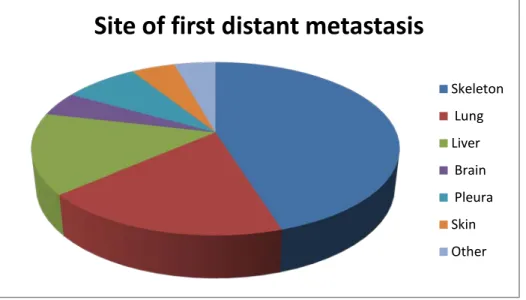
women developing distant metastasis. In our material the most common site of first distant metastasis after breast cancer is the skeleton, followed by lung and liver, see Figure 2. It is presently unknown how prognosis is affected by the site of first distant metastasis.
Figure 2: Site of first distant metastasis, based on the 5002 patients in the Stockholm breast cancer register diagnosed with distant metastasis and for whom the site of metastasis was recorded (=90% of all patients with distant metastasis).
Tumor- or patients characteristics that predict prognosis; prognosticators, are
important, primarily to decide the adjuvant therapy regimen. Among the important prognosticators for breast cancer are TNM-stage53, age at diagnosis (which is an
U-shaped relationship where both very young and old women have worse prognosis)54, histological grade55, estrogen- and progesterone-receptor over
expression56 , Her2-neu over expression57 and S-phase58. Over expression of
Site of first distant metastasis
Skeleton Lung Liver Brain Pleura Skin Other
estrogen receptor and HER2-neu are also predictors, i.e. factors that predict the
response of a certain therapy59, 60. The hunt for new prognosticators and predictors
is an important, very large and quickly changing field of research, which it unfortunately is impossible to give an account of in the thesis.
2.2 CONTRALATERAL BREAST CANCER
The reason lightning doesn't strike twice in the same place is that the same place isn't there the second time.
Willie Tyler (1940-), comedian Cancer is not evenly distributed in the population and having had one cancer does not protect against being diagnosed with another. Rather the opposite actually; people who have had one cancer are at increased risk of having yet another cancer
61. Secondary malignancies is naturally most apparent among patients with a first
cancer of favorable prognosis, so it should not be surprising that one fourth of all second primary cancers arise among breast cancer patients 61. Breast cancer
patients have about 25% increased risk of non-breast secondary primary cancers compared to healthy persons 62, and their risk to develop yet another breast cancer
is much higher, approximately twofold 63.
In 1921 Alson R. Kilgore (1987-1959) published the first scientific paper reporting on CBC, called “The Incidence of Cancer in the Second Breast After Radical Removal of One Breast for Cancer”64. The introduction states his two reasons for studying this
disease and the main obstacle; the women developing CBC is of interest as they seem to constitute a susceptible subpopulation and, in practice, the risk need to be
determined in order to give recommendations for prophylactic mastectomy, which was the only prevention available at the time. The main obstacle was to determine whether the cancer in the opposite breast was a new primary or a distant metastasis of the first cancer. (An attempt to answer this question using modern literature will be offered in Background - Occurrence 2.2.1.1)
The two cancers of CBC-patients might be diagnosed at the same time; synchronous
CBC, or separated in time; metachronous CBC. What these definitions are meant to
do is to separate the cancers that developed simultaneously from those that did not,
unfortunately we do not know when the cancers developed, we only know when they were diagnosed. This gives rise to the need for a cutoff, for allowing for the possibility that one of the cancers were more difficult to diagnose/took longer to become registered so that the two cancers were diagnosed separated in time even though they did developed simultaneously.
This cutoff has been set quite differently in the literature of CBC, from one month65, 66 of latency time to 1 year67, 68, but 3 or 6 months is most commonly seen. We chose
to use the cutoff of 3 months (92 days), mainly due to two reasons; in the unified health care system of Sweden there is good routines both for diagnostic procedures and registering of cases into the cancer register, so we do not see any reason for a prolonged cutoff due to fear of delay in reporting. At the same time it has been of interest to study the effect of treatment of the first cancer on the metachronous
0 10 20 30 40 50 60 70 1 4 7 10 13 16 21 24 27 30 37 41 46 49 52 56 59 62 65 69 72 75 80 83 86 90 95 100 N umb er o f CB C c as es
Latency time (in weeks)
Figure 3: Number of CBC-cases in relation to time between cancers
*303 cases diagnosed the first week. Our cutoff was set at 12 weeks (92 days) of latency time. cancer, and in those instances we want to have a cutoff that is sufficiently long to allow such effects to take place. All taken together we ended up with the Swedish
lagom (translates to intermediate, just right) alterative of 3 months. When
investigating the number of CBCs diagnosed per week of latency time in our CBC-cohort we found that the absolute majority of the synchronous CBC cases are diagnosed the same week as the first cancer, the number of CBC diagnosed then rapidly drops and from the ninth week the number of diagnosed CBC are down to a level which is kept for the two years we investigated. See Figure 3.
2.2.1 Occurrence
The risk of CBC has repeatedly been shown to be 0.6-0.7% yearly 65, 69-73 translating
into approximately 10-15% of all breast cancer patients being diagnosed with CBC during the first 20 years after initial diagnosis 71, 74. This risk has been consistently
shown during 50 years of CBC research, but comparison between these studies might be difficult due to methodological issues, e.g. different definitions of CBC or of synchronous/metachronous CBC, as outlined in a review by Chen et al75.
In most of the studies investigating the risk or incidence of CBC there has been some attempt to exclude the synchronous cancers, since the population at risk for
synchronous cancer is healthy women, while the population at risk for
metachronous CBC is breast cancer patients (see Discussion-CBC specific challenges 7.1.1). We can consequently regard the risk of 0.7% or the incidence of 7/1000
person years to be the incidence of metachronous CBC. For synchronous CBC the incidence is much lower; 1.6/100 000 person years76 and constitutes between 0.9 –
2.2.1.1 Temporal trends
Metachronous CBC have gotten attention as an emerging clinical problem, since the population at risk is increasing, as a consequence of the steeply increasing breast cancer incidence of the past decades, and improving breast cancer mortality. Despite increasing absolute numbers of CBC-patients, studies both in Sweden and in the US have observed decreasing incidence of metachronous CBC over calendar period76, 81.
In the US study this decrease was shown to be mainly seen after ER-positive first cancer, and the authors suggest that this might be due to the widespread use of endocrine therapy.
Regarding synchronous CBC, on the other hand, our material show an increasing incidence over calendar time, in accordance with the increasing incidence of unilateral breast cancer (the population at risk for unilateral breast cancer and synchronous CBC is the same). These two opposing temporal trends of synchronous and metachronous CBC is a tentative explanation for the constant CBC-risk
mentioned in the previous paragraph and was the rational for Study I.
2.2.1.2 Age trends
The age trend for synchronous CBC follows the same pattern as for unilateral breast cancer (and indeed for most other cancers); synchronous CBC increase steeply with age82.
For metachronous CBC the story is somewhat more complicated; the risk for metachronous CBC is shown to be constant72, 83, 84 (or almost constant85) from
diagnosis of the first cancer during the rest of the patient’s life. Many studies show that the level of risk is determined by the age at diagnosis of the first cancer72, 83, 85, 86
(though this is not un-disputed87, 88); patients diagnosed with their first cancer when
they were young are at a constantly higher risk than patients who had their first cancer when they were older.
This leads to the observation that younger patient groups have a higher incidence of CBC than older patient groups63, 89. Even if all of the individual patients are at their
constant risk, the younger patient group will constitute only of patients with a high risk, while a older patient group will constitute of a group with mixed risks; higher for those that have lived a long time since their first cancer (i.e. got the first cancer when they were young) and lower for those who just had their first cancer (i.e. got the first cancer when they were old).
2.2.1.3 The more we look, the more we find?
As previously mentioned, 0.9 – 1.9% of all women with breast cancer are found (by palpation and/or mammography) to have cancer also in the other breast at time of first diagnosis; synchronous CBC. However, it seems the more we look the more we find; if the opposite breast is investigated with ultrasound in addition to palpation and mammography, an additional 2% is found90, and if MRI is added to palpation
and mammography an additional 4% is found91 (including both invasive and in-situ
lesions). Further, two studies have made on so called blind biopsies, where a sample of 2-3cm3 breast tissue was surgically removed from the contralateral breast (that
was clinically and mammographically free of cancer) at the time of the primary breast cancer surgery. In 6-7% of the patients invasive or in-situ cancers were found92, 93 and the one study that also included benign lesions of precancerous type
could diagnose an additional 9% of the investigated patients with this condition. An autopsy study of pre/peri-menopausal women showed that close to 20% had clinically undetected (invasive/in-situ) breast cancer94.
2.2.1.4 Is CBC a true primary cancer?
A central question in CBC-research is whether the second cancer is actually a new primary cancer, or if it is a metastasis from the first cancer. According to the ‘seed and soil’ hypothesis, first proposed by Fuchs and Paget95, cancers will tend to
metastasize in organs that are similar to that where the first tumor developed, hence metastases to the contralateral breast would be a very plausible idea. However, today most agree that the most likely scenario is a combination of the “seed and soil” hypothesis and another hypothesis, proposed by James Ewing96, that suggests that
the organs to which the cancer metastasize are determined by anatomical and mechanical routes. In light of that hypothesis it is of interest to note that, in contrast to sites to which breast cancer commonly metastasizes, the contralateral breast is not highly vascularized. Traditionally, criteria of the presence of an in-situ component in the second cancer or of different histology at the two cancers, have been used to distinguish between true second primary cancers and metastases, this method however has serious drawbacks; since 80% of all breast tumors in general are of ductal histology97 and 40% lacks in-situ component98, the risk of
falsely categorized tumors as metastasis is very high.
Several studies have been performed to answer this question by molecular means, unfortunately they are all quite small, but the message is, despite this, rather clear. Janschek et al. compared mutations in the p53-gene in 33 patients with
synchronous and metachronous CBC, for 13 patients this analysis was informative and among these only one patient had the same mutation99. Imyaitov et al.
investigated the allelic imbalance profiles in 28 synchronous and metachronous CBCs, they found evidence for different origin of the two tumors in 23 cases, among the remaining 5 cases the clinical and histopathological information indicated different origin of the two tumors in 4 cases, leaving one case unanswered100.
Brommesson et al investigated 8 synchronous CBC tumors by comparative genomic hybridization and found 1 case that had genomic similarities. Indications from the epidemiological perspective also speak against that metastases should contribute significantly to the group of CBC cancers. If the second cancer would be a metastasis then TNM-stage of the first cancer should be the most important risk factor for CBC, but despite extensive research of the question there is still no distinct conclusion whether higher TNM-stage is at all a risk factor for CBC. Further, the prognosis of CBC is worse than for unilateral breast cancer, but the prognosis for patients with metastasized breast cancer is much worse still (See Background –Prognosis 2.1.4). There is no obvious reason
that a metastasis in the contralateral breast should be so much less harmful than a metastasis in e.g. the skin or the skeleton. Finally, among patients with distant spread, metastasis in only one site is rarely seen, therefore, if CBC was a metastasis
to the opposite breast one would expect that CBC without distant metastasis also was a rare scenario, which it is not.
In conclusion, evidence from different areas of medical research taken together indicate that CBC, in the overwhelming majority of the cases, indeed is a new primary cancer, though the phenomenon that one breast cancer metastasize to the opposite breast does occur. Judging from the genetic studies referred to above (albeit small) the proportion of CBC that are not true primaries are approximately 6% of all CBCs diagnosed, and probably less if using the same definition of CBC as in the studies included in this thesis; that the second cancer should be diagnosed without distant metastasis.
2.2.2 Risk factors
Only three risk factors have been consistently shown to increase the risk of CBC; young age at the first cancer (See Background- Age trends 2.2.1.2), family history of
breast cancer and lobular histology of the first cancer.
Having at least one first-degree relative with breast cancer approximately doubles the risk of CBC, and having both mother and sister affected by breast cancer confers a 5 times increased risk101. Some studies also indicate that the risk is increased if the
relative had early disease onset102.
About 85% of all invasive breast tumors arise in the milk ducts, so called ductal
cancer, 10% arise in the lobuli, so called lobular cancer, the remaining 5% have a
mixed, or other, histology103. Breast cancers of lobular histology have a more
favorable prognosis than ducal cancers104, but also have approximately double the
risk of CBC87.
An intuitive hypothesis when it comes to lifestyle risk factors for CBC could be referred to as more of the same; that women with CBC have more of the risk factors
that have been characterized for unilateral breast cancer. This has been researched extensively; studies investigating established hormonal risk factors for unilateral breast cancer; age at menopause, breast feeding, hormone replacement therapy and oral contraceptives have consistently shown no association with risk of CBC 87, 105-109,
while the results have been inconstant for number of children86, 87, 106-109, age at
parity86, 87, 106, 107, 109, 110 and age at menarche87, 106, 107, 109, 110. Regarding other life
style risk factors as smoking, alcohol consumption, obesity and social-economical status results are also mixed 87, 106, 108, 110-112, but indications are that alcohol
consumption is a risk factor for CBC 108, 111, 112 and smoking is not87, 106, 112.
As previously mentioned (See Background –Risk factors 2.12), the breast is sensitive
to ionizing radiation, therefore it has been a concern for many years whether adjuvant radiotherapy for breast cancer increases the risk of CBC. Overall, the results indicate that the risk is not increased in the general population of breast cancer patients71, 113, but a risk increase can be detected among patients that were
2.2.3 Diagnosis and treatment
Since the high risk of CBC for breast cancer patients is well recognized in the
healthcare system the contralateral breast is closely examined at time of first breast cancer diagnosis. After the initial treatment period all breast cancer patients in Sweden are followed-up with clinical examinations and mammography during 5 years, one of the main aims of this follow-up program is the early detection of potential CBCs38.
The close follow-up (in particular by mammography) together with the heighted awareness among the patients are a plausible explanation for the fact that the second cancer of metachronous CBC is more likely to be diagnosed at smaller tumor size and at a more favorable stage, compared to unilateral breast cancer116.
CBC-patients who have their second cancer detected by mammography have been shown to have better survival than patients with the second cancer diagnosed by clinical examination117, this seem to be mostly, but not entirely, due to the cancers being
diagnosed at a better stage and younger age118.
Regarding synchronous CBC, since they are diagnosed, and presumably developed, simultaneously, it is not possible to distinguish which cancer was the ‘first’ and which was the ‘second’. However, oftentimes the larger and/or more spread cancer will give rise to symptoms that will lead to the diagnosis of that cancer and also a sub-clinical cancer in the opposite breast. Therefore, the tumor that is recorded as ‘second’ cancer is often smaller and of more favorable stage also when the cancers are diagnosed only days apart, this could however be seen as an artifact.
In general, there is a lack of specific treatment directives when it comes to adjuvant therapy after metachronous CBC, the patients will most likely be treated as any (unilateral) breast cancer patient, while naturally taking potential earlier therapy into account when calculating maximum dose of chemotherapy or the fields of radiotherapy. Neither for synchronous CBC exists any specific directives, most commonly the adjuvant therapy regimen will be decided by the aggressiveness of the most advanced cancer, however, endocrine therapy will probably be given if any of the cancers are estrogen receptor positive.
2.2.4 Prognosis
During many years the question of whether CBC-patients had worse prognosis than unilateral breast cancer patients remained unanswered, most likely due to inherited problems in studying survival after secondary cancers (See Discussion -CBC-specific challenges 7.1.3). Though still not completely uncontroversial, we can now say with
some certainty that the prognosis of CBC is indeed worse than for unilateral breast cancer, but that this is highly dependent on the latency time76. Figure 4 shows a
Kaplan-Meier plot of percentage survival over time (in years) since CBC-diagnosis for patients with synchronous CBC (red), CBC with short latency time (<5 years) (green) and CBC-patients with long latency time (>5years) (black), this can be compared to the survival for unilateral breast cancer patients (blue). This plot is based on the cohort of CBC-patients included in this thesis, and is in close agreement to what has been show by Hartman et al.76, though it has lower power.
Figure 4: Kaplan-Meier plot of survival after breast cancer (blue) and CBC. Synchronous CBC (red), CBC with short latency time (<5 years) (green) and CBC-patients with long latency time (>5years) (black). The time scale is in years. Given that breast cancer metastasis might be diagnosed 20 or 30 years after initial breast cancer diagnosis, in the case of a CBC-patient dying in breast cancer, how do we know which cancer the metastases came from? The answer is that we do not. Metastasis are seldom biopsied, if they were, it might be possible to distinguish a larger genomic similarity with one of the primary cancers, though it is not certain, since the cancer is likely to have mutated further from initial diagnosis until diagnosis of the metastases.
For synchronous cancers it is more likely that the larger/more advanced cancer is the cancer setting metastases, for metachronous cancer, the situation is somewhat more complicated. Even though breast cancer can metastasize long after initial diagnosis, it is still more likely within the first few years50, thus it should be less
likely that the first cancer is the metastasizing cancer. This indication is further enhanced by the fact that to be able to get a second cancer, the patient has to survive the first cancer long enough, it therefore follows that the first cancer is less
3
AIMS OF THIS THESIS
Leave this world a little better than you found it.
Sir Robert Baden-Powell (1857-1941), founder of the Scout Movement
The overall aim of this thesis has been to increase the biological understanding and clinically applicable knowledge about contralateral breast cancer.
More specifically, the aims of the four studies were to investigate the following: • To what extent the increasing proportion of synchronous CBCs can be
explained by improvements in the diagnostic work-up of breast cancer patients.
• If adjuvant radiotherapy for the first cancer can affect the aggressiveness of the second cancer and explain the bad prognosis for CBC patients with short latency time.
• How estrogen receptor status of the first and second cancer relates, and how these estrogen receptor patterns affect the prognosis after CBC.
• Whether mammographic density at diagnosis of the first cancer, or its changes over time, can be used to predict the risk of CBC.
4
MATERIALS
"Data! Data! Data!" he cried impatiently. "I can't make bricks without clay."
Sir Arthur Conan Doyle (1859–1930), author of Sherlock Holmes
4.1 REGISTERS
One of the often brought up advantages about epidemiological research in Sweden is the many high-quality health registers. Sweden has five main health registers, one of these registers was used in this thesis, namely the Swedish cancer register. The Swedish cancer register has close to complete coverage of the population and close to complete follow-up119, 120. Sweden also has a number of smaller registers for
specific conditions, usually not covering the whole country and constructed primary for internal quality control. These registers constitute a hugely underused source of information for epidemiological research. Information from the Stockholm breast cancer register (SBCR) was used extensively in this thesis.
4.1.1 Swedish cancer register
Since 1958 all cancers diagnosed in Sweden are reported to the Swedish cancer register, every tumors is reported separately, resulting in that the same person might have several posts in the register, if that person suffered from more than one primary cancer. Also, each cancer is reported both by the diagnosing
pathology/cytology laboratory and by the treating physician, to ensure completeness. The information recorded in the register is the International Classification of Disease (ICD)-code, the date of diagnosis and the personal
identification number and vital/emigration status. From the ICD-code the laterality of the breast cancer can be derived, which is of great importance in this thesis, as CBC is defined as two primary cancers in opposite breasts. From the personal
identification number date of birth and sex can be derived (additionally; the personal identification number can be used to link the patients to the other health registers and to medical records, though this functionality has not been used in this thesis)
4.1.2 Stockholm breast cancer register (SBCR)
The Stockholm Breast Cancer Register (SBCR) was started 1970 to register breast cancer patients that participated in clinical trials. Gradually the registry was expanded to cover all breast cancer patients and from 1976 there is a close to complete coverage of all breast cancer patients in the Stockholm-Gotland Health care region (which presently has a catchment area of 1.9 million people). As of December 2008 SBCR contained information on 32 153 breast cancer patients. The register is maintained by the Oncological Center in Stockholm and the aim is quality control of the breast cancer care in the region, even though SBCR also has been used somewhat for breast cancer research.
Information recorded in SBCR includes personal identification number, date of diagnosis, family history of breast cancer, tumors characteristics, details of surgery and, from 1990, adjuvant therapy. The register is continuously updated with
information on local recurrence and distant metastasis, as the patients are clinically followed up, and SBCR and the Swedish Cause of Death Register are regularly linked so that information on cause of death for the deceased patients is available.
4.2 CBC-COHORT 4.2.1 Inclusions
When collection of medical records for this thesis started we made a selection of all patients with contralateral breast cancer (CBC) in the SBCR that fulfilled our criteria for CBC-diagnosis. The following inclusion criteria were used:
• Diagnosed with two primary, invasive breast cancers, one in each breast. • Not diagnosed with any other malignancy before the second breast cancer. • No distant metastasis diagnosed at time of the first breast cancer diagnosis.
The last two criteria was set primarily to increase the probability that the second breast cancer was indeed a new primary cancer, and not metastatic spread from the first breast cancer or any other malignancy, to the second breast. It has also been of importance when studying effect of treatment of the first cancer, to ensure that the patients have not received cancer therapy for any other cancer.
As the selection was made from SBCR the same restrictions in time and place as for that register applies also to the CBC-cohort;
• Both breast cancers diagnosed in the Stockholm-Gotland health care region. • Both cancers diagnosed during 1970-2005.
4.2.2 Exclusions
When the medical records were collected we applied additional criteria for the CBC-cohort; we had selected all CBC-patients from the SBCR, but since the register was only complete from 1976 we decided to not include CBC-patients diagnosed before then.
Also, obviously, patients for whom we could not find the medical record could not be included, but these were mercifully few, only 17 patients (1%). Further we excluded patients who were diagnosed with distant metastasis before diagnosis of the second breast cancer and patients who had the second breast cancer diagnosed via axilla lymph node metastases. This was done to decrease the probability that the second cancer was a metastasis of the first breast cancer. Finally, for some patients the information in the medical records was different than in the registers, e.g. 19 patients had a malignancy at another site recorded in the medical record, for 4
patients the cancer recorded in the medical record was non-invasive (in-situ). See Figure 5 for exclusions.
Figure 5: CBC cohort, with exclusions.
4.2.3 Populations for study I-IV
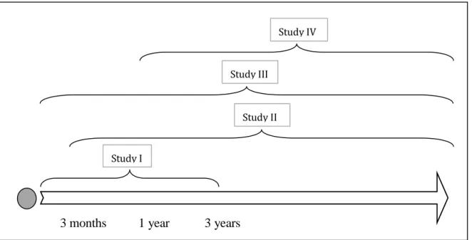
The four studies in this thesis all used the CBC-cohort, but due to the different research questions different subsets were used, as explained in Figure 6.
Study I included all CBC-cases with three years or less between the cancers, with the aim of including all CBCs that were likely to have been sub clinically present in the
17 (1%) patients for which medical records could not be found 18 (1%) patients diagnosed before 1976.
1295 (90%) of original selection was included in final cohort
1440 patients
selected from SBCR
110 (8%) patients excluded
69 patients excluded due to distant metastasis before second breast cancer
19 patients excluded due to malignancy at other site before second breast cancer 7 patients excluded since the second cancer was diagnosed though axilla lymph node metastases
4 patients excluded due to any of the breast cancer not being invasive 11 patients excluded due to miscellaneous reasons
opposite breast at time of the first diagnosis. Study II included all CBC-cases with more than three months between the cancers, with the aim of including all CBCs that might potentially have been affected by adjuvant therapy for the first cancer. Study III used all CBC-cases, here the aim was to evaluate the impact of estrogen receptor status and compare synchronous and metachronous CBC. Finally, Study IV used all CBC-cases with more than one year between the cancers, with the aim of including all CBCs (and selected controls with unilateral breast cancer) with at least two mammograms far enough apart for a potential change of mammographic density to be possible to evaluate.
Figure 6: Populations used for Studies I-IV.
4.2.4 Medical records
Medical records were collected from the archives of the oncology and surgical clinics of the hospitals in the Stockholm region. The retrieval rate was very good (99%), a fact that is most likely attributable to the unified health care system of Sweden. The medical records were read and the predefined variables were extracted, the
pathological-anatomical reports from all breast cancer surgeries were also copied and collected. The variables extracted were anthropological measures, preexisting diseases (thyroid disease, diabetes, benign breast disease and schizophrenia), breast cancer family history and hormonal factors (e.g. pregnancy, parity, oral
contraceptives use, menopause, hormonal replacement therapy) at time of the first breast cancer. For both cancers we also collected mode of detection, all available tumor characteristics (size, hormone receptor status, histological grade, and lymph node status), type of surgery and type of adjuvant therapy (with details on dose and duration). We further collected information on local/regional recurrence and date and site of distant metastasis, but no treatment or follow-up after diagnosis of distant metastasis. For some of the variables, comparison was possible between the Stockholm Breast Cancer Register and the data collected from medical records; overall the concordance was good (e.g. menopause status:96%, estrogen receptor
Study I
Study III
Study II
Study IV
3 months 1 year 3 years
however regarded as the gold standard. Regarding the adjuvant treatment a significant difference is that the information in SBCR is from the time of treatment conference (so called ‘intention to treat’) while the information collected from the medical records was the actual treatment given.
4.2.5 Mammograms
A mammogram is radiological picture of a breast, the breast is compressed and the mammogram is taken either in cranio-caudal fashion (vertically), media-lateral (from the side) or media-lateral-oblique (diagonally), these are different so-called
views. The media-lateral-oblique (MLO) view has been shown to be the most
useful121, partly since it better includes the upper-lateral quadrant of the breast,
where many breast cancers occur122. For breast cancer screening the MLO view was
predominantly used, while for diagnosis and follow-up (the setting mostly relevant to Study IV) both the cranio-caudal (CC) and MLO view were used, but of the above mentioned reasons we preferentially used the MLO view. In routine follow-up after breast cancer mammograms are oftentimes taken every 12 months, after 5 years the patient is usually referred back to the screening program, where mammograms are taken every 18-24 months until the patient is no longer in the screened age interval.
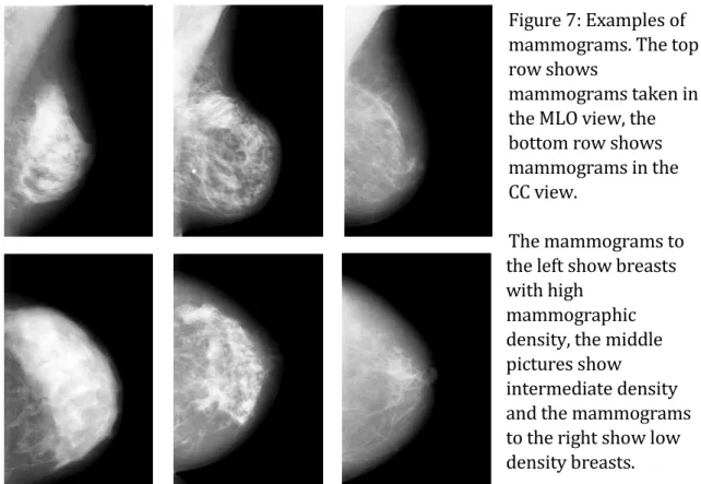
Figure 7: Examples of mammograms. The top row shows
mammograms taken in the MLO view, the bottom row shows mammograms in the CC view.
The mammograms to the left show breasts with high
mammographic density, the middle pictures show
intermediate density and the mammograms to the right show low density breasts. For Study IV we collected mammograms from the time of diagnosis of the first cancer and until 6 months before the second cancer for the CBC-cases. In this study we also selected controls (patients with unilateral breast cancer), five unilateral breast cancer patients were selected to each case. The controls were individually matched to the cases on calendar period and age of diagnosis, adjuvant therapy for the first cancer and follow-up time. The reason for selecting five controls when we only planned to collect the mammograms for one of them was to allow for the event
that no mammograms were found for a particular patient. For the controls
mammograms were collected from time of breast cancer diagnosis until 6 months before the cutoff date (the date corresponding to second breast cancer diagnosis for the individually matched case). To be able to investigate the effect of factors as hormone replacement therapy, estrogen receptor status at first cancer, menopause, etc, this information were collected from the medical records of the controls, this was done after collection of the mammograms. The collected variables were
however only a small selection of the variables collected from the medical records of the CBC-patients. The retrieval rate of mammograms was markedly worse than for medical records, partly due to different regulations regarding archiving.
The mammograms were digitized using an Array 2905HD Laser Film Digitizer (Array Corporation, Tokyo, Japan). Mammographic density was measured using an automated thresholding method123 which incorporates the knowledge of a trained
observer, by using measurements obtained by an established user-assisted
threshold method - Cumulus124 - as training data. The externally validated results
showed a high correspondence between our automated method and the established used-assisted thresholding method; Cumulus (rpercent mammographic density = 0.88 (95% CI:
0.87-0.89). Kallenberg et al125 have developed a similar automated method, which
also extracted a number of features from the pixels in the mammograms and used them to train, and validate, a measurement of mammographic density against Cumulus (the latter taken as the ground truth); this automated method has performed similarly (rpercent mammographic density =0.90 with Cumulus).
5
METHODS
In respect of military method, we have, firstly, Measurement; secondly, Estimation of quantity; thirdly, Calculation; fourthly, Balancing of chances; fifthly, Victory.
Sun Tzu (~500 BC), author of The Art of War 5.1 DESIGN AND STATISTICAL ANALYSIS
Do not try to bend the spoon — that's impossible. Instead, only try to realize the truth: there is no spoon.
The movie Matrix (1999)
Epidemiological studies are either experimental studies, in which the researcher
influence the investigated exposure, e.g. as in a randomized clinical trial, or
observational studies, where the exposure and outcome is merely observed and
causal interpretation is attempted through adjustments. Observational studies are either retrospective or prospective, in a retrospective study information about the
exposure is gathered after the outcome has occurred, this might be a problematic approach since it is, at least theoretically, possible that the case-control status influences the assessment of exposure, e.g. due to recall bias126. In a prospective
study the exposure is assessed before the outcome occurs, this can be done either by actually starting the study before the outcome happens, or by going back to records of the exposure which were documented before the outcome (