e-Publications@Marquette
Psychology Faculty Research and Publications
Psychology, Department of
1-1-2013
Semantic Memory Functional MRI and Cognitive
Function After Exercise Intervention in Mild
Cognitive Impairment
J Carson Smith
University of Maryland - College Park
Kristy A. Nielson
Marquette University, [email protected]
Piero Antuono
Medical College of Wisconsin
Jeri-Annette Lyons
University of Wisconsin - Milwaukee
Ryan J. Hanson
University of Wisconsin - Milwaukee See next page for additional authors
Accepted version.
Journal of Alzheimer's Disease
, Vol. 37, No. 1 (2013): 197-215.
DOI
. © 2013 IOS
Press. Used with permission.
Hantke, and Matthew D. Verber
Journal of Alzheimer’s Disease, Vol 37, No. 1 (2013): pg. 197-215. DOI. This article is © IOS Press and permission has been granted for this version to appear in e-Publications@Marquette. IOS Press does not grant permission for this article to be further copied/distributed or hosted elsewhere without the express permission from IOS Press.
1
Semantic Memory fMRI and
Cognitive Function After Exercise
Intervention in Mild Cognitive
Impairment
J. Carson Smith
Department of Kinesiology, School of Public Health, University of
Maryland
College Park, MD
Department of Neurology, Medical College of Wisconsin,
Milwaukee, WI
Kristy A. Nielson
Department of Psychology, Marquette University,
Department of Neurology, Medical College of Wisconsin
Milwaukee, WI
Piero Antuono
Department of Neurology, Medical College of Wisconsin
Milwaukee, WI
Jeri-Annette Lyons
Department of Biomedical Sciences,
University of Wisconsin-Milwaukee,
Journal of Alzheimer’s Disease, Vol 37, No. 1 (2013): pg. 197-215. DOI. This article is © IOS Press and permission has been granted for this version to appear in e-Publications@Marquette. IOS Press does not grant permission for this article to be further copied/distributed or hosted elsewhere without the express permission from IOS Press.
2
Ryan J. Hanson
Department of Psychology, University of Wisconsin-Milwaukee,
Milwaukee, WI
Alissa M. Butts
Department of Psychology, Marquette University,
Milwaukee, WI
Nathan C. Hantke
Department of Psychology, Marquette University,
Milwaukee, WI
Matthew D. Verber
Department of Kinesiology, University of Maryland,
College Park, MD
Abstract: Mild cognitive impairment (MCI) is associated with early memory loss, Alzheimer neuropathology, inefficient or ineffective neural processing,
and increased risk for Alzheimer’s disease (AD). Unfortunately, treatments aimed at improving clinical symptoms or markers of brain function generally have been of limited value. Physical exercise is often recommended for people diagnosed with MCI, primarily because of its widely reported cognitive
benefits in healthy older adults. However, it is unknown if exercise actually benefits brain function during memory retrieval in MCI. Here, we examined the effects of exercise training on semantic memory activation during functional magnetic resonance imaging. Seventeen MCI participants and 18 cognitively intact controls, similar in sex, age, education, genetic risk, and medication use, volunteered for a 12-week exercise intervention consisting of supervised treadmill walking at a moderate intensity. Both MCI and control participants significantly increased their cardiorespiratory fitness by
approximately 10% on a treadmill exercise test. Before and after the exercise intervention, participants completed a fMRI famous name discrimination task and a neuropsychological battery, Performance on Trial 1 of a list-learning task significantly improved in the MCI participants. Eleven brain regions activated during the semantic memory task showed a significant decrease in activation intensity following the intervention that was similar between groups (p-values ranged .048 to .0001). These findings suggest exercise may
improve neural efficiency during semantic memory retrieval in MCI and cognitively intact older adults, and may lead to improvement in cognitive function. Clinical trials are needed to determine if exercise is effective to delay conversion to AD.
Journal of Alzheimer’s Disease, Vol 37, No. 1 (2013): pg. 197-215. DOI. This article is © IOS Press and permission has been granted for this version to appear in e-Publications@Marquette. IOS Press does not grant permission for this article to be further copied/distributed or hosted elsewhere without the express permission from IOS Press.
3 Keywords:Alzheimer’s Disease, Dementia, Exercise, Magnetic Resonance Imaging, Non-Pharmacologic Treatment, Physical Activity, Physical Fitness, Memory
Introduction
There is an urgent need to identify effective treatments that may improve cognitive function and brain function in those most at
risk for Alzheimer’s disease (AD).1 One of the greatest predictors of AD
is a diagnosis of mild cognitive impairment (MCI), with 40% of individuals diagnosed with MCI progressing to AD over a 4-year
period2 and approximately 60% exhibiting autopsy-verified AD
post-mortem.3 Recent diagnostic criteria now include the term ‘MCI due to
AD’, underscoring the probable decades-long neuropathologic history
that eventually leads to clinically observable symptoms and a MCI
diagnosis.4,5 In addition to impaired episodic memory function, one of
the first observable symptoms of MCI due to AD is the inability to remember familiar names, the most common memory complaint
among older adults.6 Indeed, the semantic memory system, in
particular, is vulnerable to the earliest stages of AD.7,8
In addition to episodic and semantic memory impairments, individuals diagnosed with MCI have been shown to exhibit alterations
in cerebral perfusion9 and functional activation of brain networks,10-12
as well as increased brain amyloid,13,14 cortical thinning,15 and atrophy
of the hippocampus.16 It has been hypothesized that reduced
functional connectivity within the default mode network may be related to increased amyloid retention, and further, that greater task-activated
neuronal activity may exacerbate amyloid deposition.17,18 Greater
task-induced brain activation during successful memory retrieval may
reflect a compensatory response19,20 or utilization of cognitive reserve21
that helps to sustain normal function. Alternatively, it is possible that greater task-related neural activation is a sign of underlying
neuropathology and neural inefficiency or overload that may be
indicative of impending cognitive decline.22,23 For example, it has been
suggested that a successful intervention for cognitive decline will result in more efficient neural processing, perhaps via reduced neural
interference,23 and thus, a reduction in the neural activation required
Journal of Alzheimer’s Disease, Vol 37, No. 1 (2013): pg. 197-215. DOI. This article is © IOS Press and permission has been granted for this version to appear in e-Publications@Marquette. IOS Press does not grant permission for this article to be further copied/distributed or hosted elsewhere without the express permission from IOS Press.
4
Despite the need for effective treatments for early memory loss, a recent NIH consensus panel concluded there was no solid evidence for any intervention or modifiable factor to improve memory or brain
function in MCI.24 It was noted, however, that physical exercise is one
intervention that has shown considerable promise. Previous studies in cognitively intact healthy older adults have shown that exercise
training25 or greater self-reported physical activity26 was associated with an increase or greater fMRI activation during a cognitive task. Although exercise is known to produce cognitive and hippocampal
benefits in healthy older adults,27,28 we know very little regarding how,
or if, exercise may affect brain function in patients diagnosed with
MCI.1 Two clinical trials have shown that an exercise intervention leads
to limited improvement in cognitive function in MCI participants29,30
and older adults with subjective memory complaints.31 Another study
found greater caudate activation during semantic memory retrieval in
physically active compared to physically inactive MCI participants.32
Greater grey matter volume was reported in early-stage AD patients who had greater cardiorespiratory fitness compared to those with
lower fitness.33 However, it is unknown if exercise training alters
neural processing during memory retrieval in individuals diagnosed with MCI. The purpose of the current study was to determine if a 12-week walking exercise intervention affects semantic memory fMRI activation and neuropsychological outcomes in individuals diagnosed with MCI compared to cognitively intact older adults. Based on the previous effects of exercise on task-activated fMRI, we hypothesized that exercise training would lead to an increase in semantic memory-related activation in both MCI participants and healthy controls.
Materials and Methods
Participants and Pre-Screening
Community dwelling older adults, ages 60 to 88 years, were recruited from in-person informational sessions at retirement
communities and community recreation centers, through newspaper and other local advertisements, and through referrals from local
physicians. Participants were pre-screened with a structured telephone interview to determine eligibility. Eligible volunteers then provided written informed consent, physician approval for moderate intensity
Journal of Alzheimer’s Disease, Vol 37, No. 1 (2013): pg. 197-215. DOI. This article is © IOS Press and permission has been granted for this version to appear in e-Publications@Marquette. IOS Press does not grant permission for this article to be further copied/distributed or hosted elsewhere without the express permission from IOS Press.
5
exercise was obtained, and a neurological evaluation was conducted to further determine eligibility. On a separate day, prior to baseline
neuropsychological or exercise testing, eligible participants underwent a mock MRI scan session and practiced the fMRI task. Participants who completed the baseline neuropsychological and exercise testing
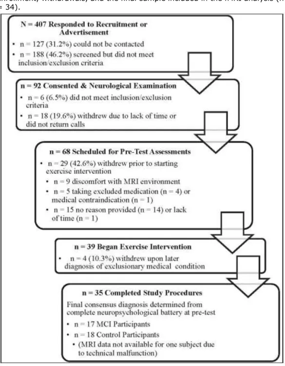
sessions were paid for their participation. Figure 1 describes the flow of participants from initial recruitment to the completion of the study. This study was conducted according to the Helsinki Declaration of 1975 and was approved by the Institutional Review Board at the Medical College of Wisconsin.
Inclusion and Exclusion Criteria
Volunteers who indicated they engaged in less than 3 days/week of moderate intensity physical activity for the past 6 months were included. Participants were excluded if they reported a history or evidence of: 1) neurological illnesses/conditions, such as head trauma with significant loss of consciousness (>30 min), cerebral ischemia, vascular headache, carotid artery disease, cerebral palsy, epilepsy, brain tumor, chronic meningitis, multiple sclerosis, pernicious
anemia, normal-pressure hydrocephalus, HIV infection, Parkinson’s
disease, or Huntington's disease; 2) medical illnesses/conditions that may affect brain function, such as untreated hypertension, glaucoma, and chronic obstructive pulmonary disease; 3) current untreated Axis I psychiatric disturbance meeting DSM-IV Axis I criteria, including
severe depressive symptoms and substance abuse or dependence; 4) exclusion criteria specific to MR scanning: pregnancy, weight
inappropriate for height, ferrous objects within the body, and a history
of claustrophobia; 5) left-handedness (laterality quotient [LQ] < 50);34
6) current use of psychoactive medications, except stable doses of SSRI and SNRI antidepressants; 7) any unstable or severe
cardiovascular disease or asthmatic condition; and 8) history of transient ischemic attack or > 4 on the modified Hachinski ischemic
scale. The Geriatric Depression Scale (GDS)35 and the Lawton and
Brody Self-Maintaining and Instrumental Activities of Daily Living
(ADL) Scale36 were also administered. Participants were excluded if
they scored > 15 on the GDS, or showed relatively impaired ADLs. Participants taking acetylcholinesterase inhibitors (AChEI), as well as beta-blockers or other anti-hypertensive medications, were included as
Journal of Alzheimer’s Disease, Vol 37, No. 1 (2013): pg. 197-215. DOI. This article is © IOS Press and permission has been granted for this version to appear in e-Publications@Marquette. IOS Press does not grant permission for this article to be further copied/distributed or hosted elsewhere without the express permission from IOS Press.
6
long as their medication dosage was stable for at least one month prior to and during the intervention. There were no significant differences between the groups in AChEI use.
Neuropsychological Test Battery and Clinical Criteria for
MCI
A comprehensive neuropsychological test battery was
administered before and after the exercise intervention, between 0700 hrs and 1100 hrs, prior to the exercise test and on a different day than the MRI scan session. The battery included the Mini-Mental State
Exam,37 Mattis Dementia Rating Scale – 2,38 Rey Auditory Verbal
Learning Test (AVLT),39 the Logical Memory and Letter-Number
Sequencing subtests of the Wechsler Memory Scale-III,40 Symbol-Digit
Modalities Test,41 Controlled Oral Word Association Test,42 animal
fluency, and the Clock Drawing Test.43 Alternate forms of the AVLT and
the DRS were used at the pre- and post-intervention test sessions. Determination of cognitive status (MCI or healthy control) was
determined using the core clinical criteria set forth by the recent
NIH-Alzheimer’s Association workgroup on the diagnosis of MCI due to AD.4
MCI was defined by: 1) subjective concern regarding change in cognition; 2) impairment in one or more cognitive domains; 3) preservation of independence in activities of daily living; and 4) not demented.
Exercise Test
Before and after the exercise intervention, participants completed a submaximal exercise test to estimate peak aerobic
capacity (VO2peak). Prior to each exercise test, the metabolic cart
system was calibrated against known concentrations of O2 and C O2.
The exercise test was conducted on a motorized treadmill (General Electric, Milwaukee, WI) using a modified Balke-Ware protocol (2.0 mi/hr (3.2 km/hr) at 0° grade, with grade increase of 1° per minute), in accordance with the American College of Sports Medicine
Guidelines.44 Each test began and ended with 3-5 min of level walking
at 1-2 mi/hr (1.6-3.2 km/hr). Heart rate, blood pressure (every 2 minutes), ratings of perceived exertion (every minute), and expired air
Journal of Alzheimer’s Disease, Vol 37, No. 1 (2013): pg. 197-215. DOI. This article is © IOS Press and permission has been granted for this version to appear in e-Publications@Marquette. IOS Press does not grant permission for this article to be further copied/distributed or hosted elsewhere without the express permission from IOS Press.
7
(ParvoMedics, Sandy, UT). The exercise test was terminated when the
participant’s heart rate reached 85% of heart rate reserve, if there was
an abnormal blood pressure response (e.g., raise in diastolic blood pressure > 110 mmHg), or the participant indicated a desire to stop the test. Heart rate reserve was defined as age-predicted maximal
heart rate (220−age) minus resting heart rate determined after 10
minutes of supine rest prior to the exercise test. VO2peak (ml/kg/min at
STPD) was estimated from the highest VO2 value obtained during the
Test.44
Exercise Intervention
Participants completed a 12-week treadmill walking exercise intervention (44 total sessions). A qualified personal fitness trainer or exercise physiologist supervised each session of exercise, which was conducted individually or in a group of no more than two participants at a fitness center location near their home or within their community. The exercise intensity, session duration, and weekly frequency was gradually increased during the first 4 weeks until the participants were walking 30 minutes per session, 4 sessions per week, at an intensity approximately 50-60% of heart-rate reserve (HRR) during weeks 5- 12. The intervention was tailored to each individual based on baseline exercise capacity. The treadmill grade and/or speed were modified each session based on the heart rate and perception of effort of the participant (not more than 15 on the Borg 6-20 RPE scale) and was designed to moderately challenge the participant and to increase cardiorespiratory fitness. Each session began and ended with 10
minutes of very light walking and flexibility exercise. Participants wore a Polar R heart rate monitor and also provided subjective ratings of
perceived exertion throughout each exercise session.45,46
Functional MRI Famous Name Recognition Task
Participants underwent an MRI session before and after (within 3-5 days) the intervention on a day that neuropsychological testing or exercise was not performed. The fMRI task stimuli consisted of 30 names of easily recognized famous persons (e.g., Frank Sinatra) and 30 names of non-famous individuals chosen from a local phone book. Only names with a high rate of identification (> 90% correct for
Journal of Alzheimer’s Disease, Vol 37, No. 1 (2013): pg. 197-215. DOI. This article is © IOS Press and permission has been granted for this version to appear in e-Publications@Marquette. IOS Press does not grant permission for this article to be further copied/distributed or hosted elsewhere without the express permission from IOS Press.
8
targets and foils) were selected from an original pool of 784 names.47
A trial consisted of the visual presentation of a single name for 4 s. Participants were instructed to make a right index finger key press if the name was famous and a right middle finger key press if the name was non-famous. Both accuracy (% correct) and reaction time (in ms) were recorded. The 60 name trials were randomly interspersed with 20 4-s trials in which the participant was instructed to fixate on a single centrally placed crosshair in order to introduce “jitter” into the fMRI time course. The imaging run began and ended with 12 s of fixation. Total time for the single imaging run was 5 min 44 s.
fMRI Acquisition
Whole-brain, event-related fMRI was conducted on a General Electric (Waukesha, WI) 3.0 Tesla scanner equipped with a quad split quadrature transmit/receive head coil. Images were collected using an echoplanar pulse sequence (TE = 25 ms; flip angle = 77 degrees; field of view (FOV) = 240 mm; matrix size = 64 x 64). Thirty-six
contiguous axial 4-mmthick slices were selected to provide coverage of the entire brain (voxel size = 3.75 x 3.75 x 4 mm). The interscan interval (TR) was 2 s. High-resolution, three-dimensional spoiled gradient-recalled at steady-state (SPGR) anatomic images were acquired (TE = 3.9 ms; TR = 9.6 ms; inversion recovery (IR) preparation time = 450 ms; flip angle = 12 degrees; number of excitations (NEX) = 1; slice thickness = 1.0 mm; FOV = 240 mm; resolution = 256 x 224). Foam padding was used to reduce head movement within the coil.
Image Analysis
During image analysis, the analyst was blind to participant
group. Functional images were analyzed with the Analysis of Functional
NeuroImages (AFNI) software package.48 For each image time series,
the first 5 TRs were excluded, and each subsequent point was time-shifted to the beginning of the TR. The time series were spatially registered to reduce the effects of head motion, aligned to the
participant’s high resolution anatomical image, transformed into
standard stereotaxic space,49 spatially smoothed with a 4 mm
Journal of Alzheimer’s Disease, Vol 37, No. 1 (2013): pg. 197-215. DOI. This article is © IOS Press and permission has been granted for this version to appear in e-Publications@Marquette. IOS Press does not grant permission for this article to be further copied/distributed or hosted elsewhere without the express permission from IOS Press.
9
change. A deconvolution analysis was used to extract separate
hemodynamic response functions (HRFs) for famous and non-famous names from the time-series. HRFs were modeled for the 0-16 s period post-stimulus onset. Motion parameters were incorporated into the model as nuisance regressors. Despite the high task accuracy rate (see Table 2), estimation of the HRFs for identification of famous names and rejection of non-famous names was restricted to correct trials. Area under the curve (AUC) was calculated by summing the hemodynamic responses at time points 4, 6, and 8 s post trial onset.
Spatial Extent Analysis
This analysis was performed to examine between group differences in the spatial extent of activation comparing the Famous and Non-famous name conditions. For each group, statistical
parametric maps were generated to identify voxels where the AUC for famous names differed significantly from the AUC for non-famous names. An individual voxel probability threshold (t (16) = 3.25, p = .005) was coupled with a minimum cluster volume threshold of 0.343 ml (8 contiguous voxels). This combination of individual voxel
probability and minimum cluster size thresholds is equivalent to a whole brain family-wise error threshold of p < .05 based on 100,000
Monte Carlo simulations.50
Functional Region of Interest (fROI) Analysis
A fROI analysis was conducted to evaluate potential group differences in the magnitude of the BOLD response in functionally
active regions.12,26,32 A fROI map was generated with a disjunction
mask by conjoining the activated regions identified in the spatial extent analysis across the four groups. Any voxel deemed significantly activated by the Famous-Non-famous name subtraction in at least one of the four groups contributed to the final fROI map. For each
participant, an average AUC was calculated from all voxels within each fROI.
Journal of Alzheimer’s Disease, Vol 37, No. 1 (2013): pg. 197-215. DOI. This article is © IOS Press and permission has been granted for this version to appear in e-Publications@Marquette. IOS Press does not grant permission for this article to be further copied/distributed or hosted elsewhere without the express permission from IOS Press.
10
APOE Genotyping
APOE genotype was determined from a venous blood sample
using a PCR method described by Saunders et al.,51,52 with
modification. DNA was isolated from 300ul whole blood using the UltraClean Blood DNA Isolation kit (non-spin) (MoBio, Carlsbad, CA), according to manufacturer’s instructions. Isolated DNA (2ul) was amplified with primers specific for APOE2, APOE3, and APOE4 in separate reactions with FAST SYBR Green master mix (Life
Technologies, Grand Island, NY) using a StepOne Plus real-time PCR system (Life Technologies). All reactions were examined for
amplification and genotype resolved by melt peak analysis.
Statistical Analysis
AUC (4, 6, and 8 s post stimulus onset) in each fROI served as the dependent variable in a 2 Group (MCI vs. Healthy Controls) X 2 Time (Pre-Exercise vs. Post-Exercise) repeated measures analysis of variance (ANOVA) (SPSS 19) to examine main effects of Group and Time, and the interaction between Group and Time. The false
discovery rate (FDR) was calculated to control the family-wise error rate for the multiple repeated measures ANOVAs conducted on the fROIs. Similar 2 Group X 2 Time repeated measures ANOVAs were conducted for the neuropsychological test outcomes and measure of
cardiorespiratory fitness (VO2peak). Group demographic variables were
compared before the exercise intervention using independent samples t-tests. Significance was determined by a two-tailed alpha < .05.
Results
Participant Baseline Characteristics
The MCI and Control groups did not differ prior to the exercise
intervention in age, education, sex, proportion of APOE-ε4 carriers,
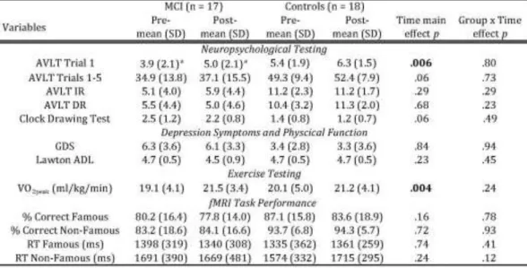
and ADL scores (see Table 1). As expected, the Controls performed significantly better than the MCI participants on all of the
neuropsychological measures, except DRS-Construction (see Table 1). The MCI participants had slightly greater symptoms of depression,
Journal of Alzheimer’s Disease, Vol 37, No. 1 (2013): pg. 197-215. DOI. This article is © IOS Press and permission has been granted for this version to appear in e-Publications@Marquette. IOS Press does not grant permission for this article to be further copied/distributed or hosted elsewhere without the express permission from IOS Press.
11
however the mean scores for both groups were in the normal range.35
The groups were intact and did not differ on activities of daily living.
Exercise Intervention Fidelity
The mean (±SD) number of exercise sessions completed, which did not differ between groups, was 42.3 (2.2) out of 44 total sessions, and the mean (±SD) adherence rate was 96.1 (5.0%) of the total exercise sessions. The mean (±SD) intensity of the exercise, which also did not differ between groups, during the first four weeks and weeks 5-12 was 46.9 (7.1%) HRR and 54.7 (11.0%) HRR. The mean (±SD) rating of perceived exertion (RPE) was 10.6 (1.8) and 10.8 (2.0), respectively,
which is most closely associated with the verbal descriptor of “Light”
exertion. The exercise intervention resulted in a significant mean
increase in VO2peak by 2.0 ml/kg/min, an approximately 10.6%
increase in cardiorespiratory fitness. Although the MCI group appeared
to show a greater increase in VO2peak, the change in cardiorespiratory
fitness over time did not significantly differ between the groups (see Table 2), and the groups did not differ in fitness at baseline.
Semantic Memory fMRI Task Behavioral Performance
As shown in Table 2, performance of the famous name
recognition task was similar in both the MCI and Control groups; the mean (±SD) percent correct for famous and non-famous names was 80.2 (16.4) and 83.2 (18.6), respectively. The groups did not differ in percent correct famous name recognition or in reaction time for either name category at both pre- and post-exercise intervention, and there were no significant changes over time in task performance in either group. The Control group performed better than the MCI group on percent correct rejection of non-famous names (94.0% vs. 83.6%; p < .05). Only correct trials were included in the analysis of the fMRI data.
Journal of Alzheimer’s Disease, Vol 37, No. 1 (2013): pg. 197-215. DOI. This article is © IOS Press and permission has been granted for this version to appear in e-Publications@Marquette. IOS Press does not grant permission for this article to be further copied/distributed or hosted elsewhere without the express permission from IOS Press.
12
Semantic Memory fMRI Activation
–
Spatial Extent
Analysis
Maps showing regions that were activated in the comparison of famous and non-famous name conditions in the MCI and control
groups at the pre- and post- exercise intervention scans are presented in Figure 2 (see Table 3 for activation loci and volumes). The Famous > Non-famous subtraction (shown in red Figure 2) resulted in a
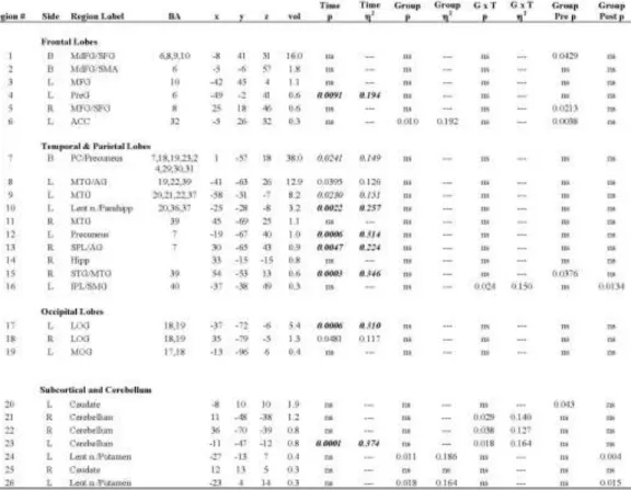
greater volume of semantic processing-related activation in the Control group (77.0 ml) compared to the MCI group (19.1 ml) pre-exercise. The volume of activated tissue decreased post-exercise in both groups (to 23.0 ml and 11.3 ml, respectively). Twenty-six regions (i.e., fROIs) showed significant activation in at least one of the groups at one of the time points (shown in Figure 3).
Semantic Memory fMRI Activation
–
fROI Analysis
The analysis of mean activation intensity (%AUC) in the 26 fROIs revealed significant main effects of Time in 11 regions, significant main effects of Group in three regions, and significant Group x Time interactions in three regions (see Table 4). Seven regions survived the FDR threshold among the main effects of Time; however, none of the main effects for Group or the Group x Time interactions survived the FDR threshold. Figure 4 shows the seven fROIs that survived the FDR threshold for the main effect of Time, plus two additional regions with p-values that were just above the FDR threshold. In all regions and in both groups, semantic memory-related activation significantly decreased from pre- to post-exercise
intervention.
Neuropsychological Test Performance
As shown in Table 2, Trial 1 learning on the AVLT significantly improved from pre- to post-exercise intervention in both the MCI and Control groups. There was also nearly significant improvement on Trials 1-5 learning on the AVLT (p = .06) and the Clock test (p = .06). With the exception of ADLs, which were intact and did not differ
Journal of Alzheimer’s Disease, Vol 37, No. 1 (2013): pg. 197-215. DOI. This article is © IOS Press and permission has been granted for this version to appear in e-Publications@Marquette. IOS Press does not grant permission for this article to be further copied/distributed or hosted elsewhere without the express permission from IOS Press.
13
on the neuropsychological diagnostic and outcome measures before and after the intervention, and there were no significant Group x Time interactions for the neuropsychological outcomes.
Discussion
A 12-week walking exercise intervention led to a 10% increase
in maximal aerobic capacity and an associated decrease in semantic
memory retrieval-related fMRI activation in MCI participants, and cognitively intact older adults. Single trial list-learning significantly improved in both groups, and learning through repetition (AVLT Trial 1-5 sum) improved by approximately two words in the MCI
participants and by approximately three words in cognitively intact elders. While these cognitive improvements did not differ statistically between the groups, the quality of the cognitive improvement in MCI
participants was remarkable given their history of cognitive decline
and likelihood for future cognitive decline. Despite the use of alternate forms, some improvement due to familiarity with the test format might be expected in cognitively intact participants. Yet, the familiarity effect in persons with impaired cognition is typically observed as a stable performance over time. Although the controls continued to outperform MCIs, the cognitive improvement in this MCI sample exceeds what
could be expected with repeated test administration.53 While we did
not have a non-exercise control condition, this observation
nonetheless represents a sizeable treatment effect in the context of baseline performance. Indeed, the achievement of cognitive stability in
MCI is considered a marker of treatment success in pharmacologic,54
cognitive training,55 and exercise interventions.31 The cognitive
improvement in the MCI participants in the current study is supported by the changes in task-activated fMRI that were concurrently
observed. This supports the hypothesis that exercise may impact on the neural networks related to memory retrieval.
Our hypothesis of increased fMRI activation after the exercise intervention was not confirmed. We based our prediction on the few previous studies among healthy older adults that reported increased task-activated fMRI activation after exercise training or in more physically active participants. In cross-sectional comparisons of
Journal of Alzheimer’s Disease, Vol 37, No. 1 (2013): pg. 197-215. DOI. This article is © IOS Press and permission has been granted for this version to appear in e-Publications@Marquette. IOS Press does not grant permission for this article to be further copied/distributed or hosted elsewhere without the express permission from IOS Press.
14
showed greater fMRI activation26 and were less likely to exhibit
cognitive decline than those who were less active or noncarriers.56 In
the only other exercise intervention to examine effects on task-activated fMRI, cognitively intact older adults exhibited improved inhibitory control (flanker) task performance and, as a consequence of the intervention, increased fMRI activation in middle and superior frontal gyri and the superior parietal lobule, and decreased activation
in the anterior cingulate cortex.25 Thus, because the intervention led to
both a change in behavioral task performance and currently recorded neural activation, the effects of the intervention on brain networks are difficult to isolate. As such, these results are difficult to compare with the current study where task performance was constant. In our study, behavioral performance on the alternate forms of a famous name task was always high in both groups, did not change over time, and we only included correct trials in the fMRI analysis. Thus, we examined the effects of exercise on successful semantic memory retrieval at both the pre- and post-intervention measurements, without the confounding effects of improved task performance. Given the consistent memory retrieval performance, our results suggest that semantic memory retrieval-related neural activation became more efficient in both MCI and cognitively intact participants from before to after the exercise intervention.
These effects highlight one of the primary theoretical debates in the fields of cognitive aging and dementia. While greater neural
activation during memory retrieval may reflect successful
compensation or neural reserve, as suggested for example by the
STAC theory,19 compensation resulting in greater neural activation is
also paradoxically associated with increased brain amyloid
accumulation and greater probability of MCI diagnosis. This suggests the compensatory response, while promoting a preservation of
function, may ultimately lead to greater AD pathology. Our previous cross-sectional26,32 and longitudinal work56 is consistent with the idea that greater neural activation, as measured by greater extent and intensity of fMRI activation during fame recognition, is associated with cognitive stability over time. However, our current prospective
intervention data suggest that exercise training may enhance cognitive and neural reserve in MCI, not through a greater capacity to activate
Journal of Alzheimer’s Disease, Vol 37, No. 1 (2013): pg. 197-215. DOI. This article is © IOS Press and permission has been granted for this version to appear in e-Publications@Marquette. IOS Press does not grant permission for this article to be further copied/distributed or hosted elsewhere without the express permission from IOS Press.
15 neural workload during successful engagement of semantic memory networks. Whether or not exercise training alters the underlying clinical or neuropathological trajectory of AD remains to be determined.
We selected a semantic memory fMRI task as the primary outcome. While previous studies have shown that fMRI activation
during episodic memory tasks may predict future cognitive decline,11,57
others have reported that semantic memory fMRI activation may be better predictors of longitudinal cognitive change than episodic
memory fMRI tasks.58 The famous name recognition task we used has
several advantages over episodic memory encoding or recognition memory tasks. First, the famous name task requires little effort and both memory impaired and cognitively intact persons can perform the task with a high degree of accuracy. Moreover, the event-related fMRI design permits the exclusion of incorrect trials and thus limits the analysis to only successful memory performance. Blocked fMRI
designs, for example the comparison of a memory encoding block to a
rest block,11 do not allow one to distinguish correct from incorrect task
performance and thus groups differences may reflect activation due to greater effort or task difficulty in the cognitively impaired group. The high performance in both groups within the current study, paired with the inclusion of only correct trials, removes any confounding influence of task difficulty or performance differences between groups.
Moreover, famous name task performance (by design) did not change over time in either the MCI or cognitively intact elders. Thus, the effects we observed cannot be attributed to improved task
performance. Rather, our results suggest exercise training resulted in a reduced neural response to perform consistently successful semantic memory retrieval, an effect observed in both controls and MCI
participants.
One interesting exception to this effect occurred in the
precuneus and posterior cingulate cortex (PCC). These regions, which show a reduction in BOLD signal intensity during non-semantic tasks
compared to the resting state,59 are activated by a number of semantic
tasks.60 The precuneus and PCC activation in the current study, along
with the other regions activated by the famous name task, overlaps with the semantic memory system and the ‘default mode network’,
Journal of Alzheimer’s Disease, Vol 37, No. 1 (2013): pg. 197-215. DOI. This article is © IOS Press and permission has been granted for this version to appear in e-Publications@Marquette. IOS Press does not grant permission for this article to be further copied/distributed or hosted elsewhere without the express permission from IOS Press.
16
consistent with data indicating the resting state reflects ongoing
semantic processing.60 Importantly, these are regions that show
hyperactivation and resistance to deactivation during non-semantic tasks in MCI, and also exhibit early signs of amyloid retention in both
MCI and healthy APOE-e4 carriers.10 As MCI progresses to AD,
precuneus/PCC hypometabolism and reduced functional connectivity
have been observed.17,18 One hypothesis is that precuneus/PCC
hyperactivation in MCI is a compensatory response to hippocampal
neurodegeneration.10,17,19 In our sample, while the Group x Time
interaction in the bilateral precuneus/PCC was not significant (region 7 in Figure 4, p = .08), an exploratory post-hoc analysis showed that the decrease over time was significant in the control group (p = .002), but not in the MCI group (p > .7). It is possible that the effects of exercise training on precuneus/PCC activation were blunted in the MCI
participants in order to preserve compensatory activation in the face of early hippocampal neurodegenerative processes. This is consistent with our previous study that found no difference in precuneus/PCC activation during fame discrimination between physically active and physically inactive MCI participants.32 In the cognitively intact controls, there may have been room to improve on the efficiency in the
precuneus/PCC region because it was not necessary for this region to simultaneously compensate for medial temporal lobe dysfunction. However, this interpretation is speculative and should be viewed with caution, as we did not measure AD-related neuropathology in our participants.
Previous exercise clinical trials in MCI participants have been
limited to outcomes from neuropsychological testing.1 Two studies that
reported a failure of improvement in neurocognitive performance in MCI after an exercise intervention suffer from somewhat questionable
efficacy of the intervention.61,62 In contrast, another study reported
improved verbal fluency, Stroop Color-Word Interference and Symbol-digit Modalities Test performance after a 6-month exercise
intervention, which evidenced a comparable increase in fitness as achieved in the current study (11.5% vs. 10.6% in the current
study).29 In contrast to the current study, their effects were observed
only in women, whereas we did not find a difference between men and women on any outcome. Finally, a study of elders with MCI and elders with subjective memory complaints not meeting criteria for MCI
Journal of Alzheimer’s Disease, Vol 37, No. 1 (2013): pg. 197-215. DOI. This article is © IOS Press and permission has been granted for this version to appear in e-Publications@Marquette. IOS Press does not grant permission for this article to be further copied/distributed or hosted elsewhere without the express permission from IOS Press.
17
demonstrated stable cognition in those who participated in a 6-month home-based exercise program, compared with those who did not exercise, who declined significantly over the same period (change of
−0.26 points, 95% CI −0.89–0.54).31 These results also add support
for our interpretation of the current study that improved cognition in MCI over a 3-month exercise intervention is remarkable.
Memory training interventions have demonstrated effectiveness to improve training-related and non-training-related cognitive task
performance in healthy younger and cognitively intact older adults.63
The changes in task-related fMRI activation after memory training interventions, however, have been inconsistent, with some studies
reporting decreased activation and some increased activation.63 In one
study, fMRI activation increased in both older adults diagnosed with MCI (mean age 70 years) and healthy controls after a 6-week, one
session per week, memory training intervention.64 That study
employed an episodic memory task in a blocked fMRI design, where task performance differed between groups and both groups exhibited task improvement after training. As with some exercise interventions, this leaves unclear the foundations for activation changes. In the
current study, our MCI and control participants did not differ or change over time on the activation task and only correct trials were included in the analysis, thereby precluding the likelihood that the effects could be due to inherent group differences or task difficulty. Moreover, despite the primary effect of reduced magnitude of semantic memory activation, MCI participants also showed new areas of activation after the intervention (see Table 3, Figure 2), particularly in left superior
frontal gyrus, medial frontal gyrus (Brodmann’s areas 6 & 8), and left
lateral occipital and fusiform gyri (Brodmann’s areas 18 & 19). These
results are consistent with the findings of Belleville et al. and partly support theories that hypothesize cognitive improvement or
compensation will result in the recruitment of new neural
circuits.20,21,65 Although the comparative and potential synergistic
effects of exercise with cognitive training interventions are of great
interest,56 direct comparisons of the combined treatments have not yet
been made.
One limitation of this study is that we did not include a ‘no
Journal of Alzheimer’s Disease, Vol 37, No. 1 (2013): pg. 197-215. DOI. This article is © IOS Press and permission has been granted for this version to appear in e-Publications@Marquette. IOS Press does not grant permission for this article to be further copied/distributed or hosted elsewhere without the express permission from IOS Press.
18
account for the effects that may be due to the passage of time or practice. Some have suggested that repeated fMRI scans result in reduced task activation due to familiarity with the task or scanner
environment.66 However, such fMRI practice effects also are
accompanied by moderate to large task related learning effects from the first to the second session. Thus, it is possible the fMRI changes over time reflect changes in task difficulty, which could have resulted in greater error (in blocked designs) or effort-related activation on the first scan. Yet, when a well learned task is repeated after several weeks, the changes in fMRI activation are very minimal and
bidirectional.67 This is a critical distinction when making comparisons
across studies.58 Our famous name recognition task is not subject to
task-related practice effects. The participants enter the scanner with decades of experience recognizing famous names and so perform the task with ease, even with existing episodic memory impairments. Moreover, task performance did not improve over time. We also have shown (unpublished data), using the same famous name task in a
sample of 16 cognitively intact APOE-ε4 carriers, that the patterns and
intensity of fMRI activation did not change over an 18-month interval during which little or no regular physical activity occurred, in the
absence of any intervention.68 In regard to changes over time on the
AVLT, stable cognitive function among healthy older adults is often denoted by a small increase in cognitive test performance on alternate
forms due to familiarity with the test format.69 Among those with MCI,
this familiarity effect often manifests as equivalent performance, not
increased performance, over repeated test administrations.53 Thus,
given the expectation that those with MCI, in the absence of intervention, are likely to exhibit cognitive decline, the measured cognitive improvement in the MCI group suggests a real treatment effect that can be attributed to the exercise intervention. We
acknowledge, however, that additional randomized controlled trials are needed.
We used a sound experimental manipulation of cardiorespiratory fitness in response to a well-supervised and well-attended exercise intervention to determine if neural function during memory retrieval could be affected in participants with MCI. Our results provide the first evidence that exercise may induce neural plasticity in individuals diagnosed with MCI. However, the rigor necessary to achieve a similar
Journal of Alzheimer’s Disease, Vol 37, No. 1 (2013): pg. 197-215. DOI. This article is © IOS Press and permission has been granted for this version to appear in e-Publications@Marquette. IOS Press does not grant permission for this article to be further copied/distributed or hosted elsewhere without the express permission from IOS Press.
19
degree of integrity in an exercise intervention may not be feasible in larger clinical trials, and thus the effects we report may be attenuated in community-based or unsupervised exercise programs. Another limitation is that our sample was primarily Caucasian and well educated, and so it will be important to determine the effects of exercise in multiple ethnic groups. Lastly, our MCI group was by definition not demented and had intact ADLs. These effects may not generalize to individuals diagnosed with AD, or to those with greater clinical symptoms or functional impairments.
Regarding potential mechanisms, one can only speculate due to the pleiotropic effects of exercise. In rodents, exercise has been well documented to stimulate the transcription, translation, and release of neurotrophic factors (e.g., brain derived neurotrophic factor, insulin-like growth factor-1) and to promote neurogenesis, particularly in the
dentate gyrus of the hippocampal formation.70-72 There is also
preliminary evidence that exercise may produce these neurotrophic
effects in the hippocampal formation in healthy adults73 and healthy
older adults.27 It is also possible that the cholinergic effects of exercise may increase cerebral perfusion, possibly affecting the neurodynamics
of the blood-oxygen level dependent fMRI signal74 that may reflect
improved network efficiency.23 There is preliminary evidence that
exercise may attenuate the accumulation of beta-amyloid in rodents75
and cognitively intact older adult APOE-ε4 carriers.76 However, it is unknown if these potential mechanisms are engaged by exercise in
individuals diagnosed with MCI or others at increased AD risk.1
The current findings are consistent with the effects of greater
left caudate activation in more physically active MCI participants.32
Dopaminergic projections within frontal-striatal networks, which are
agonized by exercise77 and by working memory training
interventions,78 may dampen neural interference and improve the gain
on task relevant neural signals during semantic memory retrieval.23,79
Moreover, noradrenergic activation in response to single sessions of
exercise has been shown to benefit memory consolidation in MCI,80
suggesting that the long-term cognitive benefits may accumulate with repeated bouts of exercise.
Journal of Alzheimer’s Disease, Vol 37, No. 1 (2013): pg. 197-215. DOI. This article is © IOS Press and permission has been granted for this version to appear in e-Publications@Marquette. IOS Press does not grant permission for this article to be further copied/distributed or hosted elsewhere without the express permission from IOS Press.
20
In conclusion, a 12-week walking exercise intervention in MCI participants resulted in improved single trial list-learning performance and a reduction in fMRI activation during a semantic memory retrieval task. Decreased fMRI semantic memory-related activation after the exercise intervention, coupled with improved episodic memory
performance, suggests exercise training may improve neural efficiency during successful memory retrieval in MCI. This further suggests the large-scale manipulations of cardiovascular and brain neurotrophic systems with exercise promote neural plasticity and improved brain function in MCI. Whether or not these effects can delay or prevent AD conversion, and how exercise compares to pharmacologic, cognitive training, or other interventions, remains to be determined.
Acknowledgements
We thank all of the participants for their time and effort, and gratefully acknowledge the cooperation and collaboration of the community and commercial fitness centers where the participants exercised. We also appreciate the dedication of our excellent research staff, including study coordinator Karen Outzen; exercise physiologists and personal fitness trainers Andrew Mattes, Karlee Sweere, Lynn Wheeler, Rebecca Ohmen, Bethany Brooks, Ed Possing, Bradley Kohl, David Cornell, Jacqueline Geib, David Johnson, Rong Tang, and William Massey; and the nurses and MRI technicians at the CTSI Translational Research Unit at the Medical College of Wisconsin. Additionally, we acknowledge Kevin Regner, M.D., Jeanne Palmer, M.D., Elizabeth Cogbill, M.D., Tinoy Kizhakekuttu, M.D., and Kodlipet Dharmashankar, M.D., for their collegial provision of medical coverage during exercise and MRI test sessions; and Erin Browning for her technical expertise and help in MRI data collection. Technical support for APOE genotyping was provided by Stacy Meyer and Erin Koester. This work was
supported by a grant from the Graduate School Research Growth Initiative at the University of Wisconsin-Milwaukee; and by the Clinical and Translational Science Award (CTSA) program of the National Center for Research Resources, National Institutes of Health [grant number UL1RR031973].
Journal of Alzheimer’s Disease, Vol 37, No. 1 (2013): pg. 197-215. DOI. This article is © IOS Press and permission has been granted for this version to appear in e-Publications@Marquette. IOS Press does not grant permission for this article to be further copied/distributed or hosted elsewhere without the express permission from IOS Press.
21
References
1Smith JC, Nielson KA, Woodard JL, Seidenberg M, Rao SM (2013) Physical
activity and brain function in older adults at increased risk for
Alzheimer's disease. Brain Sciences 3, 54-83.
2Petersen RC (2000) Mild cognitive impairment: transition between aging and
Alzheimer's disease. Neurologia 15, 93-101.
3DeKosky ST, Marek K (2003) Looking backward to move forward: early
detection of neurodegenerative disorders. Science 302, 830-834.
4Albert MS, DeKosky ST, Dickson D, Dubois B, Feldman HH, Fox NC, Gamst A,
Holtzman DM, Jagust WJ, Petersen RC, Snyder PJ, Carrillo MC, Thies B, Phelps CH (2011) The diagnosis of mild cognitive impairment due to Alzheimer's disease: recommendations from the National Institute on Aging-Alzheimer's Association workgroups on diagnostic guidelines for
Alzheimer's disease. Alzheimers Dement 7, 270-279.
5Sperling RA, Aisen PS, Beckett LA, Bennett DA, Craft S, Fagan AM, Iwatsubo
T, Jack CR, Jr., Kaye J, Montine TJ, Park DC, Reiman EM, Rowe CC, Siemers E, Stern Y, Yaffe K, Carrillo MC, Thies B, Morrison-Bogorad M, Wagster MV, Phelps CH (2011) Toward defining the preclinical stages of Alzheimer's disease: recommendations from the National Institute on Aging-Alzheimer's Association workgroups on diagnostic guidelines
for Alzheimer's disease. Alzheimers Dement 7, 280-292.
6Jonker C, Geerlings MI, Schmand B (2000) Are memory complaints
predictive for dementia? A review of clinical and population-based
studies. Int J Geriatr Psychiatry 15, 983-991.
7Lonie JA, Herrmann LL, Tierney KM, Donaghey C, O'Carroll R, Lee A, Ebmeier
KP (2009) Lexical and semantic fluency discrepancy scores in aMCI
and early Alzheimer's disease. J Neuropsychol 3, 79-92.
8Henry JD, Crawford JR, Phillips LH (2004) Verbal fluency performance in
dementia of the Alzheimer's type: a meta-analysis. Neuropsychologia
42, 1212-1222.
9Xu G, Antuono PG, Jones J, Xu Y, Wu G, Ward D, Li SJ (2007) Perfusion fMRI
detects deficits in regional CBF during memory-encoding tasks in MCI
subjects. Neurology 69, 1650-1656.
10Sperling RA, Dickerson BC, Pihlajamaki M, Vannini P, Laviolette PS, Vitolo
OV, Hedden T, Becker JA, Rentz DM, Selkoe DJ, Johnson KA (2010) Functional Alterations in Memory Networks in Early Alzheimer's
Disease. Neuromolecular Med 12, 27-43.
11Bookheimer SY, Strojwas MH, Cohen MS, Saunders AM, Pericak-Vance MA,
Mazziotta JC, Small GW (2000) Patterns of brain activation in people at
risk for Alzheimer's disease. N.Engl.J.Med. 343, 450-456.
12Woodard JL, Seidenberg M, Nielson KA, Antuono P, Guidotti L, Durgerian S,
Journal of Alzheimer’s Disease, Vol 37, No. 1 (2013): pg. 197-215. DOI. This article is © IOS Press and permission has been granted for this version to appear in e-Publications@Marquette. IOS Press does not grant permission for this article to be further copied/distributed or hosted elsewhere without the express permission from IOS Press.
22
memory activation in amnestic mild cognitive impairment. Brain 132,
2068-2078.
13Mufson EJ, Chen EY, Cochran EJ, Beckett LA, Bennett DA, Kordower JH
(1999) Entorhinal cortex beta-amyloid load in individuals with mild
cognitive impairment. Exp Neurol 158, 469-490.
14Quigley H, Colloby SJ, O'Brien JT (2011) PET imaging of brain amyloid in
dementia: a review. Int J Geriatr Psychiatry 26, 991-999.
15Dickerson BC, Bakkour A, Salat DH, Feczko E, Pacheco J, Greve DN,
Grodstein F, Wright CI, Blacker D, Rosas HD, Sperling RA, Atri A, Growdon JH, Hyman BT, Morris JC, Fischl B, Buckner RL (2009) The cortical signature of Alzheimer's disease: regionally specific cortical thinning relates to symptom severity in very mild to mild AD dementia
and is detectable in asymptomatic amyloid-positive individuals. Cereb
Cortex 19, 497-510.
16De Leon MJ, George AE, Golomb J, Tarshish C, Convit A, Kluger A, De Santi
S, McRae T, Ferris SH, Reisberg B, Ince C, Rusinek H, Bobinski M, Quinn B, Miller DC, Wisniewski HM (1997) Frequency of hippocampal
formation atrophy in normal aging and Alzheimer's disease. Neurobiol
Aging 18, 1-11.
17Buckner RL, Snyder AZ, Shannon BJ, LaRossa G, Sachs R, Fotenos AF,
Sheline YI, Klunk WE, Mathis CA, Morris JC, Mintun MA (2005) Molecular, structural, and functional characterization of Alzheimer's disease: evidence for a relationship between default activity, amyloid,
and memory. J Neurosci 25, 7709-7717.
18Sperling RA, Laviolette PS, O'Keefe K, O'Brien J, Rentz DM, Pihlajamaki M,
Marshall G, Hyman BT, Selkoe DJ, Hedden T, Buckner RL, Becker JA, Johnson KA (2009) Amyloid deposition is associated with impaired
default network function in older persons without dementia. Neuron
63, 178-188.
19Park DC, Reuter-Lorenz P (2009) The adaptive brain: aging and
neurocognitive scaffolding. Annu Rev Psychol 60, 173-196.
20Cabeza R (2002) Hemispheric asymmetry reduction in older adults: the
HAROLD model. Psychol Aging 17, 85-100.
21Stern Y (2002) What is cognitive reserve? Theory and research application
of the reserve concept. J Int Neuropsychol Soc 8, 448-460.
22Driscoll I, Resnick SM, Troncoso JC, An Y, O'Brien R, Zonderman AB (2006)
Impact of Alzheimer's pathology on cognitive trajectories in
nondemented elderly. Ann Neurol 60, 688-695.
23Li SC, Lindenberger U, Sikstrom S (2001) Aging cognition: from
neuromodulation to representation. Trends Cogn Sci 5, 479-486.
24Daviglus ML, Bell CC, Berrettini W, Bowen PE, Connolly ES, Cox NJ,
Dunbar-Jacob JM, Granieri EC, Hunt G, McGarry K, Patel D, Potosky AL, Sanders-Bush E, Silberberg D, Trevisan M (2010) NIH
State-of-the-Journal of Alzheimer’s Disease, Vol 37, No. 1 (2013): pg. 197-215. DOI. This article is © IOS Press and permission has been granted for this version to appear in e-Publications@Marquette. IOS Press does not grant permission for this article to be further copied/distributed or hosted elsewhere without the express permission from IOS Press.
23 Science Conference Statement: Preventing Alzheimer's Disease and
Cognitive Decline. NIH Consens State Sci Statements 27, 1-30.
25Colcombe SJ, Kramer AF, Erickson KI, Scalf P, McAuley E, Cohen NJ, Webb
A, Jerome GJ, Marquez DX, Elavsky S (2004) Cardiovascular fitness,
cortical plasticity, and aging. Proc Natl Acad Sci U S A 101,
3316-3321.
26Smith JC, Nielson KA, Woodard JL, Seidenberg M, Durgerian S, Antuono P,
Butts AM, Hantke NC, Lancaster MA, Rao SM (2011) Interactive effects of physical activity and APOEepsilon4 on BOLD semantic memory
activation in healthy elders. Neuroimage 54, 635-644.
27Erickson KI, Voss MW, Prakash RS, Basak C, Szabo A, Chaddock L, Kim JS,
Heo S, Alves H, White SM, Wojcicki TR, Mailey E, Vieira VJ, Martin SA, Pence BD, Woods JA, McAuley E, Kramer AF (2011) Exercise training
increases size of hippocampus and improves memory. Proc Natl Acad
Sci U S A 108, 3017-3022.
28Etnier JL (2009) Chronic exercise and cognition in older adults In Exercise
and Cognitive Function, McMorris T, Tomporowski PD, Audiffren M, eds. Wiley-Blackwell, Chichester, pp. 227-248.
29Baker LD, Frank LL, Foster-Schubert K, Green PS, Wilkinson CW, McTiernan
A, Plymate SR, Fishel MA, Watson GS, Cholerton BA, Duncan GE, Mehta PD, Craft S (2010) Effects of aerobic exercise on mild cognitive
impairment: a controlled trial. Archives of Neurology 67, 71-79.
30Suzuki T, Shimada H, Makizako H, Doi T, Yoshida D, Tsutsumimoto K, Anan
Y, Uemura K, Lee S, Park H (2012) Effects of multicomponent exercise on cognitive function in older adults with amnestic mild cognitive
impairment: a randomized controlled trial. BMC Neurol 12, 128.
31Lautenschlager NT, Cox KL, Flicker L, Foster JK, van Bockxmeer FM, Xiao J,
Greenop KR, Almeida OP (2008) Effect of physical activity on cognitive function in older adults at risk for Alzheimer disease: a randomized
trial. Journal of the American Medical Association 300, 1027-1037.
32Smith JC, Nielson KA, Woodard JL, Seidenberg M, Verber MD, Durgerian S,
Antuono P, Butts AM, Hantke NC, Lancaster MA, Rao SM (2011) Does physical activity influence semantic memory activation in amnestic
mild cognitive impairment? Psychiatry Res 193, 60-62.
33Burns JM, Cronk BB, Anderson HS, Donnelly JE, Thomas GP, Harsha A,
Brooks WM, Swerdlow RH (2008) Cardiorespiratory fitness and brain
atrophy in early Alzheimer disease. Neurology 71, 210-216.
34Oldfield RC (1971) The assessment and analysis of handedness: the
Edinburgh inventory. Neuropsychologia 9, 97-113.
35Yesavage JA (1988) Geriatric Depression Scale. Psychopharmacol Bull 24,
709-711.
36Lawton MP, Brody EM (1969) Assessment of older people: self-maintaining
Journal of Alzheimer’s Disease, Vol 37, No. 1 (2013): pg. 197-215. DOI. This article is © IOS Press and permission has been granted for this version to appear in e-Publications@Marquette. IOS Press does not grant permission for this article to be further copied/distributed or hosted elsewhere without the express permission from IOS Press.
24
37Folstein MF, Folstein SE, McHugh PR (1975) "Mini-mental state". A practical
method for grading the cognitive state of patients for the clinician. J
Psychiatr Res 12, 189-198.
38Jurica PJ, Leitten CL, Mattis S (2001) Dementia Rating Scale-2 professional
manual, Psychological Assessment Resources, Lutz, FL.
39Rey A (1964) L'examen clinique en psychologie, Presses Universitaires de
France Paris.
40Wechsler D (1997) WAIS-III/WMS-III technical manual, Psychological
Corporation, San Antonio.
41Smith AR (1991) Symbol Digit Modalities Test, Western Psychological
Services, Los Angeles.
42Benton AL, Hamsher K (1978), Iowa City, IA.
43Cosentino S, Jefferson A, Chute DL, Kaplan E, Libon DJ (2004) Clock
drawing errors in dementia: neuropsychological and neuroanatomical
considerations. Cogn Behav Neurol 17, 74-84.
44ACSM (2006) American College of Sports Medicine Guidelines for Exercise
Testing and Prescription, Lippincott, Williams & Wilkins.
45Borg G (1998) Borg's Perceived Exertion and Pain Scales, Human Kinetics,
Champaign, IL.
46Cook DB, O'Connor PJ, Eubanks SA, Smith JC, Lee M (1997) Naturally
occurring muscle pain during exercise: assessment and experimental
evidence. Med Sci Sports Exerc 29, 999-1012.
47Douville K, Woodard JL, Seidenberg M, Miller SK, Leveroni CL, Nielson KA,
Franczak M, Antuono P, Rao SM (2005) Medial temporal lobe activity for recognition of recent and remote famous names: an event-related
fMRI study. Neuropsychologia 43, 693-703.
48Cox RW (1996) AFNI: software for analysis and visualization of functional
magnetic resonance neuroimages. Computers and Biomedical
Research 29, 162-173.
49Talairach J, Tournoux P (1988) A co-planar stereotaxic atlas of the human
brain, Thieme, Stuttgart.
50Ward BD (2000) in In, Analysis of Functional NeuroImages (AFNI) software
NIH, Bethesda, MD.
51Mayeux R, Saunders AM, Shea S, Mirra S, Evans D, Roses AD, Hyman BT,
Crain B, Tang MX, Phelps CH (1998) Utility of the apolipoprotein E genotype in the diagnosis of Alzheimer's disease. Alzheimer's Disease
Centers Consortium on Apolipoprotein E and Alzheimer's Disease. N
Engl J Med 338, 506-511.
52Saunders AM, Hulette O, Welsh-Bohmer KA, Schmechel DE, Crain B, Burke
JR, Alberts MJ, Strittmatter WJ, Breitner JC, Rosenberg C (1996) Specificity, sensitivity, and predictive value of apolipoprotein-E
Journal of Alzheimer’s Disease, Vol 37, No. 1 (2013): pg. 197-215. DOI. This article is © IOS Press and permission has been granted for this version to appear in e-Publications@Marquette. IOS Press does not grant permission for this article to be further copied/distributed or hosted elsewhere without the express permission from IOS Press.
25
53Devanand DP, Liu X, Brown PJ, Huey ED, Stern Y, Pelton GH (2012) A
two-study comparison of clinical and MRI markers of transition from mild
cognitive impairment to Alzheimer's disease. Int J Alzheimers Dis
2012, 483469.
54Farlow MR (2009) Treatment of mild cognitive impairment (MCI). Curr
Alzheimer Res 6, 362-367.
55Stott J, Spector A (2011) A review of the effectiveness of memory
interventions in mild cognitive impairment (MCI). Int Psychogeriatr
23, 526-538.
56Woodard JL, Sugarman MA, Nielson KA, Smith JC, Seidenberg M, Durgerian
S, Butts A, Hantke N, Lancaster M, Matthews MA, Rao SM (2012) Lifestyle and genetic contributions to cognitive decline and
hippocampal structure and function in healthy aging. Curr Alzheimer
Res 9, 436-446.
57O'Brien JL, O'Keefe KM, LaViolette PS, DeLuca AN, Blacker D, Dickerson BC,
Sperling RA (2010) Longitudinal fMRI in elderly reveals loss of
hippocampal activation with clinical decline. Neurology 74, 1969-1976.
58Hantke N, Nielson KA, Woodard JL, Breting LM, Butts A, Seidenberg M,
Carson Smith J, Durgerian S, Lancaster M, Matthews M, Sugarman MA, Rao SM (2013) Comparison of semantic and episodic memory BOLD
fMRI activation in predicting cognitive decline in older adults. J Int
Neuropsychol Soc 19, 11-21.
59Binder JR, Frost JA, Hammeke TA, Bellgowan PS, Rao SM, Cox RW (1999)
Conceptual processing during the conscious resting state. A functional
MRI study. J Cogn Neurosci 11, 80-95.
60Binder JR, Desai RH, Graves WW, Conant LL (2009) Where is the semantic
system? A critical review and meta-analysis of 120 functional
neuroimaging studies. Cereb Cortex 19, 2767-2796.
61Scherder EJ, Van Paasschen J, Deijen JB, Van Der Knokke S, Orlebeke JF,
Burgers I, Devriese PP, Swaab DF, Sergeant JA (2005) Physical activity and executive functions in the elderly with mild cognitive impairment. Aging Ment Health 9, 272-280.
62Miller LA, Spitznagel MB, Busko S, Potter V, Juvancic-Heltzel J, Istenes N,
Glickman E, Gunstad J (2011) Structured exercise does not stabilize cognitive function in individuals with mild cognitive impairment
residing in a structured living facility. Int J Neurosci 121, 218-223.
63Buschkuehl M, Jaeggi SM, Jonides J (2012) Neuronal effects following
working memory training. Dev Cogn Neurosci 2 Suppl 1, S167-179.
64Belleville S, Clement F, Mellah S, Gilbert B, Fontaine F, Gauthier S (2011)
Training-related brain plasticity in subjects at risk of developing
Alzheimer's disease. Brain 134, 1623-1634.
65Reuter-Lorenz PA, Lustig C (2005) Brain aging: reorganizing discoveries
Journal of Alzheimer’s Disease, Vol 37, No. 1 (2013): pg. 197-215. DOI. This article is © IOS Press and permission has been granted for this version to appear in e-Publications@Marquette. IOS Press does not grant permission for this article to be further copied/distributed or hosted elsewhere without the express permission from IOS Press.
26
66Kelly AM, Garavan H (2005) Human functional neuroimaging of brain
changes associated with practice. Cereb Cortex 15, 1089-1102.
67Loubinoux I, Carel C, Alary F, Boulanouar K, Viallard G, Manelfe C, Rascol O,
Celsis P, Chollet F (2001) Within-session and between-session
reproducibility of cerebral sensorimotor activation: a test--retest effect
evidenced with functional magnetic resonance imaging. J Cereb Blood
Flow Metab 21, 592-607.
68Smith JC, Durgerian S, Woodard JL, Nielson KA, Butts AM, Hantke NC,
Seidenberg M, Lancaster MA, Matthews MA, Sugarman MA, Rao SM (2012) Physical activity and brain function in older adults at genetic
risk for Alzheimer’s disease. Med Sci Sports Exerc 44, S104.
69Shapiro DM, Harrison DW (1990) Alternate forms of the AVLT: a procedure
and test of form equivalency. Arch Clin Neuropsychol 5, 405-410.
70van Praag H, Shubert T, Zhao C, Gage FH (2005) Exercise enhances
learning and hippocampal neurogenesis in aged mice. J.Neurosci. 25,
8680-8685.
71Trejo JL, Carro E, Torres-Aleman I (2001) Circulating insulin-like growth
factor I mediates exercise-induced increases in the number of new
neurons in the adult hippocampus. J Neurosci 21, 1628-1634.
72Intlekofer KA, Cotman CW (in press) Exercise counteracts declining
hippocampal function in aging and Alzheimer's disease. Neurobiol Dis.
73Pereira AC, Huddleston DE, Brickman AM, Sosunov AA, Hen R, McKhann GM,
Sloan R, Gage FH, Brown TR, Small SA (2007) An in vivo correlate of
exercise-induced neurogenesis in the adult dentate gyrus. Proceedings
of the National Academy of Science U.S.A. 104, 5638-5643.
74Smith JC, Paulson ES, Cook DB, Verber MD, Tian Q (2010) Detecting
changes in human cerebral blood flow after acute exercise using
arterial spin labeling: implications for fMRI. J Neurosci Methods 191,
258-262.
75Adlard PA, Perreau VM, Pop V, Cotman CW (2005) Voluntary exercise
decreases amyloid load in a transgenic model of Alzheimer's disease. J
Neurosci 25, 4217-4221.
76Head D, Bugg JM, Goate AM, Fagan AM, Mintun MA, Benzinger T, Holtzman
DM, Morris JC (2012) Exercise Engagement as a Moderator of the
Effects of APOE Genotype on Amyloid Deposition. Arch Neurol 69,
636-643.
77de Castro JM, Duncan G (1985) Operantly conditioned running: effects on
brain catecholamine concentrations and receptor densities in the rat. Pharmacology Biochemistry & Behavior 23, 495-500.
78Backman L, Nyberg L, Soveri A, Johansson J, Andersson M, Dahlin E, Neely
AS, Virta J, Laine M, Rinne JO (2011) Effects of working-memory
Journal of Alzheimer’s Disease, Vol 37, No. 1 (2013): pg. 197-215. DOI. This article is © IOS Press and permission has been granted for this version to appear in e-Publications@Marquette. IOS Press does not grant permission for this article to be further copied/distributed or hosted els