Mental Practice as a Gateway to Modified
Constraint-Induced Movement Therapy:
A Promising Combination to Improve Function
KEY WORDS • function • mental priming
• modified constraint-induced movement therapy (mCIMT) • stroke
S
troke, the leading cause of disability in the United States (American Heart Asso-ciation, 2004), causes motor deficits that compromise quality of life (Bamford, Dennis, Sandercock, Burn, & Warlow, 1990) and lead to considerable care costs (Dobkin, 1995). However, increased use and function of the more-affected arm have been reported after people with chronic stroke (>1 year after stroke) partici-pated in constraint-induced movement therapy (CIMT) (Miltner, Bauder, Sommer, Dettmers, & Taub, 1999; Taub et al., 1993; Van der Lee et al., 1999; Wolf, LeCraw, Barton, & Jann, 1989). The most well-recognized variant of CIMT has emphasized massed practice with the more-affected upper limb in two ways: (a) participants’ less-affected upper limbs are restricted during 90% of waking hours of a 2-week period, and (b) participants engage in 6-hr activity sessions using their more-affected limbs on the 10 weekdays of the same 2-week period. However, CIMT may be difficult to implement clinically because of its demanding contact time and expense and patients’ lack of compliance and interest (Page, Levine, Sisto, Bond, & Johnston, 2002). Consistent with these concerns, a CIMT trial by Van der Lee and colleagues (1999) stated that most participants found CIMT participation strenuous. Simple shortening of CIMT contact time has proven to be a straightforward, efficacious solution to CIMT limitations (Pierce et al., 2003; Sterr et al., 2002). The most well-studied example has been modified constraint-induced movement therapy (mCIMT), which combines structured, 30-min, functional practice ses-sions using the more-affected arm, with restriction of the less-affected upper arm 5 days per week for 5 hr per day, both during a 10-week period. Besides its abilityStephen J. Page, PhD, FAHA,is Director of Research and Associate Professor, University of Cincinnati Academic Medical Center, and Director, Neuromotor Recovery and Rehabilitation Laboratory, Drake Rehabilitation Center, Cincinnati. Mailing address: Department of Rehabilitation Sciences, University of Cincinnati, 3202 Eden Avenue, Suite 275, Cincinnati, OH 45267-0394;
Peter Levine, PTA,is Senior Research Assistant, University of Cincinnati Academic Medical Center, and Co-Director, Neuromotor Recovery and Rehabilitation Laboratory, Drake Rehabilitation Center, Cincinnati.
Valerie Hill, MS, OTR/L,is Research Occupational Therapist, University of Cincinnati Academic Medical Center.
Modified constraint-induced movement therapy (mCIMT) is a reimbursable regimen that improves the use and function of more-affected arms in patients who have had a stroke. To participate in this regimen, however, patients must exhibit active extension of the more-affected wrists and fingers, which renders many people inel-igible. This study determined the efficacy of a mental practice program that preceded mCIMT in improving more-affected arm function in 4 patients with a stroke. Patients received therapy emphasizing activities of daily living (ADLs), followed by sessions of mental practice of the ADL. One week after completing mental practice, patients participated in mCIMT. After mental practice, patients exhibited marked changes on assessments and increased active wrist and finger extension, which qualified them for mCIMT. After mCIMT, participants exhib-ited additional functional gains, sustained 3 months later. Data suggest that mental practice provides a path-way whereby patients can participate in mCIMT, realize additional gains, and again perform valued ADLs. Page, S. J., Levine, P., & Hill, V. (2007). Mental practice as a gateway to modified constraint-induced movement therapy: A
promising combination to improve function. American Journal of Occupational Therapy, 61,321–327.
to be implemented on an outpatient basis, the mCIMT treatment effect is robust in case studies (Page, Sisto, John-ston, & Levine, 2002; Page, Sisto, & Levine, 2002), in ran-domized controlled pilot studies that enrolled people who had had acute stroke (Page, Levine, & Leonard, 2005) and subacute stroke (Page, Sisto, Johnston, Levine, & Hughes, 2001, 2002), and in a randomized controlled study that enrolled people with chronic effects from stroke (Page, Sisto, Levine, & McGrath, 2004). Data suggest that corti-cal reorganizations, brought about by increased arm use during mCIMT, are responsible for these motor changes (Szaflarski, Page, Kissela, Levine, & Lee, 2005). The clini-cal portion of mCIMT also has been successfully reim-bursed using existing CPT ™ (Current Procedural Termi-nology) codes (e.g., ADL training).
Despite some recent deviations (e.g., Bonifer, Ander-son, & Arciniegas, 2005), CIMT and mCIMT protocols have typically required that participants exhibit active extension in their more-affected wrists and fingers to be effi-cacious. Consequently, patients exhibiting active wrist flex-ion, but no extension in their affected wrists or fingers, are ineligible for mCIMT programs and often are discharged with residual motor deficits. Recently, mental practice has been shown to increase use of the more-affected arm (Page & Levine, 2005) and function (Page, 2000; Page, Levine, Sisto, & Johnston, 2001a, 2001b) in patients who have had a stroke. Importantly, the above mentioned mental practice studies enrolled patients exhibiting some active flexion in their affected wrists, but active extension in the more-affected wrist and fingers was not a requirement. On the basis of the positive outcomes reported in previous mental practice studies, we wondered whether mental practice could be an efficacious “gateway” therapy whereby patients could participate in mental practice, regain some motor function, and then participate in mCIMT.
Previously, mCIMT has been combined with surface electromyography–triggered neuromuscular stimulation (Page & Levine, 2006) to produce gains in patients who would otherwise not be eligible for mCIMT. This study reports on a group of patients with chronic effects from stroke who participated in a mental practice program that increased their more-affected wrist and finger extension. These patients were then eligible for mCIMT and, after mCIMT participation, exhibited additional functional gains.
Method
Participants
Participants in the present study were enrolled in a larger clinical trial examining mental practice. They are reported
in this article because of the motor changes they gained from participating in the present study, which resulted in subsequent mCIMT participation and additional motor changes. To initially participate in the mental practice trial, participants had to meet the following inclusion criteria: (a) stroke experienced >1 year before study enrollment, (b) 10° of active flexion in the more-affected wrist, and (c) age >18 years. We also applied the following exclusion criteria: (a) excessive pain in the more-affected arm or wrist as measured by a score of ≥5 on a visual analog scale; (b) being
admin-istered medication that impairs neuromuscular perfor-mance (e.g., botulinum toxin A); (c) cognitive deficits, as evidenced by a score ≤69 points on the Modified
Mini-Mental Status Examination (Teng & Chui, 1987); (d) excessive spasticity in the more-affected upper limb, as defined as a score of ≥4 on the Modified Ashworth
Spas-ticity Scale (Bohannon & Smith, 1987); (e) ability to actively extend the affected wrist; (f ) current enrollment in any form of physical rehabilitation; and (g) participation in any other experimental studies.
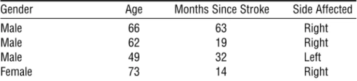
On the basis of these criteria, 4 participants (3 men, 1 woman) were included (mean age of 62.5 years; range 49–73 years; mean time since stroke = 32.0 months, range 14–63 months; and 4 strokes exhibiting hemiparesis on the dominant side; see Table 1).
Instruments
The Fugl-Meyer Assessment (Fugl-Meyer, Jaasko, Ley-man, Olsson, & Steglind, 1975) evaluates several dimen-sions of impairment, including range of motion, pain, sensation, upper extremity, lower extremity, and balance. Data arise from a 3-point ordinal scale (0 = cannot per-form; 2 = can perform fully) applied to each item, and the
items are summed to provide a maximum score of 226. The upper-extremity motor component, which consists of 66 points, was used in this study. The Fugl-Meyer Assess-ment has been shown to have impressive test–retest relia-bility (total = .98–.99; subtests = .87–1.00), interrater reli-ability, and construct validity (DiFabio & Badke, 1990; Duncan, Propst, & Nelson, 1983). The Fugl-Meyer Assessment has been used extensively in studies measuring functional recovery in patients who have had a stroke and, according to Gladstone, Danells, and Black (2002), is highly recommended for “use in clinical trials designed to
Table 1. Participant Characteristics
Gender Age Months Since Stroke Side Affected
Male 66 63 Right
Male 62 19 Right
Male 49 32 Left
evaluate changes in motor impairment following stroke” (p. 239).
The Action Research Arm Test (ARA) (Lyle, 1981) is a 19-item test divided into four categories (grasp, grip, pinch, and gross movement), with each item graded on a 4-point ordinal scale (0 = can perform no part of the test; 1 = performs test partially; 2 = completes test but takes abnormally long time or has great difficulty; 3 = performs test normally) for a total
score of 57. The test is hierarchical in that, if the patient is able to perform the most difficult skill in each category, he or she will be able to perform the other items within the cat-egory and, thus, they need not be tested. The ARA has high intrarater (r = .99) and retest (r = .98) reliability and
valid-ity (Lyle, 1981; Van der Lee et al., 2001). Design, Pretesting, and Intervention
A single-blinded, pre- and posttest, case series design was applied. After participants had signed approved consent forms, a research team member administered the Fugl-Meyer scale and the ARA.
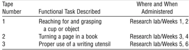
Combining physical and mental practice of ADLs is more effective than either mental practice or physical prac-tice alone (McBride & Rothstein, 1979). Therefore, 1 week after the final pretesting session, participants practiced the same set of ADLs, both via physical practice (i.e., therapy for the more-affected arm) and mental practice. Specifically, all participants received therapy for their more-affected arms 2 times per week in 30-min segments for 6 weeks. During therapy sessions, emphasis was placed on perform-ing the ADLs listed in Table 2 bimanually through the entire range of motion, with each activity practiced during the week(s) specified in Table 2. This ADL practice domi-nated the sessions, and stretching and compensatory exer-cises were provided as needed to assist patients with the abil-ity to perform the ADLs.
After therapy, participants received the appropriate, tape-recorded mental practice intervention corresponding to the week of therapy in which they were currently engaged. For example, during weeks 1 and 2, patients were practicing reaching for and grasping a cup in therapy while mentally practicing the same ADLs at home and in our lab-oratory. All mental practice interventions were on audio-tape, read by a male psychologist with 10 years’ experience
in this area. The interventions consisted of relaxation in the opening 5 min: asking patients to imagine themselves in a warm, relaxing place (e.g., a beach), then asking them to contract and relax their muscles (i.e., progressive relax-ation). This portion of the tapes was followed by sugges-tions for internal, cognitive polysensory images (Paivio, 1985) related to using the more-affected arm in functional tasks listed in Table 2. Using such images, the patient would imagine himself or herself performing the tasks from a “first-person” perspective (as opposed to seeing oneself from a third-person perspective as an outside observer might) while engaging as many senses as possible (e.g., what the movement feels like, what it looks like, what sounds one might hear during the movement). The final 3–5 min allowed patients to refocus into the room. The mental prac-tice tapes were administered directly after each therapy ses-sion in our laboratory, with each sesses-sion of mental practice occurring on days of therapy.
All patients received therapy in the same environment, in the same fashion, and from the same therapists from whom they had received outpatient therapy. Patients had been discharged from this therapy because of a perceived performance “plateau” in which they were not responding to this therapy regimen. Thus, any improvements exhib-ited in this study would be attributable to the mental prac-tice regimen.
Results
Before intervention, participants could minimally flex their wrists and were unable to extend their more-affected wrists or functionally use their more-affected wrists and fingers. Indeed, an ARA mean score of 32.9 was observed before intervention (Table 3), which primarily reflected ability to slowly pick up small items (although insufficient extension prevented grasping larger items on the ARA) and to per-form gross movements. Similarly, a mean score of 42.5 on the Fugl-Meyer Assessment was observed before interven-tion (see Table 3), which reflected intact reflexes; shoulder and elbow movements in synergy; selected flexion and extensor synergies; and partial, selected, volitional move-ments with little synergy dependence, such as shoulder abduction to 90°. All participants’ more-affected arm func-tion had not changed since outpatient occupafunc-tional therapy discharge, per their medical records and as confirmed by their physicians.
After mental practice, participants exhibited new active extension in the more-affected wrists and fingers. This movement gain manifested in increased ability to perform ARA items, such as stretching the fingers around objects, and resulted in a new mean ARA score of 37.8 (mean Table 2. Sequences on Each Audiotape and Where
and When Tape Was Used
Tape Where and When
Number Functional Task Described Administered 1 Reaching for and grasping Research lab/Weeks 1, 2
a cup or object
2 Turning a page in a book Research lab/Weeks 3, 4 3 Proper use of a writing utensil Research lab/Weeks 5, 6
change = +4.9). Patients also exhibited new ability to per-form items on the Fugl-Meyer Assessment, such as mass extension (mean score = 46.3; change score = +3.8). Patients and their caregivers each reported high compliance with the mental practice protocol. These claims were sup-ported by participants’ home use diaries administered after pretesting and checked weekly during therapy visits and by weekly telephone calls to their households. Among the 4 participants, only two missed therapy visits were reported, which were due to weather, and no missed mental practice sessions were reported, with mental practice performed for the prescribed duration at home (i.e., patients did not turn their tapes off early or skip portions of the home-based mental practice regimen).
mCIMT Eligibility
Each participant now exhibited adequate active wrist exten-sion (20° at the wrist), as well as the requisite 10° active extension in fingers of the more-affected hand, such that they were eligible for mCIMT. Thus, 1 week after mental practice posttesting, mCIMT was described and all partici-pants signed approved consent forms for an ongoing mCIMT trial. The Fugl-Meyer Assessment and ARA were next administered by the same rater who did so before and after mental practice. As in previous mCIMT studies, the clinical portion of mCIMT consisted of 30-min sessions of more-affected arm therapy, concentrating on limb use in functional tasks largely chosen by patients and their treating therapists. Shaping techniques were used, with two to three upper-limb activities chosen by the patients. During the same 10-week period, participants’ less-affected arms were restrained every weekday for 5 hr that had been identified as a time of frequent arm use. Their arms were restrained using a cotton hemi-sling, and their hands were placed in mesh, polystyrene-filled mitts with hook-and-loop fasten-ing straps around the wrist. After 10 weeks, all participants returned to the laboratory, where they were again adminis-tered the Fugl-Meyer scale and ARA by the same rater who performed the pretesting and mental practice testing.
Participants’ Fugl-Meyer Assessment and ARA scores before mCIMT were nominally different from Fugl-Meyer and ARA scores at mental practice posttesting, which had
occurred just 1 week earlier (see Table 3). After mCIMT participation, patients displayed improved ability to per-form Fugl-Meyer Assessment wrist items and hand items, such as hook grasp and 1st and 2nd digit pulpa approxima-tion. As a result, after mCIMT testing, participants exhib-ited a Fugl-Meyer score of 56.1 (change score = +9.9). A new ability to fully or partially execute ARA items—such as picking up and holding small objects between the thumb and third finger—resulted in a mean change of 8.7 points on the ARA, for a post-mCIMT score of 46.7. Function-ally, patients reported new ability to perform valued activi-ties, such as grasping a favorite cup and bringing it to their mouths, playing notes on a piano keyboard, and writing. Patients also returned 3 months after completing the mCIMT intervention; these motor changes were sustained when the Fugl-Meyer Assessment and ARA were again administered.
Discussion
Even years after a stroke has occurred, mCIMT increases more-affected arm use and function. Patients, however, must exhibit some active wrist and finger extension to par-ticipate, meaning that people who are more impaired are not eligible for this therapy. In other medical disciplines (e.g., cancer, AIDS management), singularly efficacious treatment strategies often are combined to realize a greater effect. The current study examines the efficacy of combin-ing two motor therapy strategies and should increase the number of patients recovering from stroke who could potentially be served by mCIMT.
In our study, before mental practice, patients were only able to actively flex their more-affected wrists; they exhib-ited no active finger or wrist extension. All of the partici-pants had not received therapy for their more-affected arms in months (and, in 2 cases, years), had been discharged owing to a perceived plateau, and had been told that addi-tional motor improvements were not expected. Participants then practiced the ADL listed in Table 2 during therapy and as part of the mental practice regimen described previously. This regimen resulted in new active movement in the more-affected limb, as shown on the ARA (+4.9) and Fugl-Meyer Table 3. Participant Scores Before and After Each Intervention and 3 Months After Intervention
Mental Practice mCIMT
Before After* Change Before* After Change 3-Month Follow-up
FM 42.5 46.3 +3.8 46.2 56.1 +9.9 58.0
ARA 32.9 37.8 +4.9 38.0 46.7 +8.7 50.2
*These testing sessions occurred 1 week apart.
Note. mCIMT = modified constraint-induced movement therapy; FM = Fugl-Meyer Assessment (Fugl-Meyer, Jaasko, Leyman, Olsson, & Steglind, 1975); ARA =
Action Research Arm Test (Lyle, 1981); Change = mean change scores, which were computed using the following formula:
POST mean –PRE-1 + PRE-2 .
Assessment (+3.8), which qualified patients for mCIMT. After participating in a 10-week mCIMT regimen, partici-pants showed additional motor changes (+9.9 on the Fugl-Meyer Assessment; +8.7 on the ARA), resulting in new abil-ity to perform valued ADLs. These changes were sustained 3 months after mCIMT completion.
On the basis of results of previous studies (Page & Levine, 2005; Szaflarski et al., 2005), we hypothesize that participation in mental practice and mCIMT produced increased use patterns in the more-affected limbs, causing cortical reorganizations and subsequent functional changes. Indeed, after intervention, patients reported performing ADLs with their more-affected hands that they had not done in months, such as manipulating eating and writing utensils and completing various grooming activities (e.g., using a toothbrush).
With these changes noted, the present study would have been strengthened by using additional measures of more-affected limb activity, such as activity monitors. Activity monitors provide objective, quantified measure-ment of affected limb activity and have measured more-affected limb activity with previous stroke interventions (e.g., Page, Sisto, & Levine, 2002). To overcome this short-coming, we are examining the functional and neural effects of mental practice and mCIMT using functional magnetic resonance imaging, as well as the impact of these interven-tions on affected limb use.
Although mCIMT appears promising, the optimal duration of the mental practice component needs to be identified in future work. Indeed, it seems reasonable that stroke patients who exhibit less active movement may require a longer mental practice program with more ses-sions of longer durations than less impaired patients. Also, it is believed that most motor recovery occurs during the first 6–12 months after stroke (Jorgenson et al., 1995; Parker, Wade, & Langton-Hewer, 1986). However, in this and other studies, patients with chronic effects from a stroke have responded favorably to novel interventions, bringing this precept into question. Clinicians should be cognizant of these findings and should consider combining singularly efficacious modalities, as was done in this study, before discharging patients who have had a stroke.
Finally, although outcomes were marked and in accord with those observed in previous studies of mCIMT and mental practice, a small sample and lack of control group are minor limitations. The lack of a control group is less prob-lematic; patients had supposedly “plateaued” and were not undergoing any other regimens. Given the dramatic changes that they exhibited in a relatively short time, the changes are most likely attributable to the interventions described here. However, it would be beneficial to test this combined
tech-nique with more people, which would demonstrate more clearly on which individuals the treatment effect occurs, and how generalizable the treatment effect is to various etiologies of people recovering from stroke.
Clinical Application
After they have had a stroke, patients rely on therapists to assist them with recovery of function. Despite the fact that stroke is the leading cause of disability in the United States, there is limited evidence to support that current therapy interventions are, in fact, benefiting patients. As discussed previously, some evidence supports the precept that patients recover function with clinically practical, outpatient proto-cols, such as mCIMT and mental practice, which accentu-ates why these treatments need to be implemented into stroke therapy protocols. Therapists can refer to the research for best practice techniques and also support payment for a patient’s continued treatment if using mCIMT and mental practice.
Neither mCIMT nor mental practice is expensive, inconvenient, or absolutely reliant on an occupational ther-apist for a patient to participate. A patient can use the tech-niques during treatment as well as at home. Often patients are willing to work longer and harder than facilities or pay-ment providers allow; therefore, these interventions provide the patient the opportunity to do so without these barriers. These interventions can be used at any phase of the recov-ery process.
Conclusion
Although mCIMT is a reimbursable regimen improving use and function in more-affected arms, patients must exhibit active extension in the more-affected wrist and fin-gers to participate in the program, rendering many patients ineligible. Data from this study suggest that mental prac-tice can engender active wrist and finger extension in patients who initially exhibit only limited wrist flexion. In doing so, mental practice can provide a pathway whereby patients can participate in mCIMT; realize additional, sus-tained motor gains; and again perform valued activities. The present study is one of the first to effectively combine two singularly efficacious regimens to realize greater func-tional effect in impaired patients who are recovering from stroke. ▲
References
American Heart Association. (2004). Heart and stroke statistical
Bamford, J., Dennis, M., Sandercock, P., Burn, J., & Warlow, C. (1990). The frequency, causes, and timing of death within 30 days of a first stroke: The Oxfordshire community stroke project. Journal of Neurology, Neurosurgery, and Psychiatry, 53, 824–829.
Bohannon, R. W., & Smith, M. B. (1987). Interrater reliability of a modified Ashworth scale of muscle spasticity. Physical
Therapy, 67, 206–207.
Bonifer, N. M., Anderson, K. M., & Arciniegas, D. B. (2005). Constraint-induced movement therapy after stroke: Efficacy for patients with minimal upper-extremity motor ability.
Archives of Physical Medicine and Rehabilitation, 86,
1867–1873.
DiFabio, R. P., & Badke, R. B. (1990). Relationship of sensory organization to balance function in patients with hemiplegia.
Physical Therapy, 70(9), 542–548.
Dobkin, B. H. (1995). The economic impact of stroke.
Neurol-ogy, 45, S6–S9.
Duncan, P. W., Propst, M., & Nelson, S. G. (1983). Reliability of the Fugl-Meyer Assessment of sensorimotor recovery follow-ing cerebrovascular accident. Physical Therapy, 63, 1606–1610.
Fugl-Meyer, A. R., Jaasko, L., Leyman, I., Olsson, S., & Steglind, S. (1975). The post-stroke hemiplegic patient. I. A method for evaluation of physical performance. Scandinavian
Jour-nal of Rehabilitation Medicine, 7, 13–31.
Gladstone, D. J., Danells, C. J., & Black, S. E. (2002). The Fugl-Meyer Assessment of motor recovery after stroke: A critical review of its measurement properties. Neurorehabilitation
Neural Repair, 16(3), 232–240.
Jorgenson, H. S., Nakayama, H., Raaschou, H., Vive-Larsen, J., Stoier, M., & Olsen, T. (1995). Outcome and time course of recovery in stroke. Part II: Time course of recovery. The Copenhagen stroke study. Archives of Physical Medicine and
Rehabilitation, 76, 406–412.
Lyle, R. C. (1981). A performance test for assessment of upper limb function in physical rehabilitation treatment and research. International Journal of Rehabilitation Research, 4, 483–492.
McBride, E., & Rothstein, A. (1979). Mental and physical prac-tice and the learning and retention of open and closed motor skills. Perceptual and Motor Skills, 49, 359–365.
Miltner, W., Bauder, H., Sommer, M., Dettmers, C., & Taub, E. (1999). Effects of constraint-induced movement therapy on patients with chronic motor deficits after stroke: A replica-tion. Stroke, 30, 586–592.
Page, S. J. (2000). Imagery improves motor function in chronic stroke patients with hemiplegia: A pilot study. Occupational
Therapy Journal of Research, 20, 200–215.
Page, S. J., & Levine, P. (2005). Effects of mental practice on affected limb use and function in chronic stroke. Archives of
Physical Medicine and Rehabilitation, 86, 399–402.
Page, S. J., & Levine, P. (2006). Back from the brink: EMG trig-gered stimulation combined with modified constraint induced therapy in chronic stroke. Archives of Physical
Medicine and Rehabilitation 87, 27–31.
Page, S. J., Levine, P., & Leonard, A. C. (2005). Modified constraint-induced therapy in acute stroke: A randomized
controlled pilot study. Neurorehabilitation Neural Repair,
19(1), 27–32.
Page, S. J., Levine, P., Sisto, S., Bond, Q., & Johnston, M. V. (2002). Stroke patients’ and therapists’ opinions of constraint-induced movement therapy. Clinical
Rehabilita-tion, 16, 55–60.
Page, S. J., Levine, P., Sisto, S., & Johnston, M. (2001a). Imagery combined with physical practice for upper limb motor deficit in sub-acute stroke: A case report. Physical Therapy,
81, 1455–1462.
Page, S. J., Levine, P., Sisto, S., & Johnston, M. (2001b). A ran-domized, efficacy and feasibility study of imagery in acute stroke. Clinical Rehabilitation, 15, 233–240.
Page, S. J., Sisto, S., Johnston, M., & Levine, P. (2002). Modified constraint-induced therapy after subacute stroke: A pre-liminary study. Neurorehabilitation Neural Repair, 16(3), 223–228.
Page, S. J., Sisto, S. A., Johnston, M. V., Levine, P., & Hughes, M. (2001). Modified constraint induced therapy: A ran-domized, feasibility and efficacy study. Journal of
Rehabilita-tion Research Development, 38(5), 583–590.
Page, S. J., Sisto, S. A., Johnston, M. V., Levine, P., & Hughes, M. (2002). Modified constraint induced therapy in subacute stroke: A case study. Archives of Physical Medicine and
Reha-bilitation, 83, 286–290.
Page, S. J., Sisto, S. A., & Levine, P. (2002). Modified constraint-induced therapy in chronic stroke. American Journal of
Phys-ical Medicine and Rehabilitation, 81, 870–875.
Page, S. J., Sisto, S., Levine, P., & McGrath, R. (2004). Efficacy of modified constraint-induced therapy in chronic stroke: A single blinded randomized controlled trial. Archives of
Physi-cal Medicine and Rehabilitation, 85, 14–18.
Paivio, A. (1985). Cognitive and motivational functions of imagery in human performance. Journal of Applied Sport and
Science, 10(4), 22–28.
Parker, V. M., Wade, D. T., & Langton-Hewer, R. (1986). Loss of arm function after stroke: Measurement, frequency, and recovery. International Journal of Rehabilitation Medicine, 8, 69–73.
Pierce, S. R., Gallagher, K. G., Schaumburg, S. W., Gershkoff, A. M., Gaughan, J. P., & Shutter, L. (2003). Home forced use in an outpatient rehabilitation program for adults with hemiplegia: A pilot study.Neurorehabilitation Neural Repair, 17(4), 214–219.
Sterr, A., Elbert, T., Berthold, I., Kolbel, S., Rockstroh, B., & Taub, E. (2002). Longer versus shorter daily constraint-induced movement therapy of chronic hemiparesis: An exploratory study. Archives of Physical Medicine and
Rehabil-itation, 10, 1374–1377.
Szaflarski, J., Page, S. J., Kissela, B., Levine, P., & Lee, J. (2005). Use-dependent cortical reorganization after modified constraint-induced therapy [Abstract]. Stroke, 36(2), 422. Taub, E., Miller, N. E., Novack, T. A., Cook, E. W., Fleming,
W. C., Nepomuceno, C. S., et al. (1993). Technique to improve chronic motor deficit after stroke. Archives of
Physi-cal Medicine and Rehabilitation, 74, 347–354.
Teng, E. L., & Chui, H. C. (1987). The Modified Mini-Mental State Exam. Journal of Clinical Psychiatry, 48(8), 314–318.
Van der Lee, J. H., De Groot, V., Beckerman, H., Wagenaar, R. C., Lankhorst, G. J., & Bouter, L. M. (2001). The intra- and interrater reliability of the Action Research Arm Test: A prac-tical test of upper extremity function in patients with stroke.
Archives of Physical Medicine and Rehabilitation, 82, 14–19.
Van der Lee, J. H., Wagenaar, R. C., Lankhorst, G. J., Vogelaar, T. W., Deville, W. L., & Bouter, L. M. (1999). Forced use of
the upper extremity in chronic individuals with stroke: Results from a single-blind randomized clinical trial. Stroke,
11, 2369–2375.
Wolf, S., LeCraw, D. E., Barton, L. A., & Jann, B. B. (1989). Forced use of hemiplegic upper extremities to reverse the effect of learned nonuse among chronic stroke and head-injured patients. Experimental Neurologist, 104, 125–132.