Critical Steps in the Production of Polyclonal and Monoclonal Antibodies:
Evaluation and Recommendations
Marlies Leenaars and Coenraad F. M. Hendriksen
Abstract
Antibodies are valuable tools in the laboratory and clinic. Antibodies include those secreted by a single clone of B lymphocytes, termed monoclonal antibodies, and those pro-duced by a mixture of various B lymphocyte clones, termed polyclonal antibodies. Both products have become essential instruments in fundamental immunological research, immu-nohistochemistry, diagnostic testing, and vaccine quality control. Antibody production requires a substantial number of animals, and the animals are subjected to a number of invasive procedures such as antigen injection and blood collection. However, by carefully designing an immuniza-tion protocol and by optimizing the immunizaimmuniza-tion response, it is possible to minimize animals’ pain and distress while obtaining optimal immune responses. In this article, the critical steps in the production of polyclonal and monoclo-nal antibodies are described, specifically including selection of the animal species and its age, injection protocol, and ascites tapping. Recommendations are provided for opti-mizing the immunization response.
Key Words: critical steps; guidelines; immunization protocols; monoclonal antibodies; polyclonal antibodies; production
General Introduction: Protocols for
Antibody Production
A
ntibodies are serum immunoglobulins (Igs1) that have binding specificity for particular antigens. An-tibodies are therefore of enormous utility in applica-tions such as experimental biology, medicine, biomedical research, diagnostic testing, and therapy. Polyclonal anti-bodies (PAbs1) and monoclonal antibodies (MAbs1) can be used for these purposes, although the production of these antibodies requires the use of substantial numbers of ani-mals with considerable animal welfare consequences. In thecase of PAbs, animals are given injections of antigen or antigen/adjuvant mixtures for the induction of effective an-tibody responses, and it is usually necessary to collect blood to monitor antibody response during the experiment and to obtain the antibodies. In the case of MAbs, animals are given injections of antigen or antigen/adjuvant mixtures to induce specific B cells that are obtained from the spleen or lymph nodes to establish hybridomas. When in vitro produc-tion is not feasible, it is necessary to use animals for the pro-duction of antibodies by hybridomas in the abdomen cavity.
In making a choice between producing PAbs or MAbs, the desired application of the antibody and the time and money available for production should be considered. The fact that a polyclonal antiserum can be obtained within a short time (4-8 wk) with little financial investment favors its use, whereas it takes about 3 to 6 mo to produce MAbs. Many research questions can be answered by using a poly-clonal antiserum. MAbs are specific for an epitope, which can be essential in specific cases. For additional informa-tion, we refer readers to the detailed comparison of PAbs and MAbs elsewhere in this issue ofILAR Journal(Lipman et al. 2005).
In the production of polyclonal and monoclonal anti-bodies, a number of critical steps can be identified that may influence the outcome of the animal experiment (immuno-logical results and the pain and suffering of the animals). The goal of this article is to evaluate critical steps in the production of these antibodies, and to provide recommen-dations to optimize production protocols that will ultimately result in effective antibody responses and minimal pain and suffering for the animals.
Evaluation of Critical Steps in
PAb Production
The several critical steps involved in the production of PAbs include the following: (1) preparation of the antigen, (2) selection of the animal species, (3) selection and prepa-ration of the adjuvant, (4) injection protocol, (5) postinjec-tion observapostinjec-tion, and (6) collecpostinjec-tion of the antibodies. Each step is described in the text below.
Preparation of the Antigen
When antibodies are produced, it is important to consider antigen features, which include the quality and quantity of
Marlies Leenaars, Ph.D., Animal Welfare Officer, and Coenraad F. M. Hendriksen, D.V.M., Ph.D., Animal Welfare Officer and Senior Scientist, are with the Netherlands Vaccine Institute, Bilthoven, The Netherlands.
1Abbreviations used in this article: AVMA, American Veterinary Medicine
Association; FCA, Freund’s complete adjuvant; FIA, Freund’s incomplete adjuvant; Ig, immunoglobulin; MAb, monoclonal antibody; PAb, poly-clonal antibody.
the antigen and the antigen preparation. The specificity of the immune response obtained depends on the purity of the antigen applied. Minute impurities (<1%) may prove to be immunodominant (e.g., with many bacterial antigens) and may result in antibodies that have more activity against the impurity than against the antigen of interest. Purification of antigen is time consuming and laborious but is potentially very worthwhile. Purification results in an increased num-ber of specific antibodies and the ability to avoid removing many unwanted antibodies by extensive absorption proce-dures (Leenaars et al. 1997).
Before proceeding with the immunization, the investi-gator should consider the toxicity of the antigen preparation due to, for example, contamination with endotoxins such as lipopolysaccharide or chemical residues used to inactivate the microorganism, or an extreme pH level (Hendriksen and Hau 2003). The diluents should be endotoxin free, and the pH should be adjusted within physiological limits. Upon administration, these factors are important because they may have a negative effect on the welfare of the animal as well as on the immunological results. Other clearly impor-tant factors include the working conditions during antigen preparation, which must be sterile, animal preparation, and the quality control of injection product. The quantity of injected antigen determines the immune response that is evoked. Too much or too little antigen may induce suppres-sion, sensitization, tolerance, or other unwanted immuno-modulation (Hanly et al. 1995). The antigen quantity depends on the inherent properties of the antigen, whether the antigen is purified or a component in a mixture of an-tigens, the animal species to be immunized, the adjuvant used, and the route and frequency of injection. In general terms, microgram to milligram quantities of protein antigen are needed in conjunction with an adjuvant to elicit high-titer serum responses in laboratory animals (Harlow and Lane 1988). As Hanly and coworkers (1995) have reported, the usual dose of a soluble protein administered with Freund’s adjuvant for rabbits is in the range of 50 to 1000g; for mice, 10 to 200g; for goats and sheep, 250 to 5000g.
Preinjection Recommendations:
• Carefully prepare the antigen.
• Maintain quality control of the antigen (antigen/ adjuvant mixture).
Selection of the Animal Species
When selecting the animal species for PAb production, it is important to consider the following: (1) the amount of PAb needed, (2) the ease of obtaining blood samples, (3) the phylogenetic relationship between the antigen and the ani-mal species, and (4) the intended use of the PAb.
The most frequently used animal species for PAb induc-tion in the laboratory setting are the rabbit, mouse, rat, hamster, guinea pig, goat, sheep, and chicken (Hanly et al. 1995). For the production of PAbs, rabbits are used most
often because of their convenient size, ease of handling and bleeding, relatively long life span, and adequate production of high-titer, high-affinity, precipitating antiserum (Stills 1994). When larger amounts of PAbs are needed, farm ani-mals such as sheep, goats, and horses are usually used. In some cases requiring large amounts of PAbs, chickens may be used (Erhard et al. 2000; Schade et al. 1996). After the immunization of chickens, antibodies pass from the blood to the egg yolk. Chicken egg antibodies (IgY) can be extracted from the egg to concentrations of approximately 100 to 250 mg of IgY/egg (CCAC 2002; Erhard et al. 2000). Com-pared with the IgG productivity in rabbits (approximately 250 mg per bleed), more IgY can be obtained from chickens due to the continuous secretion of IgY in the eggs. We refer readers to the detailed information on IgY production else-where in this issue (Hau and Hendriksen 2005).
From a practical as well as an animal welfare point of view, the ease of obtaining blood samples influences the selection of an animal species. When there is no identified need for a specific animal species, the animals from which samples of blood are relatively easy to obtain should be preferred over those that are difficult to bleed. In other words, the use of rabbits should be preferred over guinea pigs. Because laying hens do not require blood supplies (because the antibody is in the yolk), the stress associated with blood collection is reduced. Nevertheless, there are disadvantages of using chickens. The production of IgY is not widespread, probably due to several characteristics that include the following: specific housing requirements, lim-ited availability of conjugated antibodies, lack of investiga-tors’ experience with chickens and chicken antibodies, and isolation and purification problems (Schade et al. 1996).
The selection of an animal species is also influenced by the species from which the antigen is taken. The greater the phylogenetic distance between the animal species that is the source of the antigen and the species of the animal to be immunized, the better the immune response that will be evoked. In other words, antibodies to mouse-specific anti-gens cannot be produced easily in mice. As a result, IgY chicken PAb production might be considered because of the chicken’s phylogenetic distance to mammals.
It is very important to consider the intended use of the PAb when selecting an animal species. One example is the use of PAb in an enzyme-linked immunosorbent assay. The antibody that binds to the antigen—the primary antibody— should be from an animal species that is different from the conjugated (secondary) antibody used in the next step of the assay.
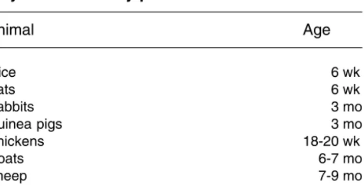
The age of the animals is another important consider-ation because that factor can influence the outcome of the immunization. It is important to use young adults, for whom the immune response is fairly robust and not affected by previous immune challenges. The robustness of the immune response decreases with age after the period of young adult-hood. When chickens are immunized, they should be of egg-laying age by the time antibody is to be harvested. Recommendations related to the age at which animals
should be used for PAb production are given in several publications (Hanly et al. 1995; Hendriksen and Hau 2003) and are summarized in Table 1.
In addition to the species, the sex of the animals must also be decided. Traditionally, female animals are used for PAb production inasmuch as these animals can be group housed more successfully than males because females are more docile and less aggressive in social interaction (Hen-driksen and Hau 2003). There are, however, no overrid-ing scientific reasons for not usoverrid-ing male animals for PAb production. Castrated male rabbits can be group housed, although this arrangement may pose ethical problems be-cause the animals have been castrated to facilitate the group housing.
Finally, it is important (albeit obvious) to consider the health status of animals used for the production of anti-bodies. Infectious agents exist that may suppress, modulate, or stimulate the immune system. The use of disease-free animals minimizes the likelihood of cross-reactivity to other antigens the animal’s immune system may have encountered.
Selection and Preparation of the Adjuvant
When the antigen to which antibodies are to be evoked is poorly immunogenic, the immune system requires a stimu-lus to induce an effective immune response. Adjuvants can be used for this purpose, and can direct an immune response against a more cellular or humoral response (Cox and Coulter 1997). More than 100 adjuvants have been de-scribed, and several reviews on adjuvants have been pub-lished (e.g., Stewart-Tull 2000; Vogel and Powell 1995). Only a few adjuvants are routinely utilized for polyclonal antibody production. Detailed information on adjuvants used for PAb production appears in thisILAR Journalissue (Stills 2005). Adjuvants used for PAb production include
Freund’s complete adjuvant (FCA1), Freund’s incomplete adjuvant (FIA1), aluminum salts (e.g., Al(OH)3, AlPO4), Quil A, Iscoms, Montanide, TiterMax™, and RIBI™.
FCA is frequently used for the production of polyclonal antibodies because high antibody titers are induced to al-most all types of antigens. However, many investigators have reported severe side effects after injection of FCA. For this reason, the available guidelines and recommendations on PAb production (e.g., CCAC 2002; Home Office 1991; NIH 1988) focus on FCA, and many universities in the United States have their own guidelines. Amyx (1987) has suggested that most undesirable effects of FCA can be eliminated by careful control of injection quantity and site selection. In an international workshop in New Orleans in 2002, it was concluded that FCA is not as problematic as a cause of pain and distress as previously suggested when injection volumes are minimized (HSUS Challenges Re-searchers to Reduce Animal Pain and Distress in Antibody Production: Satellite meeting held in conjunction with the Fourth World Congress on Alternatives and Animal Use in the Life Sciences in New Orleans, LA, August 11, 2002; C.F.M.H., personal communication) (http://www.hsus.org/ search.jsp). The fact that FCA is currently less problematic may be due to its improper former use (i.e., high volumes were used at unacceptable injection sites). In addition, ac-cording to the earlier literature, the FCA that was used was much less pure and more toxic, and it therefore induced more severe inflammatory reactions (Stewart-Tull 1995). In a more recent report (Leenaars et al. 1998), correct injection of FCA was reported to have resulted in no clinical or behavioral changes.
FCA is not the only adjuvant to induce pathological changes upon administration. Severe pathological changes have been reported after administration of TiterMax and RIBI adjuvant (Hendriksen and Hau 2003). The severity of pathological changes depends not only on the adjuvant but also on the type of antigen used. Moreover, the alterna-tive adjuvants have not often induced effecalterna-tive antibody responses.
Based on the factors described above, the selection of an alternative adjuvant for PAb production that induces mini-mal side effects and high antibody titers is not easy. When a weak immunogenic antigen is used frequently for PAb production, it may be helpful to perform a comparative study using different adjuvants combined with the antigen of interest for selecting the best adjuvant. It is important to include a pathological examination in these studies because several investigators (Johnston et al. 1991; Leenaars et al. 1998; Smith et al. 1992) have reported that although clinical and behavioral changes were not observed after injection of FCA, severe pathological changes were observed. Patho-logical changes are indeed unwanted. When antibodies to a weakly immunogenic antigen are to be produced only once, comparative studies are not recommended because the chances of success (high antibody levels) are very limited, and the presence of side effects cannot be excluded. Table 1 Recommended age of animals for
polyclonal antibody productiona
Animal Age Mice 6 wk Rats 6 wk Rabbits 3 mo Guinea pigs 3 mo Chickens 18-20 wk Goats 6-7 mo Sheep 7-9 mo
aData adapted from Leenaars PPAM, Hendriksen CFM, de Leeuw
WA, Carat F, Delahaut Ph, Fischer R, Halder M, Hanly WC, Hartinger J, Hau J, Lindblad EB, Nicklas W, Outschoorn IM, Stewart-Tull DES. 1999. The Production of Polyclonal Antibodies in Labora-tory Animal. The Report and Recommendations of ECVAM Workshop 35. ATLA 27:79-102. (Available online: http://altweb. jhsph.edu/publications/ECVAM/ecvam35.htm).
In addition to the selection of the adjuvant, the prepa-ration of the antigen/adjuvant mixtures may influence the outcome of the immunization experiment. With aseptic preparations, the first critical step is to minimize potential contamination, which may affect the quantity and quality of the antibodies as well as the welfare of the animal. The second step in the case of an emulsion (e.g., FCA and FIA are water-in-oil emulsions) is to check the stability and quality of the emulsion. When FCA or FIA is used, a thick emulsion is the result of mixing the antigen and adjuvant (Lindblad 2000). The advantage of a thick emulsion is that it effectively protects the antigen against a quick degrada-tion. The antigen is instead slowly released out of this emul-sion and is therefore present a relatively long time for endocytosis and antigen presentation. The result is a long-lasting adjuvant effect. Water-in-oil emulsions that are not properly prepared are ineffective as adjuvants (Hendriksen and Hau 2003). In the case of a water-in-oil emulsion, the stability and quality of the emulsion can be evaluated by placing a drop of the mixture on the surface of water. When the drop remains as a discrete white drop on or just below the surface, the emulsion is considered a stable water-in-oil emulsion. When the drop forms a cloud of tiny particles, it is an oil-in-water emulsion.
Antigen-Adjuvant Preparation Recommendations:
• Prepare mixtures aseptically.
• Carefully monitor the stability and quality of the emulsion. • Carefully select the injection route and volume,
espe-cially when oil adjuvants are used.
Injection Protocol
The immunization protocol can be prepared after selection of the animal species and the adjuvant. This protocol, which differs according to animal species and adjuvant, includes several critical steps that depend on each selection and are described below.
Route of Injection
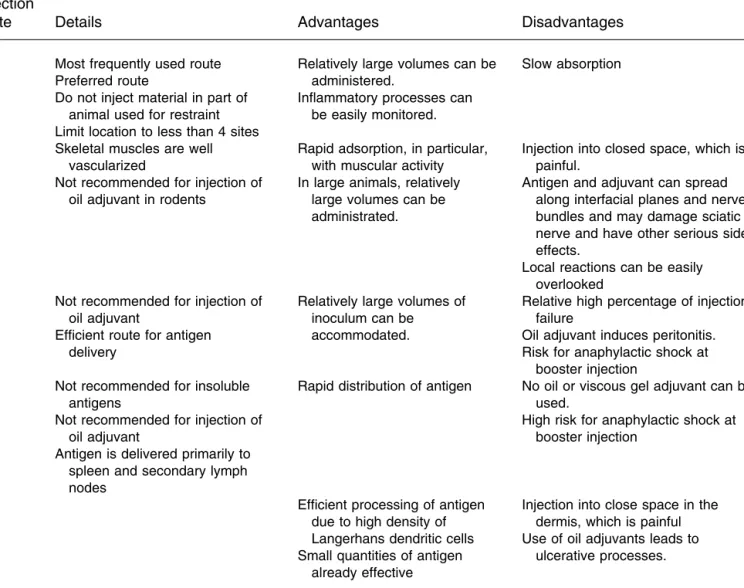
The choice of injection route is shaped to some extent by the choice of the animal species and adjuvant, as well as by the character, quantity, and volume of the antigen. The most frequently used routes of injection for PAb production are subcutaneous (s.c.), intradermal (i.d.), intramuscular (i.m.), intraperitoneal (i.p.), and intravenous (i.v.). Hendriksen and Hau (2003) have recently described the advantages and dis-advantages of the different injection routes, and this infor-mation is summarized in Table 2.
Footpad, intrasplenic, and intra-lymph node injection are usually not necessary for routine PAb production and should be justified on a case-by-case basis. The Canadian Council on Animal Care (CCAC 2002) discourages the use of the forenamed routes of injection. Indeed, because injec-tion into any closed space is painful, the choice of i.m. and i.d. routes warrants examination (CCAC 2002). The August
2002 international workshop participants mentioned above recommended discouraging the use of i.p., intrasplenic, and food pad injections, irrespective of the adjuvant, based on published literature and investigators’ personal experience (C.F.M.H., personal communication). Oil adjuvants are best administered s.c. to utilize the depot effect. Some injection routes may be eliminated by the choice of the adjuvant. The i.v. route for water-in-oil emulsions (e.g., FCA and FIA) may be lethal, and the i.v. route is not advised because of the risk of embolism for large particulate or viscous gel adju-vants (e.g., aluminum salts) (Hanly et al. 1995). For injec-tions of FCA, the i.p. route is not recommended for PAb production because it is known to induce inflamma-tion, peritonitis, and behavioral changes (CCAC 2002; Griffen et al. 2003).
The choice of animal species may also eliminate some injection routes. The i.d. route is not recommended in small rodents (mouse, rat, and hamster), and the i.p. route is not recommended in larger animals (rabbit and larger) (Hen-driksen and Hau 2003). Although no clinical or behavioral changes have been observed after s.c. injection of minimal volumes of FCA (0.1 mL in mice and 0.25 mL in rabbits), considerable pathological changes have been reported (Leenaars et al. 1998). The s.c. injection of FCA has been shown to be immunologically effective and to induce side effects that were relatively more acceptable (encapsulate depot) than i.p. (in mice) or i.m. (in rabbits) injection.
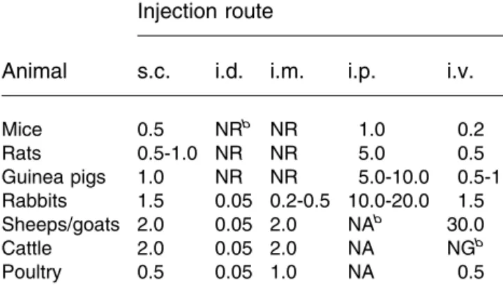
When the i.v. or i.p. route is used for booster injections, there is a risk of inducing anaphylactic shock in the animals (Hendriksen and Hau 2003). The site of injection should not interfere with subsequent handling of the animals for blood sampling. Recommended routes of injection according to animal species and adjuvant are provided in Table 3. Routes other than those identified in Table 3 warrant strong scien-tific jusscien-tification.
Volume of Injection
Regardless of the mixture to be injected, it is imperative to use the smallest possible volume that induces a sufficient antibody response. The minimal volume depends on par-ticular characteristics of the antigen. When the antigen is dissolved in a large volume (and cannot be concentrated), a large volume must be injected. The injected volume has been found to have an effect on the extent of the lesions produced (Stills and Bailey 1991); higher volumes of FCA produced larger lesions. Amyx (1987) has also suggested that the negative effects of FCA depend on the injection volume. The maximum injection volumes depend on animal species, injection route, and injection mixture. Maximum injection volumes are given in Table 4a (oil and viscous gel adjuvants) and Table 4b (no or aqueous adjuvants).
Injection Protocol Recommendations:
• Use the smallest possible volume, with the maximal limitations provided in Tables 4a and 4b.
• Carefully select the injection route (recommendations are provided in Table 3).
Number of Injection Sites
The antigen/adjuvant volume can be administered as a single injection or as multiple injections of low volumes. From the animal welfare perspective, there is a possible dilemma between the two options. Injection at one site of a large injection volume might be painful, particularly if there is limited space; however, multiple injections result in ad-ditional suffering due to the frequency of inoculation and the number of potentially painful sites. It is difficult to provide clear-cut recommendations because the available data are limited. In several European countries, a maximum of four injection sites is suggested when emulsions are used (Leenaars et al. 1999), but it is preferable to administer antigen/adjuvant mixtures at one site only.
Booster Injections
The boosting protocol can have a decisive impact on the result of the immunization. The time between two
immuni-zation steps can affect both the induction of B memory cells and the class switch of B cells. Animals can be rested for long intervals (months) between boosting. In general, a booster can be considered after the antibody titer has
Table 3 Injection route recommendations for different adjuvant types per animal speciesa
Animal Oil adjuvants and viscous gel adjuvants No or aqueous adjuvants Mice, rats, hamsters,
guinea pigs s.c. s.c., i.p., i.v.b Rabbits, sheep, goats s.c., i.d. s.c., i.d., i.m., i.v.b
Horses — s.c., i.d., i.m., i.v.b
aBased on the available guidelines and recommendations. bi.v. only with soluble antigens.
Table 2 Adjuvant injection routes for research animals: details, advantages, and disadvantagesa
Injection
route Details Advantages Disadvantages
s.c. Most frequently used route Preferred route
Do not inject material in part of animal used for restraint Limit location to less than 4 sites
Relatively large volumes can be administered.
Inflammatory processes can be easily monitored.
Slow absorption
i.m. Skeletal muscles are well vascularized
Not recommended for injection of oil adjuvant in rodents
Rapid adsorption, in particular, with muscular activity In large animals, relatively
large volumes can be administrated.
Injection into closed space, which is painful.
Antigen and adjuvant can spread along interfacial planes and nerve bundles and may damage sciatic nerve and have other serious side effects.
Local reactions can be easily overlooked
i.p. Not recommended for injection of oil adjuvant
Efficient route for antigen delivery
Relatively large volumes of inoculum can be accommodated.
Relative high percentage of injection failure
Oil adjuvant induces peritonitis. Risk for anaphylactic shock at
booster injection i.v. Not recommended for insoluble
antigens
Not recommended for injection of oil adjuvant
Antigen is delivered primarily to spleen and secondary lymph nodes
Rapid distribution of antigen No oil or viscous gel adjuvant can be used.
High risk for anaphylactic shock at booster injection
i.d. Efficient processing of antigen
due to high density of Langerhans dendritic cells Small quantities of antigen
already effective
Injection into close space in the dermis, which is painful Use of oil adjuvants leads to
ulcerative processes.
aAdapted and modified from Hendriksen C, Hau J. 2003. Production of polyclonal and monoclonal antibodies. In: Handbook of Laboratory Animal
reached a plateau or begins to decline. When the first im-munization is performed without a depot-forming adjuvant, the antibody titer usually peaks 2 to 3 wk after immuniza-tion. When a depot-forming adjuvant is used, a booster in-jection likely follows at least 4 wk after the first immunization (CCAC 2002).
An adjuvant is not always needed for booster injections. Booster injection with very small amounts of antigen may improve antibody affinity. Antibody titers after a booster injection can be measured to determine whether or not ad-ditional booster injections are needed. The number of booster injections should be limited for the welfare of the animal. Usually, a maximum of two or three booster injec-tions are recommended (Hendriksen and Hau 2003). FCA should be used only once because repeated injection of FCA (or Mycobacteriaproteins) may lead to severe tissue reac-tions. FIA should be used for booster injecreac-tions. As de-scribed above, a risk of i.v. and i.p. booster injections is the induction of anaphylactic shock in the animals.
Emulsion Use Recommendations:
• Administer the boost at least 4 wk after the prime. • Use the suggested maximum of two or three boosters.
Postinjection Observation
After immunization, animals should be monitored daily and examined for specific side effects at least three times per
week. Examination and palpation of the injection site are essential to evaluate side effects of the injected mixture. Clinical observations do not always predict severe pathol-ogy at the site of injection (Leenaars et al. 1998). Several investigators have described severe pathological changes despite the absence of observed clinical or behavioral changes. After booster injection via the i.v. or i.p. route, it is imperative to monitor the animals during subsequent hours for any anaphylactic reactions.
Monitoring Recommendation:
• Palpate the injection site to evaluate side effects of the injection.
Monitoring of the Antibody Response
In mammals, antibody responses during the experiment can be monitored by obtaining and evaluating a blood sample for antibodies in the serum. In chickens, because antibodies are excreted in the eggs, titers can be studied in the egg without an invasive action. The frequency of blood collec-tion and the maximum blood volume that can be removed safely from the animal are limited. If too much blood is withdrawn too rapidly or too frequently without replace-ment, an animal may go into short-term hypovolemic shock and become anemic.
As a rough guide, up to 10% of the circulating blood volume can be taken on a single occasion from healthy animals (BVA 1993); in practice, an amount up to 1% of total body weight can be removed safely (McGuill and Table 4b Recommended maximum volume of injection (in mL) used for injection of aqueous antigen/adjuvant mixture per injection route for different animal speciesa
Animal
Injection route
s.c. i.d. i.m. i.p. i.v.
Mice 0.5 NRb NR 1.0 0.2 Rats 0.5-1.0 NR NR 5.0 0.5 Guinea pigs 1.0 NR NR 5.0-10.0 0.5-1.0 Rabbits 1.5 0.05 0.2-0.5 10.0-20.0 1.5 Sheeps/goats 2.0 0.05 2.0 NAb 30.0 Cattle 2.0 0.05 2.0 NA NGb Poultry 0.5 0.05 1.0 NA 0.5
aAdapted and modified from the following: (1) CCAC [Canadian
Council on Animal Care]. 2002. CCAC Guidelines on: Antibody Pro-duction, Ottawa ON: CCAC. (Available online: http://www.ccac.ca/ english/gdlines/antibody/antibody.pdf); and (2) Hendriksen C, Hau J. 2003. Production of polyclonal and monoclonal antibodies. In: Hand-book of Laboratory Animal Science. 2nd ed. Boca Raton: CRC Press LLC. p 391-411.
bNR, not recommended; NA, not acceptable; NG, not given.
Table 4a Recommended maximum injection volumes (in mL) used for injection of oil and viscous gel adjuvants per injection route for different animal speciesa
Animal Injection routeb s.c. i.d. Mice 0.1 NRc Rats 0.1-0.2 NR Guinea pigs 0.2 NR Rabbits 0.1-0.25 0.025-0.05 Sheep/goats 0.5 0.05 Cattle 0.5 0.05 Poultry 0.25 0.05
aAdapted and modified from the following: (1) CCAC [Canadian
Council on Animal Care]. 2002. CCAC Guidelines on: Antibody Pro-duction, Ottawa ON: CCAC. (Available online: http://www.ccac.ca/ english/gdlines/antibody/antibody.pdf); and (2) Hendriksen C, Hau J. 2003. Production of polyclonal and monoclonal antibodies. In: Hand-book of Laboratory Animal Science. 2nd ed. Boca Raton: CRC Press LLC. p 391-411.
bi.p. and i.m., not recommended; i.v., not acceptable. cNR, not recommended.
Rowan 1989). After this maximum blood volume has been collected, animals need to rest for 3 to 4 wk (BVA 1993). Maximum blood volumes that can be collected from differ-ent animal species safely are provided in Table 5.
Collection of the Antibodies: Exsanguination
and Euthanasia
Exsanguination must be performed under general anesthesia and is best carried out by heart puncture. It should result in the death of the animal. When there is uncertainty about death, small rodents can be subjected to cervical dislocation, and larger animals can be euthanized by an overdose of an appropriate anesthetic agent. Euthanasia should be in accor-dance with the 2000 Report of the American Veterinary Medicine Association (AVMA1) panel on euthanasia (AVMA 2000).
Evaluation of Critical Steps in
MAb Production
MAbs are antibodies produced by a single clone of B cells. Köhler and Milstein (1975) discovered that these cells can be immortalized by fusion with myeloma cells, resulting in hybridoma cells that are able to produce virtually unlimited quantities of monoclonal antibodies. Since the Nobel price-winning work of these researchers, MAbs have become es-sential tools in basic research as well as in diagnostic testing and medical treatments.
The process of MAb development includes the follow-ing successive workfollow-ing phases: the generation of antigen-specific B cells, the fusion of these cells with myeloma
cells, the cloning and selection of the specific hybridoma clone by “limiting dilution,” and the up-scaling of MAb production (Johnson 1995). Traditionally, two working phases in this process are based on procedures involving the use of laboratory animals—the generation of B cells and the MAb mass production by the ascites induction method. In the text below, we focus on these two steps.
Immunization to Generate Antigen-specific
B Cells
Production of specific B cells requires immunization of the animal with the antigen under study. Critical aspects that should be considered include antigen selection, aseptic an-tigen processing, adjuvant selection, preparation of anan-tigen- antigen-adjuvant mixture, choice of immunization route and injection volume, and aseptic inoculation, as discussed above in relation to the production of polyclonal antibodies. For immunization, BALB/c mice are typically used because many of the myeloma cells available for fusion have a BALB/c origin. In specific cases, immunocompromised (e.g., severe combined immunodeficient [SCID1]) mice or other animal species such as the rat and hamster, and even human cases may also be used. Recommendations regard-ing immunization protocols for B cell generation are diffi-cult to devise because protocols differ based on the type of antigen and adjuvant used. Generally, several booster im-munizations are needed, with intervals of 14 to 28 days between boosters, although other protocols exist. It may be possible to use serum antibody titer information to guide booster protocols. To address that possibility, it is advisable to perform test bleeds before immunization. Often a final booster with antigen only (without adjuvant, i.p or i.v. route) is administered a few days before the animal is sac-rificed. B cells are harvested from the spleen, although other lymphoid tissue can also be used.
Methods to generate antigen-specific B cells after in vitro “immunization” have been described (Guzman et al. 1995; Stadler 1999) but have not been very successful to date because the vast majority of the B cells produce IgM rather than IgG antibodies (Borrebaeck et al. 1988). Anti-body-producing B cells from the spleen or other lymphoid tissue are fused with nonsecreting myeloma cells (hybrid-oma cells), and the product is then cloned and selected for antigen specificity. Selection for further growth character-istics and specificity of MAbs is crucial. Failures related to in vitro MAb production are often attributable to the im-proper selection and subcloning of hybridoma cells.
The next step, the up-scaling of MAb production, is traditionally accomplished first by administering injections of MAb-producing hybridoma cells in the abdominal cavity of mice and then by collecting the ascites that develops after the next 7 to 14 days. The abdominal cavity is indeed an optimal growth chamber for the hybridoma cells because it guarantees a constant temperature, an optimal nutrient and Table 5 Maximum blood volumes to be collected
from animals during experimentationa
Animal Maximum blood volume (mL) Rabbits 15b Mice 0.3 Rats 2 Guinea pigs 5 Hamsters 0.3 Sheeps 200-600b Goats 150-400b Horses 500-7000b
aAdapted from BVA/FRAME/RSPCA/UFAW [British Veterinary
As-sociation/Fund for the Replacement of Animals in Medical Experi-ments/Royal Society for the Prevention of Cruelty to Animals/ Universities Federation for Animal Welfare]. 1993. Joint working group on refinement: Removal of blood from laboratory mammals and birds. Lab Anim 27:1-22.
oxygen supply, and the optimal removal of CO2and
meta-bolic waste products (Hendriksen and de Leeuw 1998). Hy-bridoma cells grow to high densities, and the resulting MAb concentration levels are consequently high. Nevertheless, for several reasons, the ascites method has fallen into dis-favor. The most important reasons relate to the following: (1) Pain and distress are likely to be involved with ascites production (NRC 1999). (2) High-quality in vitro produc-tion systems are increasingly available, and new producproduc-tion approaches are progressing. (3) There is evidence of con-tamination with other immunologically active compounds and the risk of MAb contamination with viruses and other microorganisms. For further information, we refer readers to discussions of in vitro systems and methods provided elsewhere in this issue (Dewar et al. 2005; Lipman et al. 2005). Also in this issue, Peterson (2005) discusses new approaches to MAb production.
Based on various reports, the most important of which is the 1999 NRC Report, there is now consensus that ascites production should be the exception, requiring rigorous and well-documented justification. Exceptional circumstances that might justify the use of ascites production include the following: emerging therapeutic applications; existing regu-latory approval for diagnostic and therapeutic products (un-til the approval expires); and poor growth of hybridoma cells in vitro, when subcloning fails to improve the growth (Marx et al. 1997). For those limited specific cases in which the ascites method can be justified, background informa-tion is provided about the underlying procedure, and inves-tigators must conform to the standards of practice given, in the Guide for the Care and Use of Laboratory Animals (NRC 1996).
MAb Production by Ascites Induction in
Laboratory Animals
BALB/c mice are generally preferred for ascites production because this strain of animals is syngeneic for the myeloma cells most frequently used for fusion. Animals are first given an i.p. injection with a priming agent. The primer most frequently used is pristane (2,6,10,14-tetramethyl pen-tadecane), but FIA is also applied (Gillette 1987). The effect of the primer is three-fold: (1) suppression of the immune system, thus preventing impairment of hybridoma cell growth; (2) toxic irritation of peritoneum, resulting in peri-tonitis and the secretion of serous fluid; and (3) retardation of hybridoma cell clearance from the peritoneal cavity (Moore and Rajan 1994).
A total of 7 to 10 days after priming, the hybridoma cells are administered by i.p. injection. Tumor development and ascites production begin within a few days after inoculation and result in an increase of abdominal distention. Ascites induction is a life-threatening procedure due to tumor growth, metastatic spreading, infiltrative growth, and, ulti-mately, respiratory distress. Some tumor cells tend to grow rapidly and form large solid tumors in the peritoneum,
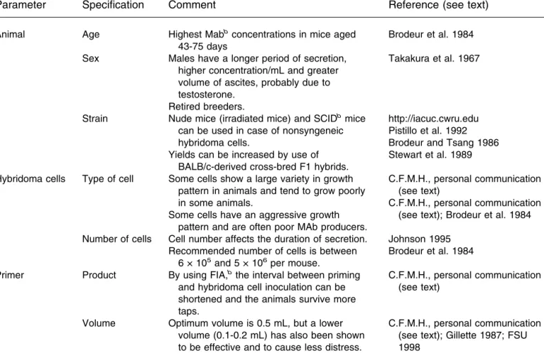
with-out significant production of ascites. Usually, however, soft tumors develop with ascites production ranging from 0 to 10 mL/mouse, with an average of 2 to 4 mL/mouse and a MAb concentration of 1 to 28 mg/mL (Hendriksen 1998). Factors that might affect the yield of ascites or the MAb concen-tration per milliliter are shown in Table 6.
During ascites development, animals should be ob-served at least three times per day for the first week and daily thereafter (including weekends and holidays) to moni-tor the degree of abdominal distention and signs of illness (http://medicine.ucsd.edu). An overview of clinical, patho-physiological, and pathological effects of ascites production is given in Table 7. It is important to note that side effects of tumor growth can be more severe due to incorrect i.p. injection of hybridoma cells as a result of inoculation of hybridoma cells in abdominal organs, such as urinary blad-der or intestines (Walvoort 1991). Peterson (2000) assessed the effects on well-being of pristane injection and ascites production using parameters such as wheel-running activity, food and water consumption, open-field box activity, clini-cal observation, and plasma corticosterone concentration. No significant evidence of distress was observed in the ani-mals studied.
Ascites Collection
Ascites fluid must be collected before the abdominal dis-tention leads to significant health problems, which usually develop 1 to 2 wk after injection of hybridoma cells. An 18-to 22-gauge syringe needle is inserted in18-to the lower abdo-men and is tapped by “massaging” the ascites. Shock from hypovoluminae may be prevented by s.c. injection of 2 to 3 mL of warm saline or lactated Ringer’s solution (http:// iacuc.cwru.edu). With each successive tap, the quantity of ascites decreases; however, while the antibody concentra-tion increases, the survival of animals decreases (from 98% for tap 1 to 35-100% for tap 3 [Jackson et al. 1999]).
Different policies exist with regard to the number of taps. According to the guidelines of the UK Co-ordinating Committee on Cancer Research (UKCCR 1998), ascites tu-mors should be drained only once. Some institutions allow removal of ascites fluid more frequently; however, repeated draining increases the risk of solid tumor deposit develop-ment, bleeding to the peritoneal cavity, and cachexia. There-fore, it is generally recommended that the number of taps should be limited to a maximum of three, or at any point if there is evidence of debilitation, pain, or distress, and that animals should be euthanized humanely according to the 2000 Report of the AVMA Panel on Euthanasia (AVMA 2000).
MAb Production: Guidelines
Due to concern for the welfare of animals, regulations and guidelines related to ascites production are now included in
the policies of individual institutes, as well as those at a national level in several countries. Ascites production is strongly discouraged in several European countries such as the United Kingdom, Germany, and The Netherlands.
Insti-tutional policies exist in the United States, and many ex-amples of policies can be found on the internet (e.g., http:// medicine.ucsd.edu and http://iacuc.cwru.edu). In 1997, a “Dear Colleague” letter sent by the US National Institutes of Table 6 Parameters affecting ascites productiona
Parameter Specification Comment Reference (see text)
Animal Age Highest Mabb
concentrations in mice aged 43-75 days
Brodeur et al. 1984 Sex Males have a longer period of secretion,
higher concentration/mL and greater volume of ascites, probably due to testosterone.
Retired breeders.
Takakura et al. 1967
Strain Nude mice (irradiated mice) and SCIDb mice can be used in case of nonsyngeneic hybridoma cells.
Yields can be increased by use of BALB/c-derived cross-bred F1 hybrids.
http://iacuc.cwru.edu Pistillo et al. 1992 Brodeur and Tsang 1986 Stewart et al. 1989 Hybridoma cells Type of cell Some cells show a large variety in growth
pattern in animals and tend to grow poorly in some animals.
Some cells have an aggressive growth pattern and are often poor MAb producers.
C.F.M.H., personal communication (see text)
C.F.M.H., personal communication (see text); Brodeur et al. 1984 Number of cells Cell number affects the duration of secretion.
Recommended number of cells is between 6 × 10⁵
and 5 × 10⁶
per mouse.
Johnson 1995 Brodeur et al. 1984
Primer Product By using FIA,b
the interval between priming and hybridoma cell inoculation can be shortened and the animals survive more taps.
C.F.M.H., personal communication (see text)
Volume Optimum volume is 0.5 mL, but a lower volume (0.1-0.2 mL) has also been shown to be effective and to cause less distress.
C.F.M.H., personal communication (see text); Gillette 1987; FSU 1998
aAdapted from Hendriksen CFM, de Leeuw W. 1998. Production of monoclonal antibodies by the ascites method in laboratory animals. Res
Immunol Forum 6 149:535-542.
bMAb, monoclonal antibody; SCID, severe combined immunodeficient; FIA, Freund’s incomplete adjuvant.
Table 7 Clinical, pathophysiological, and pathological effects of ascites productiona
Clinical effects Pathophysiological effects Pathological effects
Abdominal distension Anorexia Peritonitis
Decreased activity and body Anemia Infiltrative tumor growth
mass Dehydration Adhesions in the abdomen
Shrunken eyes Tachypnoe Enlarged abdominal organs
Difficulty with walking Circulatory shock Blood in the abdominal cavity
Hunched posture Decreased venous, arterial, and renal
Respiratory distress blood flow
Death Ascites production
Immunosuppression
aAdapted from Anon. 1989. Code of Practice for the Production of Monoclonal Antibodies. Rijswijk, The Netherlands: Veterinary Public Health
Health Office for Protection from Research Risks referred to the evidence that the ascites method causes discomfort, distress, or pain and that in vitro methods for MAb produc-tion should be preferred (NIH 1997). Based on documented evidence, in addition to the advances described in the fore-going text, it is reasonable to conclude that new develop-ments in in vitro MAb production gradually will limit the use of animals for this purpose.
References
Amyx HL. 1987. Control of animal pain and distress in antibody produc-tion and infectious disease studies. JAVMA 191:1287-1289. Anon. 1989. Code of Practice for the Production of Monoclonal
Antibod-ies. Rijswijk, The Netherlands: Veterinary Public Health Inspectorate. AVMA [American Veterinary Medicine Association]. 2000. Report of the
AVMA Panel on Euthanasia. JAVMA 218:669-696.
Borrebaeck CA, Danielsson L, Moller SA. 1988. Human monoclonal an-tibodies produced by primary in vitro immunization of peripheral blood lymphocytes. Proc Natl Acad Sci U S A 85:3995-3999.
Brodeur BR, Tsang PS. 1986. High yield monoclonal antibody production in ascites. J Immunol Methods 86:239-241.
Brodeur BR, Tsang PS, Larose Y. 1984. Parameters affecting ascites tu-mour formation in mice and monoclonal antibody production. J Immu-nol Methods 71:265-272.
BVA/FRAME/RSPCA/UFAW [British Veterinary Association/Fund for the Replacement of Animals in Medical Experiments/Royal Society for the Prevention of Cruelty to Animals/Universities Federation for Ani-mal Welfare]. 1993. Joint working group on refinement: Removal of blood from laboratory mammals and birds. Lab Anim 27:1-22. CCAC [Canadian Council on Animal Care]. 2002. CCAC Guidelines on:
Antibody Production, Ottawa ON: CCAC. (Available online: http:// www.ccac.ca/english/gdlines/antibody/antibody.pdf).
Cox JC, Coulter AR. 1997. Adjuvants—A classification and review of the modes of action. Vaccine 15:248-256.
Erhard MH, Mahn K, Schmidt P, Oltmer S, Preisinger R, Zinsmeister P, Stangassinger M. 2000. Evaluation of various immunisation procedures in laying hens to induce high amounts of specific egg yolk antibodies. ATLA 28:63-80.
Dewar V, Voet P, Denamur F, Smal J. 2005. Industrial implementation of in vitro production of monoclonal antibodies. ILAR J 46:307-313. FSU [Florida State University]. 1998. Laboratory Animal Program home
page. Monoclonal Antibody Production in the Mouse—Ascites (http:// www.fsu.edu/∼FSULAR/ascites.html).
Gillette RW. 1987. Alternatives to pristane priming for ascites fluid and monoclonal antibody production. J Immunol Methods 99:21-23. Griffen PS, Turton J, Andrews CM, Barrett P, Clarke CJ, Fung KW,
Munday MR, Roman IF, Smyth R, Walshe K, York MJ. 2003. Markers of experimental acute inflammation in the wistar han rat with particular reference to haptoglobin and C-reactive protein. Arch Toxicol 77:392-402.
Guzman J, Frei K, Nadal D. 1995. In vitro immunization: Generation of neutralizing monoclonal antibodies to human interleukin-10. J Immu-nol Methods 179:265-268.
Hanly WC, Artwohl JE, Bennett BT. 1995. Review of polyclonal antibody production procedures in mammals and poultry. ILAR J 37:93-118. Harlow E, Lane D. 1988. Adjuvants. In: Antibodies: A laboratory Manual.
Cold Spring Harbor NY: Cold Spring Harbor Laboratory. p 96-124. Hau J, Hendriksen CFM. 2005. Production of polyclonal antibodies: New
technologies. ILAR J 46:294-299.
Hendriksen C, Hau J. 2003. Production of polyclonal and monoclonal antibodies. In: Handbook of Laboratory Animal Science. 2nd ed. Boca Raton: CRC Press LLC. p 391-411.
Hendriksen CFM. 1998. A call for a European prohibition of monoclonal
antibody (MAb) production by the ascites procedure in laboratory ani-mals. ATLA 26:523-540.
Hendriksen CFM, de Leeuw W. 1998. Production of monoclonal antibod-ies by the ascites method in laboratory animals. Res Immunol 149:535-542.
Home Office. 1991. Antibody production: advice on protocols for minimal severity. In: Report of the Animal Procedures Committee for 1991. London: HMSO. p 26. (Available online: Http://iacuc.cwru.edu; Http:// www.hsus.org/search.jsp; Http://medicine.ucsd.edu).
Jackson LR, Trudel LJ, Fox JG, Lipman NS. 1999. Monoclonal antibody production in murine ascites. I. Clinical pathologic features. Lab Anim Sci 49:70–80.
Johnson DR. 1995. Murine monoclonal antibody development. In: Clifton NJ, ed. Methods in Molecular Biology. Vol 51. Totowa NJ: Humana Press. p 123-137.
Johnston BA, Eisen H, Fry D. 1991. An evaluation of several adjuvant emulsion regimens for production of polyclonal antisera in rabbits. Lab Anim Sci 41:15-21.
Köhler G, Milstein C. 1975. Continuous cultures of fused cells secreting antibody of predefined specificity. Nature 256:495-497.
Leenaars PPAM, Claassen E, Boersma WJA. 1997. Antigens and antigen presentation. In: Lefkovits I, ed. Immunology Methods Manual. Lon-don: Academic Press Ltd. p 989-1013.
Leenaars PPAM, Hendriksen CFM, de Leeuw WA, Carat F, Delahaut Ph, Fischer R, Halder M, Hanly WC, Hartinger J, Hau J, Lindblad EB, Nicklas W, Outschoorn IM, Stewart-Tull DES. 1999. The Produc-tion of Polyclonal Antibodies in Laboratory Animal. The Report and Recommendations of ECVAM Workshop 35. ATLA 27:79-102. (Available online: http://altweb.jhsph.edu/publications/ECVAM/ ecvam35.htm).
Leenaars PPAM, Koedam MA, Wester P, Baumans V, Claassen E, Hen-driksen CFM. 1998. Assessment of side effects induced by injection of different adjuvant/antigen combinations in rabbits and mice. Lab Anim 32:387-406.
Lindblad EB. 2000. Freund’s adjuvants. In: O’Hagen DT, ed. Methods in Molecular Medicine: Vaccine Adjuvants—Preparation Methods and Research Protocols. Totowa NJ: Humana Press. p 79-102.
Lipman NS, Jackson LR, Trudel LJ, Weis-Garcia F. 2005. Monoclonal versus polyclonal antibodies: Distinguishing characteristics, applica-tions, and information sources. ILAR J 46:258-268.
Marx U, Embleton MJ, Fischer R, Gruber FP, Hansson U, Heuer J, de Leeuw WA, Logtenberg T, Merz W, Portelle D, Romette J-L, Straughan DW. 1997. Monoclonal antibody production. ECVAM Workshop report. ATLA 25:121-137.
McGuill MW, Rowan AN. 1989. Biological effects of blood loss: Impli-cation of sampling volumes and techniques. ILAR News 31:5-20. Moore JM, Rajan TV. 1994. Pristane retards clearance of particulate
ma-terials from the peritoneal cavity of laboratory mice. J Immunol Meth-ods 173:273-278.
NIH [National Institutes of Health]. 1988. NIH intramural recommenda-tions for the research use of complete Freund’s adjuvant. (Grumstrupp-Scott J, Greenhouse DD). ILAR News 30:9.
NIH [National Institutes of Health]. 1997. Production of Monoclonal An-tibodies Using Mouse Ascites Method (publication 98-01, November 17, 1997). Rockville: Office for Protection from Research Risks. NRC [National Research Council]. 1996. Guide for the Care and Use of
Laboratory Animals. 7th ed. Washington DC: National Academy Press. NRC [National Research Council]. 1999. Monoclonal Antibody
Produc-tion. Washington DC: National Academy Press.
Peterson NC. 2000. Behavioral, clinical, and physiologic analysis of mice used for ascites monoclonal antibody production. Comp Med 50:516-526.
Peterson NC. 2005. Advances in monoblonal antibody technology: Genetic engineering of mice, cells, and immunoglobulins. ILAR J 46:314-319. Pistillo MP, Sguerso V, Ferrara GB. 1992. High yields of anti-HLA Human monoclonal antibodies can be provided by SCID mice. Hum Immunol 35:256-259.
Pollmann W, van Regenmortel M, Rijke E, Spielmann H, Steinbusch H, Straughan D. 1996. The production of avian (egg yolk) antibodies: IgY. In: The Report and Recommendations of ECVAM Workshop 21. ATLA 24:925-934. (Available online: http://altweb.jhsph.edu/ publications/ECVAM/ecvam21.htm).
Smith DE, O’Brien ME, Palmer VJ, Sadowski JA. 1992. The selection of an adjuvant emulsion for polyclonal antisera in rabbits. Lab Anim Sci 42:599-601.
Stadler BM. 1999. Antibody production without animals. Dev Biol Stand 101:45-48.
Stewart F, Callander A, Garwes DJ. 1989. Comparison of ascites produc-tion for monoclonal antibodies in BALB/c and BALB/c derived cross-bred mice. J Immunol Methods 119:269-275.
Stewart-Tull DES. 1995. The theory and practical application of adjuvants. Chichester UK: John Wiley and Sons.
Stewart-Tull DES. 2000. Harmful and beneficial activities of immunologi-cal adjuvants. In: O’Hagen DT, ed. Methods in Molecular Medicine: Vaccine Adjuvants-Preparation Methods and Research Protocols. Totowa NJ: Humana Press. p 29-48.
Stills HF Jr. 1994. Polyclonal antibody production. In: Manning PJ,
Ring-ler DH, Newcomer CE, eds. The Biology of the Laboratory Rabbit, 2nd ed. San Diego: Academic Press Inc. p 435-448.
Stills HF Jr. 2005. Adjuvants and antibody production: Dispelling the myths associated with Freund’s complete and other adjuvants. ILAR J 46:280-293.
Stills HF Jr, Bailey MQ. 1991. The use of Freund’s complete adjuvant. Lab Anim 20:25-30.
Takakura K, Yamada H, Weber H, Hollander VP. 1967. Studies on the pathogenesis of plasma cell tumors: Effects of sex hormones on the development of plasma cell tumors. Cancer Res 27:932-937. UKCCCR [United Kingdom Co-ordinating Committee on Cancer
Re-search]. 1998. Guidelines for the Welfare of Animals in Experimental Neoplasia. 2nd ed. Br J Cancer 77:1-10.
Vogel FR, Powell MF. 1995. A compendium of vaccine adjuvants and excipients. In: Powell MF, Newman MJ, eds. Vaccine Design: The Subunit and Adjuvant Approach., New York: Plenum Press. p 141-228. Walvoort NC. 1991. Assessment of distress through pathological exami-nation. In: Hendriksen CFM, Köeter HBWM, eds. Replacement, Re-duction and Refinement: Present Possibilities and Future Prospects. Amsterdam: Elsevier. p 265-273.