Incarcerated Diaphragmatic Hernia With
Intrathoracic Bowel Obstruction After
Right Liver Donation
Raymond A. Dieter, Jr.
1,2, Jonathon Spitz
2, George Kuzycz
21
President, Northern Illinois Center for Surgery, Naperville, Illinois, USA, and past president, International
College of Surgeons
2
Department of Surgery, Northern Illinois Center for Surgery, Naperville, Illinois, USA, and Dupage
Medical Group, Inc., Glen Ellyn, Illinois, USA
Liver transplantation has become an acceptable surgical procedure with the advancement of the technical and rejection considerations involved. Initially nonliving donors were used for transplantation procedures. However, with improved techniques, living donor procedures have become much more frequent. With this, complications involving the transplant organ donor may occur. We present 2 patients with intrathoracic bowel obstruction due to herniation of the small intestine and colon through a defect in the dome of the diaphragm with development of chest pain and gastrointestinal symptoms. Both patients were diagnosed by computerized tomography scan and had a right thoracotomy with lysis of the adhesions, reduction of the hernia, repair of the diaphragm, and mesh reinforcement of the diaphragm. Neither patient had a prior diaphragm defect. These patients, on review of the literature, represent the first 2 such reported cases and suggest the need to be aware of any potential diaphragm defects before closure of the abdomen after resection of the donor liver or if they develop appropriate symptomatology.
Key words:Intrathoracic bowel obstruction – Iatrogenic diaphragm hernia – Complications of organ donation – Hepatic/liver donor complications – Incarcerated right diaphragm hernia – Endothoracic bowel obstruction – Enterothorax
Reprint requests: Raymond A. Dieter, Jr., MD, The Center for Surgery, 475 E. Diehl Road, Naperville, IL 60563. Tel.: 630 790 1700 or 630 505 7733; Fax: 630 799 0223
H
ernia formation may occur in multiple loca-tions throughout the body and may be con-genital or acquired. The inguinal and umbilical hernias are the most commonly diagnosed defects. Other hernias may occur through the anterior abdominal wall, the diaphragm, or the perineum. Congenital diaphragmatic hernias of Morgagni and Bochdalek are seen on occasion. Acquired defects may be iatrogenic or post-traumatic. Hernias, as a result of diaphragmatic trauma, are uncommon. Iatrogenic diaphragmatic herniation after liver do-nation for transplantation is a rare occurrence. Recently, we have seen 2 patients who had donated a portion of their right liver for transplantation and postoperatively developed enterothorax difficulties requiring thoracic surgery. These patients represent the first reported cases of intrathoracic bowel obstruction after liver donation through postsurgical acquired diaphragmatic defects.Patients
Patient 1 was a 45-year-old white woman who 3 years before had donated a portion of her liver for transplantation into her 18-year-old daughter who had hepatic cirrhosis due to an alpha 1 antitrypsin deficiency. This enzyme trait defect was present in both the mother and the father of the recipient. The mother also had a history of hypertension, fibromy-algia, hyperthyroidism, and of a hiatus hernia with gastroesophageal reflux. After donation of her liver, the mother developed constipation with persistent and recurrent abdominal complaints that localized
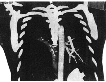
to the right side of her abdomen and chest. Because of the abdominal pain, constipation, shortness of breath with eating, and aching all over, she presented to our emergency department for evalu-ation. Chest X-rays and computerized tomography (CT) scan demonstrated abdominal gastrointestinal organs, including the right colon and loops of small bowel, herniated through the right diaphragm into the chest (Fig. 1). At right thoracotomy, the patient was found to have a hernia sac within the chest measuring the size of a large cantaloupe (15–16 cm) (Fig. 2). The hernia sac contained the right colon, the transverse colon, the omentum, and a portion of the small bowel. The lung was adherent to the hernia sac. Through a medial defect in the hernia sac there was a secondary or ‘‘daughter’’ small bowel herniation. The herniated small bowel was incarcer-ated and demonstrincarcer-ated ischemic changes with discoloration and darkened areas. There was a 7-cm diaphragmatic defect with thickened edges through which the partially strangulated and ische-mic bowel protruded. The bowel was adherent to the margins of the defect. Thus, the defect was
Fig. 1 CT scan demonstrating loops of small and large bowel incarcerated in the right chest thru a right diaphragmatic hernia defect in patient 1.
Fig. 2 Intraoperative photo demonstrating herniation of bowel into the right chest through the right diaphragm defect in patient 1.
enlarged and the adhesions to the lung, the hernia sac, the incarcerated loops, and the hernia ring were lysed. Following this, the hernia contents were reduced into the abdomen and the repair completed. Patient 2 donated the right lobe of her liver for transplantation into her sister-in-law. This donor also had a history of hypertension and being overweight. After liver donation, she was dis-charged home in 2 days. During the next several months, she had multiple abdominal complaints, which were believed to be nonsurgical in nature. The patient had a CT scan of the abdomen to evaluate her abdominal pain, which demonstrated a small left renal stone. Cervical spine X-rays, mag-netic resonance images of the spine, X-rays of the shoulder, and multiple spinal injections were ad-ministered in an attempt to relieve her symptom-atology. On arrival at our emergency room, she was complaining of nausea, vomiting, abdominal and chest pain. A CT of the abdomen demonstrated that the right colon and a portion of the small bowel were herniated into the chest through the dome of the right diaphragm. The herniated mass measured the size of a large grapefruit to a small cantaloupe (12 cm) (Fig. 3). After our consultation, she was taken to surgery for a right thoracotomy using a double lumen endotracheal tube. At surgery, an incarcerated hernia with extensive adhesions to the lung and diaphragm projected through a 7-cm diaphragmatic defect in the dome (Fig. 4). The fibrinous hernia sac overlying the adhered small bowel and cecum was opened and the adhesions divided. We were unable to reduce the herniated contents into the abdomen until the diaphragmatic
defect was enlarged and the adhesions to the hernia ring and below the diaphragm were separated.
In both patients a fashioned 3 3 6 inch (7.6 3 15.2 cm) Prolene mesh was placed on the undersur-face of the diaphragm after reduction of the hernia and sutured into place with 2-0 Prolene sutures 2 cm from the edge of the mesh and 2 cm from the defect margins in all directions (Fig. 5). A two-layer closure of the diaphragm with nonabsorbable running O-Ethibond (Ethicon, Inc., Sommerville, New Jersey) suture followed. Two chest tubes were placed through separate stab wounds and connected to underwater seal drainage. Postoperatively, the patients did well and were discharged home in a few days after removal of the chest tubes.
Discussion
Two patients are presented who developed right diaphragmatic defects with subsequent herniation of gastrointestinal structures into the chest. Both patients had previously donated a portion of their right liver to a family member in need and had simultaneous cholecystectomy. The mother under-went partial liver resection at a nearby university, and then liver transplantation was performed at the affiliated children’s hospital. Immediately postoper-atively both donors did well. Later she (the mother) developed abdominal and respiratory symptomatol-ogy. With increasing discomfort, she came to our facility in marked distress. The second patient developed abdominal and thoracic symptoms after hospital discharge for which physician evaluation
Fig. 4 Right thoracotomy demonstrating the viscerol herniation through the right diaphragm into the right chest in patient 2.
Fig. 3 CT scan demonstrates the herniated bowel in the right chest in patient 2.
and diagnostic studies failed to reveal the source of her complaints. CT scanning in our emergency room for possible pulmonary embolism demonstrated the diaphragmatic defect and the right intrathoracic incarcerated hernia. After right thoracotomy, the large herniation was reduced into the abdomen after enlargement of the diaphragmatic defect and lysis of adhesions in both patients.
Transplantation surgery is becoming more fre-quent for many of our diseased organs, including the heart, lung, kidney, liver, and gastrointestinal tract.1 Early in the development of the surgical transplantation techniques and the antirejection protocols, cadaveric organs were used. With tissue typing and increased compatibility knowledge to prevent rejection, transplantation has been more successful. Living donor programs were then developed using both related and nonrelated do-nors. Defining surgical technique for procurement of the desired organ to be removed from a living donor for subsequent transfer to the needy recipient presented a new set of potential complications in a previously uninvolved group of individuals. This meant that the organ donor may now have major
side effects of surgery, which could become life threatening.
Liver surgery has always been approached with caution due to the possibility of bleeding, bile leak, and major vascular injury. Ordinarily, the dia-phragm has not been of major concern for possible side effects of liver surgery. More commonly, except with respect to the hiatal or paraesophageal hernias, nontumorous defects of the diaphragm have been seen in the trauma patient or as a congenital lesion (Table 1). Nonhiatal diaphragmatic hernias, idio-pathic paralysis, eventration, and phrenic nerve palsy are infrequently seen and repaired either thoracoscopically or with a thoracotomy (Dieter RA Jr, Alvaz Malo OO. Thorascopic repair of paralyzed diaphragm. Video presentation at the meeting of the World Biennial ICS; November 27, 1996; Kyoto, Japan).
Iatrogenic diaphragm lesions or paralysis are less common, except with the freezing or cold therapy (ice slush) for cardiac surgery. Occasionally the phrenic nerve may be sacrificed or the diaphragm resected during primary or secondary tumor sur-gery. Iatrogenic diaphragm hernia defects, how-ever, are very uncommon.2 Infrequently, dia-phragm defects after surgery may develop after gastric or colon bypass of the esophagus. Micro-wave-assisted laparoscopic hepatectomy has result-ed in a diaphragmatic defect with herniation of the stomach.3 Diaphragm hernia formation after liver resection for transplantation in the living liver donor has rarely been reported. Hawxby et al4
reported in 2006 on such a case in a 54-year-old patient. They stated that this complication was previously unreported in living related right-sided liver donors for transplantation.4We have found no other such reports.
Table 1 Types of diaphragmatic defects I. Common Hiatal hernia Paraesophageal II. Uncommon Hernia of Morgagni Bochdalek hernia Traumatic hernia Iatrogenic hernia III. Other Eventration Paralysis—idiopathic Phrenic nerve palsy
After surgery
After cardiac surgical ice cooling
Fig. 5 Repair of right diaphragm defect with mesh after reduction of the incarcerated hernia in patient 1.
Hawxby et al4 do list a large bibliography of complications after liver donor transplantation in their article to demonstrate its rarity. However, the occurrence of diaphragmatic herniation has been reported in the pediatric liver transplant recipient patients.5 The result of this hernia defect is that abdominal organs may herniate into the chest through the defect. The result may be an enter-othorax containing stomach, colon, or small bowel. When this occurs, incarceration of the contents within the chest may lead to either large or small bowel obstruction. When recognized, early reduc-tion of the obstructed incarcerareduc-tion and surgical correction of the defect may be life saving.
Patients with Bochdalek or Morgagni hernias may incarcerate and present diagnostic and therapeutic challenges (Dieter RA Jr. Surgical emergencies in pregnancy. Paper presented at the meeting of the International College Surgeons—US Section; May 4, 1990; San Antonio, TX).6 Similarly, our 2 patients presented with symptomatology that was confusing to the previous physicians. CT scanning of the abdomen and chest was diagnostic in our patients and led to surgical correction. In both patients, a right thoracotomy was performed, and a double lumen endotracheal tube was helpful. The incarcerated and adherent hernia contents could not be returned into the abdomen until the adhesions were lysed, the hernia margins freed, and the hernia defect enlarged with a radial incision from the hernia edge. Having freed up the incarcerated hernia, the endothoracic abdominal gastrointestinal contents were readily reduced through the defect into the abdomen. Both hernia defects were in the dome of the right diaphragm. The mesh patch was allowed to extend beyond the suture line to provide greater assurance that future herniation would not develop. A chest tube was inserted and placed to an underwater seal. Both patients did well postoperatively and were discharged home for outpatient follow-up.
After liver surgery as well as donor hepatectomy, the most frequent reported donor complications include bleeding, biliary leak, wound infections, ventral hernia formation, and pleural effusion. Less frequently, vascular and biliary stricture concerns, as well as death of the donor, may be seen after living liver donation. Diaphragmatic injury and subsequent herniation with enterothorax has only been reported in 1 previous instance.4Neither of our donor patients had any knowledge of or evidence for a right diaphragmatic defect or hernia before their liver donation or our CT scans. Review of the operative procedures demonstrated no evidence for
a previous congenital diaphragm defect or trauma. On discussions with both patients they denied any history of a hernia, weakness, or trauma to the diaphragm either preoperatively or postoperatively and were not appraised of any surgical injury or the surgical techniques used during the liver donation procedure. Discussion with liver transplant sur-geons revealed no such knowledge of this compli-cation. Whether the lack of liver under the right diaphragm has a causal relationship or is a natural coexisting result is not certain. The complication has not been reported after left lobe liver donation, but has been seen in left lobe liver recipients.7,8
Associated risk factors for complications and herniation in the liver recipient may include such concerns as malnutrition, weight loss, resistance, and ability to heal after surgical intervention. Neither of these 2 patients had these problems preoperatively. We are not aware of the intraoper-ative techniques, including the use of cautery and procurement procedures, at the donor institutions. Both patients initially did well after their donation procedures. They were well nourished and healed well after their corrective thoractomy procedure.
Most traumatic diaphragmatic hernias are diag-nosed and treated within the first 3 years after injury.7,9However, we have seen these defects remain undetected for 20 or more years in the isolated patient. These 2 patients were seen within a few months of each other and had donated their livers 3 and 4 years previously. Both lesions were in the dome of the diaphragm, and we believed that neither could be reduced and repaired thorascopically—thus the open thoracotomy. Furthermore, due to the intratho-racic adhesions with incarceration, it is doubted that laparoscopic repair was a reasonable option.
Summary
Two patients developed a right-sided diaphragmat-ic hernia with herniation of abdominal contents into the chest (enterothorax) after living liver donation for liver transplantation. Both patients had donor surgery at the same institution during 3 years before our consultation and surgery. Initially, both patients had postoperative symptom evaluation and X-ray studies that failed to establish their diagnosis. Neither patient had a defect noted on preoperative X-rays nor was a defect noted in the dome of the diaphragm at the time of surgery and reported to the patients. Abdominal and chest CT in our emergency department established the diagnosis after multiple prior nondiagnostic
evaluations elsewhere for their vexing symptomol-ogy. These 2 patients present a potentially serious complication for the living right liver transplant donor and should be a consideration when appro-priate symptoms develop.
REFERENCES
1. Lauro A, Di Benedetto M, Masetti M, Cautero N, Ercolani G, Vivarelli Met al. Twenty seven consecutive international and multivisceral transplants in adult patients: a 4-year clinical experience.Transplant Proc2005;37(6):2679–2681
2. Groth SS, Whitson BA, Cunha JD, Andrade RS, Maddaus MA. Diaphragmatic hernias after sequential left ventricular assist device explantation in orthotopic heart transplant: early results of laparoscopic repair with polytetraflouralethelyne. J Thor Cardiovasc Surg2008;135(1):38–43
3. Sugita M, Nagakori K, Kudo T, Yamanaka K, Obi Y, Shizawa R et al. Diaphragmatic hernia resulting from microwave assisted laparoscopic hepatectomy. Surg Endosc 2003;17(11): 1849–1850
4. Hawxby AM, Mason DP, Klein AS. Diaphragmatic hernia after right donor hepatectomy: a rare donor complication of partial hepatectomy for transplantation. Hepatobil Pancreatic Dis Int 2006;5(3):459–461
5. Englert C, Helmke K, Richter A, Beichman M, Rogiers X, Burdelski M, et al. Diaphragmatic hernia resulting in enter-othorax following pediatric liver transplantation: a rare complication.Transplantation2006;82(4):574–576
6. Huston JM, King H, Maresh A, Liska D, Port JL, Altorki NK et al. Hernia of Morgagni. Case report.J Thor Cardiovasc Surg 2008;135(1):212–213
7. Okajima H, Iwasaki H, Seida H, Takeichi T, Uen M, Asonuma K,et al. Bowel obstruction due to diaphragmatic hernia in an elder child after pediatric liver transplantation. J Pediatric Transplant2007;11(3):324–326
8. McCabe AJ, Orr JD, Shariff K, deVille de Goyet J. Right-sided diaphragmatic hernia in infants after liver transplantation. J Pediatric Surg2005;40(7):1181–1184
9. Frame SB. Trauma. In: Dieter RA Jr, ed. Thoracoscopy for Surgeons: Diagnostic and Therapeutic. New York: Igaku Shoen, 1995:159–172