OPERATING THEATRE PROCEDURES
AND EQUIPMENT
Two things are essential for an operating theatre to run effectively and efficiently. The first and most important issterility.The purpose of all the precautions and care taken in operating theatres is to prevent infection occurring at the time of operation. This is particularly important for eye surgery where infection is not just a complication but a disaster. An additional risk in eye surgery is that intraocular damage may be caused by chemicals or inappropriate irrigating solutions entering the eye.
The second isteamwork. Surgery is not the work of one important person, “the surgeon”, with a few other people who are not so important doing what they are told. It is the work of a whole team. Everybody in the team is equally important although obviously the surgeon has had a longer training than the others.
The old proverb “the strength of a chain is its weakest link” is particularly true for operating theatre staff and procedures (see fig. 3.1). It only needs one dirty instrument to introduce infection to the eye and destroy it. It only needs one person in theatre to make a mistake with fluids which are irrigated in the eye, to cause destruction of the corneal endothelium and blindness. Everyone must share the responsibility for safety and sterility.
The reason for all good surgical practice is to make the operating theatre a safe place.
1. Safe for the patientwho must be protected from infection and other harm. (The patient is the most important person who needs protection.)
2. Safe for the stafffrom the risk of needle-stick and other injuries. These can spread Hepatitis, HIV and other infections. In those parts of the world where hepatitis and HIV infections are common, “every used needle and sharp instru-ment is as dangerous as a loaded gun.”
3. Safe for the community by careful and safe disposal of soiled materials, especially sharp instruments and needles.
In developed countries most eye surgery takes place in very sophisticated operating theatres dedicated to eye surgery alone, with staff who are fully trained. In developing countries eye surgery often takes place in general theatres, and some takes place in buildings which may have other uses and are only temporary operating theatres. Often the staff are not fully trained and accredited as nurses or theatre technicians. In spite of these short-comings many surgical teams have to
Fig . 3.1 The chain of successful surger y – if a n y one link is broken, the surger y will not be
cope with a large volume of work and often with very limited facilities.While safety and sterility are obviously the most important aspects of all theatre work, efficiency is also important because of the large volume of work.
Another aspect of safety in operating theatres concerns looking after uncon-scious patients receiving general anaesthesia, or resuscitating a patient who may have collapsed. Everyone working in a theatre team should know about “basic life support”. This is how to care for an unconscious patient or one who has collapsed. Basic life support means how to maintain a clear airway and how to give artificial respiration to a patient who has stopped breathing, and how to check the pulse and give cardiac massage to a patient who has no heart beat. All theatre staff should regularly practise these skills, so as to be ready if and when an emergency occurs. At least one member of the theatre team should be trained in “advanced life support”, which involves taking and interpreting ECG’s, the use of a defibrillator and giving appropriate intravenous fluids and drugs.
Theatre procedures depend on many different factors: the work-load; the equipment available; the choice of the surgeon; etc. Eye surgery is practised in many different ways especially in developing countries. There are some highly effective units with a massive through-put of cases. On the other hand, there are some surgeons who are only part-time ophthalmologists, and there are many mobile teams working in temporary accommodation. Therefore this chapter only discusses basic principles and guide-lines.
The Theatre Team
It is important to have an adequate number of motivated and enthusiastic staff. If they are dedicated to eye surgery they will have a better understanding of the requirements of both the surgeon and the patient. A happy relaxed atmosphere and good working relationships in the theatre team makes the work much more pleasant and mistakes less likely.
The theatre team should have a leader who takes responsibility for organising the work and making sure all the routine jobs are done regularly.This ensures both the safety and efficiency of theatre work.
For a regular operating list the basic personnel required
are:-1. A scrubbed assistant to look after the sterile instruments and assist the surgeon. 2. An anaesthetic assistant to give the local anaesthetics, prepare the patients and
if required help with any general anaesthetics.
3. A circulating nurse to clean and sterilise the instruments and apply the dressings.
4. A general assistant.
It is often appropriate that the team leader assumes a fairly basic position, such as being the general assistant, in this way he or she can supervise all the other team members.
Operating Theatre Routine
There are a variety of important jobs that must be done to keep a theatre well stocked and maintained. Many of these are rather obvious, but without mainte-nance equipment will break down, and without planning spare parts and consum-able materials will run out and take a long time to be replaced.
The general routine will include: 1. Building maintenance.
2. Cleaning.
3. Maintenance of equipment and instruments. 4. Manufacture of dressings and drapes. 5. Sterilisation and disinfection procedures. 6. Stock-keeping, storage and security.
1. Building maintenance
A good sound building is an obvious requirement for safe surgery. Eye surgery can be performed in a great variety of buildings which are not purpose built operating theatres. However the room should be as insect-proof as possible and well ventilated. It does not have to be blacked-out, although the windows should be shaded. Paint work should be in good condition and a secure water supply present. The room should have doors that can be closed during surgery. Regular inspec-tions of the insect-proofing is important.
2. Cleaning
General cleaning should be carried out regularly in addition to preparations on the day of surgery. Floors and sometimes walls and ceilings must be washed in all rooms used as part of the operating theatre suite. Any furniture including instru-ment tables, operating tables and cabinets must be wiped clean to avoid the build up of dust. Spilt blood or other debris should be wiped up as soon as possible, because once dried it may be difficult to remove. A weak solution of bleach is adequate for cleaning purposes and will kill most micro-organisms including the HIV virus.
Anyone who washes drapes and surgical instruments MUST wear gloves to protect themselves from the risk of infection.
3. Maintenance of equipment and instruments
Equipment can only function if it is regularly maintained. A schedule needs to be drawn up for items such as sterilisers, operating lights and air conditioners. The importance of having spares to enable quick repairs to be carried out locally cannot be stressed too much. Surgical instruments need to be carefully looked after and checked that they are working properly.
4. Manufacture of dressings and drapes
Eye pads.
With modern surgery and small self sealing incisions, patients are no longer routinely given an eye pad postoperatively. However many patients still require an eye pad postoperatively and outpatients may also be padded as part of their treatment. The purpose of the eye pad is to protect the eye in the immediate post-operative period. It also prevents the patient or his attendants from interfering with the eye and keeps flies out. It prevents eye-lid movements against the eye and applies gentle pressure which will encourage haemostasis and wound closure. An eye-pad is not a substitute for poor surgery, and it is rarely necessary to keep an eye padded for more than 2 days postoperatively.
The cost of buying ready-made pads is high and there are inevitable delays as a result of ordering supplies. Eye pads can be manufactured locally using cotton wool and gauze. A layer of gauze is placed on a table and onto this is put a layer of cotton wool about 2 cm thick. A further layer of gauze goes on top of the cotton wool making a cotton wool sandwich enclosed in gauze. Then, using a simple card shape as a guide, eye pads can be cut out.These are placed into an autoclave box or bin for sterilization.
The technique for applying pads is simple but important. The pad needs to be placed so that the eye-lids will not be able to open. If the eyelids open under the pad, the pad will rub against the cornea and this will harm the eye and not protect it. The pad is placed diagonally over the closed lids.
If the eye is very deeply set it is best to fold the pad in half and place it so that the folded edge fits below the eyebrow. The pad is taped firmly in place using three strips of adhesive tape, ideally 1 cm wide and positioned as shown in fig. 3.2. Added protection can be provided by a plastic eye-shield. These can be bought or made locally from old X-ray plates or cardboard (see fig. 3.3).
In most surgical cases it is safer to bandage the eye with elasticated or crepe
Fig. 3.2 Applying an eye pad Fig. 3.3 How to make a simple eye shield from X-ray plates
bandage after applying the pad. The bandage is applied across the forehead and around the head above the ear (fig. 3.4). On the second turn the bandage is taken across the eye pad and below the ear. This is repeated alternating so that the bandage passes above and below the ear on the affected side. The bandage should pass just below the occiput otherwise it will tend to slide off the head. It must not be too tight as this may damage the eye. Elasticated bandages can be reused after washing.
Swabs.
Swabs used externally on the eye can be made easily from cotton wool and gauze and then autoclaved. For intraocular surgery it is essential to use swabs which will not leave any particles inside the eye. Small sheets of absorbent cellulose material known as″Sangkeil″ sheets are available for this and they should be cut up into small triangles so that the tip can enter the anterior chamber of the eye. During surgery these are held with forceps and can be used to remove blood from the surface of the eye, the wound or even inside the eye. In particular they are used to dry the surface of the lens before applying the cryoprobe. They are also used to perform a “sponge and scissors” vitrectomy (see pages 114–5). If used inside the eye try to avoid touching the endothelial cells of the cornea. Unused sponge triangles can be resterilised at the end of surgery. They tend to become hard if sterilised too often.
Surgical drapes.
These can also be made locally out of close woven cotton preferably in a dark colour which reduces glare. The drape should be large enough to extend from the chest of the patient over the head and to the top of the operating table. The drape Fig. 3.4 To show how to apply a bandage
should have a hole in its centre for the eye about 3 × 4 cm in size. Special sterile adhesive plastic drapes can be used to cover up the eyelids and eyelashes leaving just a gap for the speculum. A much cheaper alternative is to autoclave a piece of plastic cellophane “clingfilm” spread out on paper. This can be placed across the eyelids and lashes and a small hole cut in the middle for the speculum. After use the drapes should be washed in soapy water and sun dried before packing and sterilizing.
Ideally all the theatre staff and the patient should have special clothes to wear in theatre but this may not be possible. As a minimum the surgeon should wear a theatre mask and hat and have a sterile gown and gloves for operating.
5. Sterilisation and Disinfection Procedures
The sterilising of instruments, swabs, linen and eye-pads is by far the most important step in safe surgery.Sterilisationmeans that all living micro-organisms, bacteria, viruses, fungi etc. including spores have been killed. Disinfectionmeans that bacteria which are likely to cause infection have been killed, but spores and some very resistant micro-organisms may survive disinfection. Obviously sterilisa-tion is better than disinfecsterilisa-tion. There are four common ways of sterilising or disinfecting.
Methods of Sterilisation and Disinfection
1. Autoclave 2. Dry heat oven 3. Boiling
4. Immersion in chemical solutions
Autoclaving and the dry heat oven will sterilise, and boiling and chemical solutions will only disinfect. However the methods of sterilisation may only disinfect if the treatment is not applied for long enough, and a chemical which disinfects may sterilise after a long period of immersion.
Instruments must first be cleaned before sterilising. The best time for cleaning instruments is immediately after they have been used, otherwise blood and secretions may become dried and encrusted. Dried blood and secretions are much more difficult to remove, and they prevent spores and bacteria from being killed by the sterilisation process. Instruments should be washed with soap and water using a soft brush or cloth, paying particular attention to the joints of scissors, artery forceps and needle holders. They should then be rinsed in clean water. If instru-ments are going to be stored or sterilised using dry heat or chemicals they should also be carefully dried. Theatre linen and drapes can be washed at the end of the list and left to dry in the sun.
1. Autoclave
The autoclave is by far the safest, best and most reliable way of sterilisa-tion.It is the only effective way of sterilising surgical drapes and dressings, and is the best way to sterilise surgical instruments. If used properly it is guaranteed to kill both bacteria and spores. The autoclave produces water vapour at pressures in excess of 1 atmosphere, and this enables temperatures up to 134 degrees Centi-grade to be achieved. The higher the pressure, the higher is the temperature at
which the water boils to form water vapour or steam. Steam is more efficient than either dry heat or boiling water at killing micro-organisms, and it will not damage dressings and drapes.The time to achieve sterility will vary with the temperature as shown in the following Table:
Autoclave Times
134–138 degrees at 3 minutes 126–129 degrees at 10 minutes 121–123 degrees at 15 minutes 115–116 degrees at 30 minutes(The total time for the autoclave cycle will be longer than these times, because the autoclave has to first heat up and drive out all the air, and then vent off the steam and cool down.)
The higher temperatures can safely be used for instruments, but linen, rubber gloves and cryoprobes should not be sterilised at temperatures greater than 115 degrees. Autoclaves are made in different shapes and sizes. Large ones may be very expensive and need spare parts, but simple pressure cookers are quite cheap, and the running costs for all autoclaves are very low.
Large autoclaves are used for bulk sterilisation of gowns and dressings in between lists. They can be powered by electricity, gas or kerosene.
Small electric bench top autoclaves are ideal for sterilising instruments between cases. (One very popular model is called the “little sister”).
Small pressure-cooker type autoclaves heated by a kerosene stove are easily portable and can be used where facilities are very basic.
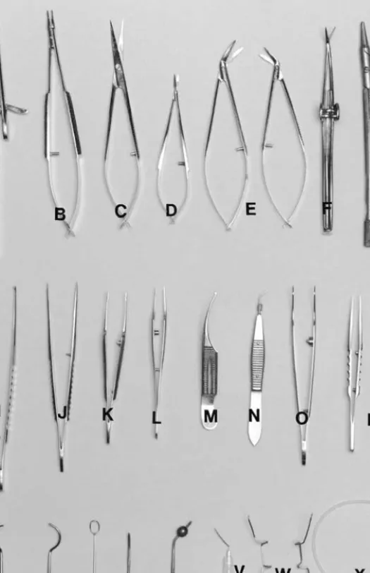
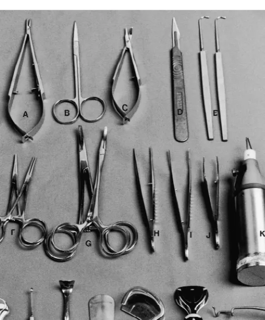
Sterility can only be achieved if steam is able to circulate around all the compart-ments and if all the air has been evacuated and none is trapped.There are specially designed autoclave bins for sterilising linen and dressings and specially designed autoclave trays for sterilising instruments. These have holes or vents in the side which can be opened to allow steam to enter and drive out all the air. At the end of the sterilisation they can be closed again so as to keep their contents sterile. It is convenient to have an intraocular tray and an extraocular tray which contain all the basic instruments for intraocular or extraocular surgery.The number of these trays will depend upon the work-load of the unit, and the exact instruments will obviously depend upon the surgeon’s preference. A basic intraocular and a basic extraocular set are illustrated in figs. 3.5 and 3.6. Setting out the instruments on trays before sterilization will limit damage to the instruments so that they will last longer. It also lessens the handling of instruments once they have been sterilised, and this will lessen the risk of infection. If it is necessary to handle instruments after sterilisation this should be done using the “no touch technique”, so that the instruments are neither lifted nor touched by the end that enters the patient’s eye. Autoclave tape may be applied to each article or alternatively to one package in a load. This special tape has stripes that change colour when the right conditions for sterilisation have been reached. It acts as a simple test for the efficient working of the autoclave.There are also autoclave indicators which change colour when the
A Heavy needle holder for large needles B Fine needle holder for fine needles C Conjunctival scissors
D Intraocular scissors (Vannas) E Right and left corneal scissors F Iridectomy scissors (De Wecker’s) G Razor blade fragment holder H Scalpel blade (Bard-Parker) I Heavy toothed forceps J Conjunctival forceps
K Fine corneo-scleral forceps with teeth at the tip (see page 34) L Fine corneo-scleral forceps with cups at the tip (see page 34) M Curved fine corneo-scleral forceps
N Intraocular forceps for handling the lens capsule (Kelman McPherson) O Capsule forceps for intracapsular extraction (Arruga)
P Fine suture tying forceps Q Right and left lens expressor R Lens loop
S Iris repositor
T Hot point cautery for use with spirit lamp (a battery cautery is shown in Fig. 3.6) U Irrigating cystitome
V Adjustable light speculum
(other patterns are shown on page 20, Figs. 2.3 and 2.4) X 2-way irrigating/aspirating cannula for extracapsular extraction Y Intraocular swabs
Z 2 cc and 5 cc syringes
A Needle holder B Straight scissors C Curved spring scissors D Scalpel blade
E Squint hooks
F Fine straight artery forceps G Curved artery forceps
H Conjunctival forceps without teeth I Heavy toothed forceps
J Fine toothed forceps K Battery cautery
L Meibomian cyst lid clamp M Small curette
N Eyelid retractor (Desmarres) O Eyelid guard
P Large eyelid clamp
Q Eyelid clamp (Cruikshank or Erhardt) R Adjustable speculum (Laing)
autoclaving has been completed satisfactorily. Special care should be taken when using an autoclave with attention to correct procedures and safety. The reservoir should be checked and kept at the right level. Only distilled or rain water should be used. Correct venting is essential although many autoclaves vent themselves at the end of the cycle. All staff using the autoclave require training and must strictly adhere to the instruction manual.
2. Dry Heat Oven
A hot air oven is quite expensive but requires very little maintenance and is a very effective way of sterilising instruments. It will preserve the edge of delicate cutting instruments better than an autoclave. However instruments must be scrupulously clean and dry first, and the process is very slow. It takes 1 hour at 180 degrees Centigrade to sterilise instruments, and then the instruments must be left to cool.
3. Boiling
The great advantage of boiling is that it is very quick, simple, easy and cheap. However boiling does not sterilise, it only disinfects. Boiling for 10 minutes will kill all bacteria but it will not kill spores. Although this is a problem in theory it is not a real problem in practice. However boiling has two particular disadvantages: 1. Repeated boiling will cause tarnishing and corrosion of instruments and will
certainly damage the cutting edge of fine instruments. Corrosion is lessened by adding 2% sodium carbonate to the solution.If possible, distilled water should be used and not tap water.This is available commercially from garages as water for car batteries. Distilled water will not cause corrosion.
2. After boiling the instruments are wet.They can be dried with a sterile gauze but this might compromise sterility and will break the “no touch technique” rule of not touching the instrument end that enters the eye. If they are used when wet, water from the handle of the instrument will drip down on to the working end and again compromise the no touch “technique”. If the instruments are placed in the boiler in a perforated rack or tray, then they will dry automatically and not require handling. In emergencies, drapes can be sterilised by boiling and then squeezed dry, although this is certainly not a technique to be recom-mended.
4. Chemical Solutions
Most chemical solutions are effective against bacteria but not against spores, and so they only disinfect but do not sterilise. However if the instruments are left in the chemical for long enough they may indeed sterilise. Chemical sterilisation and disinfection is good at preserving sharp instruments but there are two main disadvantages.
1. These chemical disinfectants are very toxic especially to the inside of the eye, and the instruments must be thoroughly rinsed in sterile water and dried very carefully after immersion in the chemical. This again will break the “no touch technique” rule.
2. With time or use, the solution may lose some of its potency, or may become chemically inactivated, or the alcohol in alcohol based chemicals may evaporate.
The most commonly used solutions are Chlorhexidine (trade name Hibitane) or ChlorhexidineandCetrimidecombined (trade name Savlon).
Hibitane is available as a 5% concentrate and needs to be diluted 10 times with 70% alcohol to make a solution which is 0.5% Chlorhexidine (Hibitane) in 70% alcohol and 30% water (i.e. one part of concentrate for 9 parts of 70% alcohol).
Savlon is available as a concentrate containing 1.5% Chlorhexidine and 15% Cetrimide and needs to be diluted thirty times in 70% alcohol (1 part of concentrate for 29 parts of 70% alcohol).
Both of these solutions should disinfect instruments after 10 minutes. After prolonged usage some of the alcohol may evaporate and the solution may lose its strength. If alcohol is not available concentrated Savlon diluted 1:30 in boiled water will disinfect instruments after 30 minutes.
Povidone Iodine is another widely used disinfecting agent, which is effective against most micro -organisms. A 10% solution can be made as follows :
Take 500ml of distilled water (cooled, boiled rain water is an alternative) Add sodium phosphate 16.6 gms and citric acid 3.4gms(these act as a chemical buffer)
Then add 50 gms of Povidone Iodine, thus making a 10% solution.This will also disinfect instruments after 10 minutes.
A 10% solution of Povidone Iodine can be used for cleaning the eyelids and the conjunctival sac preoperatively, but if it is left in the conjunctival sac for some time, some people think that 5% is safer.
Formalin vapouris an effective sterilising agent against all micro-organisms and spores. It is supplied as formalin tablets, which are placed in an airtight container at a warm room temperature. 12 hours are required for sterilisation.
The sterilisation of cryoprobes often presents a problem. They can only be autoclaved at lower temperatures and so will take half an hour for the cycle. Cryoprobes are quite often sterilised by immersion in chemical solutions between patients on the same list. As the probe actually enters the eye great care must be taken both to wash off all these chemicals, and also to make sure that the probe is not in any way contaminated. A small portable cryoprobe used with freon gas can however be safely boiled. (Do not boil the cylinder of gas itself, as it will explode!) Sterilised instruments, drapes and dressings can be safely stored and trans-ported in sealed autoclave bins and trays. If such bins or trays are not available all items must be double wrapped in linen before sterilisation and can then be safely stored or transported.
Stock keeping, Storage and Security
Good stock keeping to maintain essential supplies is often overlooked. However its importance cannot be over emphasised especially when there may be long delays between ordering supplies and their arrival. A system of monitoring stores and the rate at which consumables such as medicines, dressings, sutures etc. are used will allow for ordering and budgeting. Money is always limited so there is no place for holding excessive quantities. The only way to be aware of annual usage and any seasonal variations in consumption is to have a strict system of stock keeping.This
important part of theatre management should be the responsibility of the person in charge of the theatre.
It is obvious that equipment and supplies should be stored in a place where they will not deteriorate, and where they are safe.
Managing with Limited Resources
In many circumstances the volume of surgical work or financial limitations mean that correct theatre procedures cannot be followed strictly. In correct theatre procedures everybody who enters the operating theatre and all the staff and patients should wear a complete change of clothes. All those operating scrub up completely between cases with fresh gloves and a fresh gown. Often it may not be possible to maintain these standards. The patients may have to come to the operating theatre wearing their own clothes.The surgical staff should always scrub up completely at the beginning of a list, but may be obliged just to change their gloves in between cases or even to wipe their gloves in alcohol between cases. In order to maintain surgical through put it may be necessary to have more than one operating table in the same operating room.
To ensure safe surgery there are four areas in which “cutting corners” and compromises are strictly forbidden. These are:
1. Correct preparation of the patient for surgery, with a thorough cleaning of the face and skin preparation around the eye (see pages 57–8). This includes “marking” the eye for surgery to avoid mistakes.
2. Sterilisation of all instruments, drapes and dressings. If an item like an intraocular lens comes from the manufacturer in a sterile wrapping, then make sure that the packaging has not in any way been damaged.
3. Sterilisation and purity of all solutions used in intraocular irrigation.
4. The correct handling of instruments and dressings and the use of a “no touch technique”. Nothing that enters the eye should touch the eyelids or lashes. People working in small units, or on tour in rural areas may have to rely on boiling and chemicals as the only means of disinfecting between cases. Although this is not the best way it appears to be safe as long as everyone sticks to the rules. Some people will boil the blunt instruments and use chemicals for the sharps.
•
Don’t try to shorten the time needed for immersion in chemicals or boiling.•
Make sure that the chemical solution is fresh.•
Make sure that the instruments are dried with a sterile swab and there is no chemical residue on the instruments. This should be done by someone who understands how important this is.•
Never rely on just wiping the instruments between cases with spirit, ether or acetone. It just isn’t safe enough.Infection Control Strategy
Post-operative infections do sometimes occur, even with the most careful and well trained surgical team. Good units have an infection rate of about one case in a thousand or less. It is reasonable to assume that any infection developing within the first post-operative week has been contracted at the time of the operation. Even an isolated case of post-operative infection should make the surgeon and the surgical team review all their techniques, equipment and procedures. If several infections occur close to each other an even more radical overhaul of theatre procedures is required. Any irrigating fluids should be discarded and a new batch obtained. All made up disinfectant solutions should be discarded and new ones made, and the steriliser changed or a new method of sterilisation tried. If available an infection control officer or a microbiology department will help to trace the source of an infection.The following is a list of the more common possible sources of infection during surgery:
Sources of Infection
From the patient
Conjunctivitis. Blepharitis. Dacryocystitis.Septic lesion near the eye. From the staff Septic lesions.
Poor scrubbing up. Contaminated gloves.
Failure to observe″no touch technique″by the surgeon or assistant, or touching the lids or lashes with instruments entering the eye. Prolonged manipulation during the operation. From the theatre equipment Contaminated irrigating fluid.
Faulty steriliser.
Faulty sterilising technique.
Contamination of sterile trolleys e.g. from flies. Contamination or inactivation of disinfectant
solutions.
Broken or defective packaging for an IOL or instrument which is factory sterilised.