Delayed reconstruction of lateral complex structures of the
ankle
Gordon L Slater, Sports Foot and Ankle Surgery,Edgecliff Sydney NSW 2027, Australia
Alejandro E Pino, Hospital for Special Surgery, Department of Foot and Ankle Surgery, 535 E 70 St.,New York, NY 10021, United States
Martin O’Malley, Hospital for Special Surgery, Department of Foot and Ankle Surgery, 535 E 70th St., New York, NY 10021, United States
Author contributions: Slater GL, Pino AE and O’Malley M contributed equally to writing this work; Slater GL contributed data.
Correspondence to: Gordon L Slater, MD, Sports Foot and Ankle Surgery, Level 2, Suite 211, 204-233 New South Head Road, Edgecliff NSW 2027, Australia. [email protected] Telephone: +61-1300388778 Fax: +61-60409555 Received: December 29, 2010 Revised: April 8, 2011 Accepted: April 15, 2011
Published online: April 18, 2011
Abstract
Lateral ankle instability is one of the most common and well-recognized conditions presenting to foot and ankle surgeons. It may exist as an isolated entity or in conjunction with other concomitant pathology, making it important to appropriately diagnose and identify other conditions that may need to be addressed as part of treatment. These associated conditions may be a source of chronic pain, even when the instability has been appropriately treated, or may lead to failure of treatment by predisposing the patient to ankle inversion injuries. The primary goal of this editorial is to provide a brief summary of the common techniques used in the delayed reconstruction of lateral ankle ligamentous injuries and present a method we have successfully employed for over 15 years. We will also briefly discuss the diagnosis and treatment of the more common associated conditions, which are important to identify to achieve satisfactory results for the patient. We present the outcomes of 250 consecutive reconstructions performed over the last 10 years and
describe our operative technique for addressing lateral ankle ligamentous injuries.
© 2011 Baishideng. All rights reserved.
Key words: Lateral ankle ligaments; Ankle instability; Ankle ligament reconstruction; Anatomic ligament reconstruction; Ankle sprain
Peer reviewer: Evangelos Pappas, MD, PhD, Professor, Associate Professor, Division of Physical Therapy, Long Island University-Brooklyn Campus, University-Brooklyn, NY 11201, United States
Slater GL, Pino AE, O’Malley M. Delayed reconstruction of lateral complex structures of the ankle. World J Orthop 2011; 2(4): 31-36 Available from: URL: http://www.wjgnet. com/2218-5836/full/v2/i4/31.htm DOI: http://dx.doi. org/10.5312/wjo.v2.i4.31
INTRODUCTION
Ankle instability is one of the most common conditions treated by orthopedic surgeons, especially those specializing in conditions of the foot and ankle or sports medicine[1,2].
It is generally accepted that the majority of these injuries can be treated successfully with conservative measures, yet there remains a percentage of patients that go on to suffer from sequelae of the injury. Most patients with chronic complaints stemming from the initial event, found to be as high as 20% of individuals sustaining twisting injuries, complain of stiffness or various patterns of instability[3-5].
These chronic conditions can usually be treated with conservative modalities and are tolerated well by the majority of patients but, in the case of gross instability or high patient activity level, surgical intervention may be warranted. Recent literature has found an association between ankle instability and the development of osteoarthritis, which may be a cause of prolonged and worsening symptoms after neglected ankle instability[6,7].
Gordon L Slater, Alejandro E Pino, Martin O’Malley
BRIEF ARTICLE
Determining which patients may go on to chronic instability and the appropriate time to intervene with more aggressive forms of treatment continues to be a challenge for treating physicians.
PATHOGENESIS
Colville described reconstruction of the lateral ligamen-tous complex of the ankle after a combined inversion-plantar flexion injury of the foot, which led to rupture of the ankle joint capsule, anterior talofibular ligament (ATFL) and the calcaneofibular ligament (CFL)[8].
Brostrom, well known for his contribution to the diag-nosis and treatment of ankle instability, found that 27% of patients evaluated for ankle injuries with ATFL dis-ruption had concomitant CFL involvement. Fifty eight percent of these patients had increased inversion of the ankle when compared to the uninvolved extremity[9].
Advanced imaging may not be necessary in the evalua-tion of all patients with ankle inversion injuries but, when obtained, rarely show injury to the posterior talofibular ligament, even in the setting of ATFL and CFL disrup-tion[10]. On the other hand, MRI evidence of ATFL
dis-ruption may not be clinically significant. MRI has proven useful in the evaluation of chronic ankle discomfort after recurrent ankle injuries, especially when attempting to identify associated injuries, such as osteochondral injuries or peroneal pathology. One should keep in mind, howev-er, that a normal MRI does not preclude ankle instability.
DIAGNOSIS
Patients with ankle instability typically present for treat-ment after sustaining a severe twisting injury which has progressed to recurrent sprains. The severity of the inju-ry has been linked to the clinical presentation, such that those patients who are unable to bear weight or return to sport immediately after the injury or have difficulty walk-ing on uneven surfaces usually have, at the very least, a complete rupture of the ATFL[11].
Diagnosis is typically based on clinical findings and is made when there is increased laxity on anterior drawer and inversion testing compared to the contralateral ankle. An attempt to isolate rotational instability of the ankle helps to identify if there is an injury to the syndesmosis. Other findings in these high ankle sprains include inability to perform a single leg hop on the involved side, a posi-tive squeeze test and pain with external rotation of the ankle. Some advocate the use of stress radiographs in the workup of ankle laxity, while others question the validity of this modality[12-19]. The use of the Telos device aims to
more objectively quantify the degree of instability[16, 17].
In order to ensure optimal results in treating patients with ankle instability, it is of utmost importance to iden-tify and treat pathology that may be a result of, or lead to, ankle instability. Examples of associated pathology include osteochondral defects, peroneal injuries and
neu-rological conditions leading to muscle imbalance. The heel should be examined for proper alignment because varus malalignment may lead to recurrent injuries after treatment, or failure of ligamentous reconstruction.
Most advocate conservative treatment for acute liga-mentous injuries although there is some evidence that suggests that early surgical intervention leads to a lower incidence of chronic instability[3,9,20-22]. There is no reliable
objective criteria that we are aware of that can be used to help dictate whether one will proceed to chronic instabil-ity after an acute injury. Once a patient does present with these symptoms after failure of conservative means, sur-gery should be considered. Such complaints in patients with high activity levels, when combined with positive findings on physical exam, may best be addressed with surgical reconstruction of the injured ligaments.
SURGICAL OPTIONS
The surgical treatment of lateral ligamentous injuries can be classified as either anatomic or non-anatomic recon-structions. Brostrom popularized an anatomic technique for the repair of ligamentous injuries, while Kalsson et al found that a delayed anatomic reconstruction was also possible[9,17]. These reconstructions aim at re-establishing
continuity to both the ATFL and CFL through direct repair, which may further be augmented with an imbri-cation of the inferior retinaculum[23]. Longitudinal,
trans-verse and oblique incisions have been described and se-lection should be dictated by any associated pathology[24].
Typically on gross examination a redundant and incom-petent ligamentous complex is identified. The ligaments should be identified and directly repaired at a length that provides sufficient tension to stabilize the ankle.
Non-anatomic ligamentous reconstructions, such as the Evans, Watson-Jones and Chrisman-Snook proce-dures, have been described in the literature[10,25-32]. These
procedures involve rerouting of a slip of autogenously harvested tendon to impart stability to the ankle and sub-talar joint. Stiffness, arthritis and complications associated with tendon harvest have been attributed to these tech-niques, yet they may still have an important role in liga-mentous reconstruction. Non-anatomic reconstructions may best be suited for those patients with long-standing instability, hypermobility or a failed anatomic repair[17].
Unlike the original technique described by Brostrom, our preferred approach to lateral ankle ligament recon-struction is to begin with an arthroscopic evaluation and treatment (Figure 1) of any intraarticular pathology, when indicated. This is undertaken with a 2.7 mm arthroscope and a low-pressure calf tourniquet. The joint and syndes-mosis are inspected and articular injuries or intraarticular scar formation may be addressed. Any gross instability of the syndesmosis may need to be addressed with a separate stabilization technique, which is outside the scope of this editorial. When peroneal pathology is identified through a thorough preoperative evaluation, a posterolateral portal
may be used for tendoscopy and tendon debridement or for a more thorough evaluation when the preoperative evaluation was equivocal. This portal may later be incor-porated into a longitudinal incision for ligamentous re-construction, which allows for treatment of any peroneal pathology in an open fashion.
SURGICAL TECHNIQUE
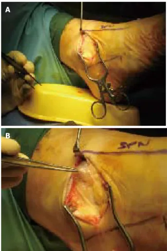
Our incision is dictated by other pathology and ensures exposure of the lateral ligamentous complex, inferior retinaculum and the peroneal tendons, if necessary (Figure 2). Dissection through the subcutaneous tissue is performed in line with the incision, protecting any branches of the superficial peroneal nerve when identified (Figures 3A and 3B). The ATFL, CFL and anterior joint capsule are identified, allowing for mobilization and retraction of the inferior retinaculum. Careful dissection is made at the level of the CFL in order to prevent injury to the peroneal tendons. The anterior joint capsule is then incised transversely, leaving a stump of capsule attached to the fibula for later repair. The ankle joint may now be
explored if arthroscopy was not first performed.
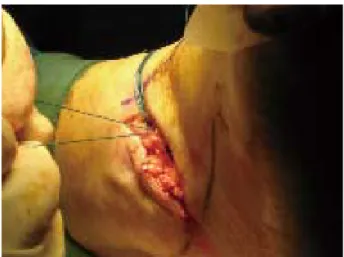
Reconstruction is then begun with placement of mul-tiple, interrupted, nonabsorbable sutures in the capsule, ATFL and CFL, allowing for later sequential imbrication (Figure 4). The sutures are tied with the foot positioned in an eversion, followed by advancement of the inferior retinaculum over the imbricated repair (Figure 5), which is secured to the fibula with suture or suture anchor. A layered closure is then performed, followed by place-ment of a soft dressing and a supportive boot or splint.
Two hundred and fifty patients who underwent surgi-cal reconstruction of their lateral ligamentous structures by a single surgeon between 1998 and 2008 were con-tacted for interview. Patients were included in the study if they underwent lateral ankle ligament reconstruction due to symptomatic instability with sporting activity and activi-ties of daily living despite a course of conservative man-agement. All patients had more than three instances of what they considered significant instability, with findings on physical exam that included ecchymosis, diffuse soft tissue swelling, pain along the course of the ATFL and CFL and an inability to bear weight immediately after the injury. The majority of patients in this study listed Aus-tralian Rules football, basketball, netball or soccer as the sporting activity during which the initial injury occurred.
B
Figure 1 Portal Placement and set-up for ankle arthroscopy.
Figure 2 Placement of a curved, anteriolateral skin incision between the superficial peroneal nerve and peroneals, just distal to the fibula.
A
Figure 3 Identification (A) and isolation (B) of a branch of the superficial peroneal nerve.
Patients with a varus heel alignment or peroneal weakness due to a neurologic condition were excluded from the study.
The most commonly encountered concomitant in-juries at the time of lateral ligamentous reconstruction included intraarticular synovitis, hypertrophy of the ATFL leading to impingement and discrete osteochondral inju-ries. Any area of synovitis or soft tissue impingement was treated with arthroscopic debridement, while osteochon-dral lesions were generally small and amenable to debride-ment and osteoplasty.
The female to male ratio was 1:2, with a mean age of 34.47 (range 9-90). The patients were asked a series of questions in regard to postoperative pain and function, with 2.25% of patients admitting to residual pain. Two of these patients had a Brostrom procedure in conjunction with internal fixation of an ankle fracture. The remainder of the patients remain pain free at their most recent evalu-ation. The overall satisfaction rate was 91%.
The majority of patients denied continued instability, with 78% returning to activities without recurrence of in-version injuries. Of the remaining patients, 13% returned to a pre-injury level of activity with some residual instabil-ity, while 9% were limited in their ability to return to activ-ity by continued instabilactiv-ity.
The delayed reconstruction of the lateral ankle liga-mentous complex is a well-established technique for the surgical management of ankle instability. The authors prefer an anatomic repair, with the addition of retinacu-lum advancement, as described by Brostrom and Gould[9, 23]. Satisfactory reports have been described with the use
of non-anatomic, tendon weaving repairs, but we feel that these techniques should be reserved for salvage cases due the associated complications, such as stiffness and sacrifice of a portion of the peroneals[25, 26, 28, 30-32]. The
pe-roneals are important stabilizers of the hindfoot and play a vital role in eversion of the foot and maintenance of the longitudinal arch. Harvesting of the peroneal tendons has been associated with loss of strength in approximately 8% of patients[33].
In order to ensure optimal results from ligament recon-struction, it is important to identify and address any fac-tors that may predispose the patient to recurrent instabil-ity, such as varus heel malalignment and peroneal injuries. Other factors that may lead to poor results after surgical intervention include unrecognized concomitant pathology, such as articular cartilage injuries, unrecognised fractures, ankle synovitis or soft tissue impingement. In our experi-ence, the addition of an arthroscopic examination aids in the diagnosis and treatment of these intraarticular condi-tions that may predispose patients to poor outcomes if not appropriately addressed.
In the current series, the most commonly encountered intraarticular injuries included areas of soft tissue impinge-ment and osteochondral lesions. A recent study investi-gated causes for residual disability after ankle sprains in an attempt to compare the ability of arthroscopy to diagnosis pathology versus standard and advanced imaging[34]. The
authors found arthroscopy to be as good in identifying intraarticular pathology as other imaging and able to iden-tify missed lesions in 14 of 72 patients. Fifty-four percent of patients had ATFL injuries, 40.3% had osteochondral lesions and 4.2% of patients had impingement due to fibrous bands. Of those patients with impingement due to fibrous bands, arthroscopy was the only means of de-tecting the abnormality. A similar study in which arthros-copy was used to detect pathology in chronically unstable ankles found osteochondral injuries in 66% of patients and elongation of the ATFL and CFL in 86% and 64% of patients respectively[35].
Ankle inversion injuries continue to be a common condition, with a minority of patients progressing to chronic pain and instability. The diagnosis and treatment of this condition is well described, but the literature is lim-ited to small series with limlim-ited follow-up. We present our experience with ankle instability in the form of disruption of the lateral ankle ligamentous complex in order to illus-trate that good long term results can be anticipated in ap-propriately selected patients with an anatomic reconstruc-tion, as long as all pathology is identified and addressed. Figure 5 Imbrication of the lateral structures with a retinacular reinforcement. Figure 4 Interrupted suture placement in the lateral ligamentous complex.
The treating physician should be knowledgeable in terms of other injuries commonly associated with lateral ankle ligamentous injuries in order to properly evaluate and care for their patients.
COMMENTS
BackgroundAnkle sprains are among the most common musculoskeletal injuries seen by physicians. The majority of these injuries have satisfactory outcomes with the use of conservative treatments, but in the face of continued instability and patient limitation in spite of such intervention, surgical reconstruction of the lateral ligament complex may be warranted. In our experience, good long- term outcomes can be obtained with the use of an anatomic reconstruction of the lateral ligaments of the ankle.
Research frontiers
There has been some debate as to wether an anatomic or nonanatomic reconstruction of the lateral ligaments yields better results. Nonanatomic reconstructions have been associated with more hindfoot stiffness, and usually sacrifice a portion of the peroneals. There has also been some controversy in regards to the use of arthroscopy in the treatment of ankle instability, which we believe is an important tool, when used appropriately, in the treatment of chronic lateral ligament dysfunction.
Innovations and breakthroughs
Many researchers have found good short-term results with the use of an anatomic reconstruction of the lateral ligaments of the ankle. We have shown that the majority of our patients continue to have a high satisfaction rate at ten-year follow-up, with 91% returning to a pre-injury functional level. We also present the directed use of arthroscopy as an important tool in the treatment of concomitant injuries associated with chronic ankle instability.
Applications
The use of an anatomic approach to the reconstruction of the lateral ligaments of the ankle in our practice, when combined with an arthroscopic treatment of any intraarticular pathology, when indicated, has led to a high patient satisfaction rate.
Terminology
Lateral ligament complex: anterior talofibular ligament (ATFL) and the calcaneofibular ligament (CFL). Modified Brostrom: Anatomic repair of the ATFL and CFL, with imbrication of the inferior retinaculum. Directed arthroscopy: Arthroscopic treatment of concomitant pathology identified preoperatively with a thorough history and physical exam, in conjunction with the use of appropriate diagnostic modalities.
Peer review
Overall, this was an interesting editorial on the author’s preferred treatment of ankle instability which will be of interest to readership.
REFERENCES
1 Brand RL, Black HM, Cox JS. The natural history of inadequately treated ankle sprain. Am J Sports Med 1977; 5: 248-249 2 Glick JM, Gordon RB, Nishimoto D. The prevention and
treatment of ankle injuries. Am J Sports Med 1976; 4: 136-141 3 Freeman MA. Treatment of ruptures of the lateral ligament
of the ankle. J Bone Joint Surg Br 1965; 47: 661-668
4 Kannus P, Renström P. Treatment for acute tears of the lateral ligaments of the ankle. Operation, cast, or early controlled mobilization. J Bone Joint Surg Am 1991; 73: 305-312
5 Pennal GF. Subluxation of the Ankle. Can Med Assoc J 1943;
49: 92-95
6 Sugimoto K, Takakura Y, Okahashi K, Samoto N, Kawate K, Iwai M. Chondral injuries of the ankle with recurrent lateral instability: an arthroscopic study. J Bone Joint Surg Am 2009;
91: 99-106
7 Valderrabano V, Hintermann B, Horisberger M, Fung TS. Ligamentous posttraumatic ankle osteoarthritis. Am J Sports Med 2006; 34: 612-620
8 Colville MR. Reconstruction of the lateral ankle ligaments.
Instr Course Lect 1995; 44: 341-348
9 Broström L. Sprained ankles. V. Treatment and prognosis in recent ligament ruptures. Acta Chir Scand 1966; 132: 537-550 10 Saltrick KR. Lateral ankle stabilization. Modified Lee and
Chrisman-Snook. Clin Podiatr Med Surg 1991; 8: 579-600 11 Balduini FC, Vegso JJ, Torg JS, Torg E. Management and
rehabilitation of ligamentous injuries to the ankle. Sports Med 1987; 4: 364-380
12 Ahovuo J, Kaartinen E, Slätis P. Diagnostic value of stress radiography in lesions of the lateral ligaments of the ankle.
Acta Radiol 1988; 29: 711-714
13 Bulucu C, Thomas KA, Halvorson TL, Cook SD. Biomechanical evaluation of the anterior drawer test: the contribution of the lateral ankle ligaments. Foot Ankle 1991; 11: 389-393 14 Laurin C, Mathieu J. Sagittal mobility of the normal ankle.
Clin Orthop Relat Res 1975; : 99-104
15 Rijke AM, Vierhout PA. Graded stress radiography in acute injury to the lateral ligaments of the ankle. Acta Radiol 1990;
31: 151-155
16 Sauser DD, Nelson RC, Lavine MH, Wu CW. Acute injuries of the lateral ligaments of the ankle: comparison of stress radiography and arthrography. Radiology 1983; 148: 653-657 17 Karlsson J, Bergsten T, Lansinger O, Peterson L. Reconstruction
of the lateral ligaments of the ankle for chronic lateral instability. J Bone Joint Surg Am 1988; 70: 581-588
18 Rijke AM, Jones B, Vierhout PA. Injury to the lateral ankle ligaments of athletes. A posttraumatic followup. Am J Sports Med 1988; 16: 256-259
19 Cox JS, Hewes TF. "Normal" talar tilt angle. Clin Orthop Relat Res 1979; 140 : 37-41
20 Anderson KJ, Lecocq JF. Operative treatment of injury to
the fibular collateral ligament of the ankle. J Bone Joint Surg Am 1954; 36-A: 825-832
21 Grønmark T, Johnsen O, Kogstad O. Rupture of the lateral ligaments of the ankle: a controlled clinical trial. Injury 1980;
11: 215-218
22 Kolind-Sorensen V. [Lesions of the lateral ligament of the ankle joint]. Ugeskr Laeger 1975; 137: 1637-1638
23 Gould N, Seligson D, Gassman J. Early and late repair of lateral ligament of the ankle. Foot Ankle 1980; 1: 84-89 24 Sobel M, Warren RF, Brourman S. Lateral ankle instability
associated with dislocation of the peroneal tendons treated by the Chrisman-Snook procedure. A case report and literature review. Am J Sports Med 1990; 18: 539-543
25 Watson-Jones R. The classic: "Fractures and Joint Injuries" by Sir Reginald Watson-Jones, taken from "Fractures and Joint Injuries," by R. Watson-Jones, Vol. II, 4th ed., Baltimore, Williams and Wilkins Company, 1955. Clin Orthop Relat Res 1974; 105 : 4-10
26 van der Rijt AJ, Evans GA. The long-term results of Watson-Jones tenodesis. J Bone Joint Surg Br 1984; 66: 371-375 27 Snook GA, Chrisman OD, Wilson TC. Long-term results
of the Chrisman-Snook operation for reconstruction of the lateral ligaments of the ankle. J Bone Joint Surg Am 1985; 67: 1-7
28 Chrisman OD, Snook GA. Reconstruction of lateral ligament tears of the ankle. An experimental study and clinical
evaluation of seven patients treated by a new modification
of the Elmslie procedure. J Bone Joint Surg Am 1969; 51: 904-912
29 EVANS DL. Recurrent instability of the ankle; a method of surgical treatment. Proc R Soc Med 1953; 46: 343-344
30 Gillespie HS, Boucher P. Watson-Jones repair of lateral instability of the ankle. J Bone Joint Surg Am 1971; 53: 920-924 31 Hedeboe J, Johannsen A. Recurrent instability of the ankle joint. Surgical repair by the Watson-Jones method. Acta Orthop Scand 1979; 50: 337-340
32 Liu SH, Baker CL. Comparison of lateral ankle ligamentous
reconstruction procedures. Am J Sports Med 1994; 22: 313-317 33 St Pierre RK, Andrews L, Allman F Jr, Fleming LL. The Cybex II evaluation of lateral ankle ligamentous reconstructions.
Am J Sports Med 1984; 12: 52-56
34 Takao M, Uchio Y, Naito K, Fukazawa I, Ochi M. Arthroscopic
assessment for intra-articular disorders in residual ankle disability after sprain. Am J Sports Med 2005; 33: 686-692 35 Hintermann B, Boss A, Schäfer D. Arthroscopic findings in
patients with chronic ankle instability. Am J Sports Med 2002;
30: 402-409