1
Effect of Pupil Size on Corneal Aberrations before and after
Standard, Custom LASIK and Corneal Refractive Therapy
António Queirós 1, César Villa-Collar 2, José Manuel González-Méijome 1, Jorge Jorge 1, Angel Ramón Gutiérrez 31
Clinical & Experimental Optometry Research Lab. Department of Physics (Optometry), School of Sciences, University of Minho, Braga, Portugal. 2
Clínica Oftalmológica Novovision, Paseo de la Castellana, Madrid, Spain. 3
Department of Ophthalmology, University of Murcia, Murcia, Spain.
Corresponding Author:
António Queirós
Clinical & Experimental Optometry Research Lab Department of Physics (Optometry)
Campus Gualtar University of Minho 4710-057 Braga – Portugal Tel.: +351 253 60 40 72 Fax : +351 253 67 89 81 E-mail: aqp@fisica.uminho.pt
ABSTRACT
PURPOSE: To evaluate the effect of changing the pupil size on the corneal first surface higher order aberrations induced by different refractive treatments (standard LASIK; custom LASIK and corneal refractive therapy).
DESIGN: Observational study.
METHODS: SETTING: Clínica Oftalmológica NovoVision, Madrid, Spain. PATIENTS: Eighty-one right eyes with a mean age of 29.94±7.5 years, of which 50 were female (61.7%), were retrospectively analyzed. Corneal videokeratographic data were used to obtain corneal first surface higher order aberrations (HOA) for aperture diameters from 3 to 8 mm using the Vol-CT software. Total Root Mean Square (RMS) and RMS for 3rd to 6th-order Zernike polynomials as well as spherical-like, coma-like,
secondary-astigmatism and spherical+coma-like were calculated.
RESULTS: We verified an increase of the HOA Total RMS after treatments of
0.014±0.025µm, 0.019±0.027µm 0.018±0.031µm for standard LASIK, custom LASIK and corneal refractive therapy, respectively for 3mm pupil diameter. For the 8mm aperture diameter changes in Total RMS increased by a factor of 50x compared with the variation for the 3mm diameter up to 0.744±0.731µm, 0.493±0.794µm,
0.973±1.055µm for standard LASIK, custom LASIK and corneal refractive therapy, respectively.
CONCLUSIONS: The three techniques increase the wavefront aberrations of the cornea and change the relative contribution of coma-like and spherical-like
aberrations. For a large aperture (>5mm), corneal refractive therapy induces more spherical-like aberrations than standard LASIK and this more than custom LASIK. However, no differences clinically or statistically significant did exist for narrower apertures. Standard LASIK and custom LASIK did not displayed statistically significant differences regarding to HOA.
3
INTRODUCTION
Laser-assisted in situ keratomileusis (LASIK) and corneal refractive therapy (CRT) for orthokeratology are two techniques that attempt a certain independency from conventional compensation by spectacles or traditional contact lenses.1-3 Both of these techniques use a similar principle to correct myopia, which is making the central corneal surface flatter, thus reducing the total power of the eye. However, they are substantially different in the way they achieve such effect. While LASIK removes stromal tissue, corneal refractive therapy produces a redistribution of the corneal thickness, affecting particularly the epithelium. In both cases the peripheral cornea remains unchanged. (Queiros et al 2009, accepted for publication).
After myopia treatment with either of the aforementioned techniques there is an increase in corneal asphericity (Q), changing from its initially prolate shape (Q<0) to an oblate contour (Q>0), being flatter in the centre than in the paracentral zone surrounding the treatment area.1,4-7 However even when the anterior corneal surface has been classically defined by a unique Q value, or two Q values corresponding to the orientations of the two principal meridians,8 corneal asphericity changes
significantly depending on the peripheral reference point taken for the calculations.9 It is also expected that these multi-aspheric concept to be more complex after corneal refractive procedures and even depending on the technique used. For example, wavefront-guided or topography-guided laser surgery, also know as customized LASIK (CL) is supposed to induce a less negative impact on quality of vision compared with standard LASIK procedures (SL).
Alterations in Q produce an increase in optical aberrations with a significant impact in the quality of vision,10 but also on contrast sensitivity11,12 and other aspects of visual function such as night vision disturbances.3 With the development of
techniques for measuring optical quality of the eye, several studies have allowed for a better knowledge of the optical quality of the corneal surface after LASIK3,12,13 or corneal refractive therapy.11,14 Both refractive techniques significantly increase higher order aberrations in the eye,1 particularly third and fourth order aberrations.14,15
These particular aberrations have shown to be those with more impact in the visual quality of the eye.16
The ocular aberrations are major determinant factors of the retinal image quality. These aberrations of the eye are the combination of aberrations of the anterior corneal surface plus those from the internal ocular media and depend on many factors and conditions: changing from person to person,17 depending on the pupil size,18 age,19 accommodation,20 retinal eccentricity21 and the refractive condition.22
Since the treatment zones vary significantly according to treatment technique and the cornea’s response to the different correction procedures,23 and because the cornea possesses different degrees of asphericity according to the corneal zone being analysed,9 it is important to study aberration values for different corneal diameters in order to fully characterize this important property that defines the post-surgical corneal contour, as well as evaluate its impact on the higher order
aberrations (HOA) induced as a consequence of such changes.
The purpose of this study was to evaluate the changes of different HOA of the corneal anterior surface as a function of diameter being analyzed after two surgical and one non-surgical refractive treatments (corneal refractive therapy, standard LASIK and custom LASIK).
5
METHODS
Subjects and inclusion criterion
The clinical records of 81 patients submitted to corneal refractive therapy (CRT, n=27), standard LASIK (SL, n=27) and customized LASIK (CL, n=27) at the ophthalmology clinic Novovision, (Madrid, Spain) have been retrospectively analyzed and their corneal topographies processed using custom Vol-CT 6.89 software (Sarver & Associates, Inc., Carbondale, Illinois, USA). Only the right eye from each patient was considered for statistical analysis in order to avoid the known correlation
between the response of both eyes from same individual to treatment. Only patients with myopia between -0.75D and -4.25D and astigmatism below -1.75D were
included in order to match the range of treatments more commonly performed in corneal refractive therapy. When the right eye did not meet the previous inclusion criterion, the left eye was used. No patient suffered from ocular disease or had been submitted to previous ocular surgery. Complete optometric and ophthalmological examinations were performed before surgical and non-surgical correction of myopia through the aforementioned techniques. A minimum of 3 months was required to guarantee that the topography was stable.24,25 After that, the patients should have demonstrated to be successfully treated regarding to residual refractive error (≤
±0.50), visual acuity (≥20/20 or higher uncorrected visual acuity), surface regularity and centering of the treatment zone (less than 0.5 mm of decentration) before being elected for this study. Another important inclusion criterion was that the
videokeratoscope examinations had been performed between 4:00 and 8:00 P.M. to minimize the influence of diurnal variations in corneal thickness26 that might
potentially influence anterior corneal topography.27
LASIK surgery
In all cases the ablation was central, with an optic zone of 6.50 mm for all LASIK treatments. A transition zone of 0.30 mm for the spherical cases in the standard LASIK group and 1.25 mm for astigmatic corrections and custom LASIK procedures was used.
Surgical routine for LASIK surgery was held according to international standards, and the commonly accepted criteria for refractive surgery procedures
were observed regarding to predictability, efficacy and safety. After creating a 120 µm, 9.5 mm diameter flap with a Hansatome microkeratome (Chiron Vision, model 2765; Bausch & Lomb, Claremont, California, USA), standard LASIK and custom LASIK ablation profiles were produced using the Allegretto Wave Eye-Q - 400 Hz - (Wavelight, Erlangen, Germany). All surgical procedures were uneventful and successful.
Corneal refractive therapy lens characteristics
The rigid gas permeable material used for the CRTTM lenses (paflufocon D, Dk=100 barrer - Paragon Vision Sciences, Mesa, AZ, USA) with parameters, base curve radius (BCR=8.52±0.22 mm [8.2,9.0]), return zone depth (RZD=531.00±23.14 [500,575] µm) and landing zone angle (LZA=32.72±1.10 degrees [31,35]). Trial lenses were derived from nomograms in the form of sliding tables produced by the manufacturer Paragon CRT sigmoid reverse geometry contact lens.28 Fitting was evaluated according to the recommendations of the manufacturer regarding fluorescein pattern, topographical evaluation, refractive and visual outcomes.
Computing corneal monochromatic HOA from corneal topography
Topographic data were obtained using the Atlas Mastervue videokeratoscope (Humphrey Zeiss Instruments, San Leandro, CA, USA). The corneal topographer was calibrated before data acquisition according to the manufacturer’s
recommendations. Corneal videokeratographic data were downloaded onto floppy disks in ASCII file format, which contained information about corneal elevation, curvature, power and position of the pupil.
HOA were expressed as Zernike polynomials Z3-3 to Z66, which comprise corneal aberrations up to the sixth order using the Calculations facility of Vol-CT 6.89 software (Sarver & Associates, Inc., Carbondale, Illinois, USA). Total HOA Root Mean Square (RMS) and RMS values for 3rd, 4th, 5th and 6th-order were calculated. Total HOA RMS (including Zernike polynomials Z3-3, Z3-1,…, Z64, Z66), spherical-like aberrations (including Zernike polynomials Z40 and Z60), coma-like aberrations (including Zernike polynomials Z3-1, Z31, Z5-1 and Z51), secondary astigmatism
(including Zernike polynomials Z4-2, Z42, Z6-2 and Z62) and another RMS resulting from the sum of spherical-like and coma-like aberrations. All aberrations were calculated
7
for a aperture diameter between 3mm to 8 mm (Sph_3mm to Sph_8mm for spherical-like aberrations; Coma_3mm to Coma_8mm for coma-spherical-like aberrations; Total_3mm to Total_8mm for total aberrations; and Astig_3mm to Astig_8mm for secondary
astigmatism).
Statistical analysis
The SPSS software package v.16 (SPSS Inc., Chicago, IL, USA) was used for statistical analysis. Kolmogorov-Smirnov Test was applied in order to assess
normality of data distribution. Kruskal-Wallis Test or ANOVA were performed to evaluate whether statistically different values were present among the clinical groups of standard LASIK, custom LASIK and corneal refractive therapy. When normality could not be assumed, Wilcoxon Signed Ranks Test was used for paired comparison between techniques and Paired Samples Test was used when normality could be assumed for pair comparisons between treatments. For statistical purposes, a P value lower than 0.05 was considered statistically significant.
RESULTS
Mean age was 29.94±7.5 years (ranging from 16 to 49) for the 81 subjects of which 50 were female (61.7%) and 31 were male (38.3%). Total preoperative spherical equivalent was -2.82±0.77 D ranged from -1.50 to -4.38D.
Table 1 presents descriptive data for demographic characteristics and refractive components M, J0 and J45 before treatment and statistical comparison between three treatment groups. No statistically significant differences were found for the comparison of spherical equivalent (P = .998, Kruskal-Wallis Test) nor for J0 or J45 between the three conditions before treatment.
Preoperative data for different RMS analyzed are presented in tables 2, 3 and 4. There were no statistically significant differences between three techniques for all RMS values at all diameters under analysis with the exception for Coma_4mm (P = .038, ANOVA), Sph_6mm (P = .032, ANOVA) and Sph_7mm (P = .041, ANOVA).
Table 2, 3 and 4 shows the values of different aberrations analyzed for the standard LASIK, custom LASIK and corneal refractive therapy before treatment, after treatment and the difference between them (mean ± standard deviation). Statistical significance is shown for the difference in all values of the aberrations studied and for the different diameters from 3 mm to 8 mm. We found statistically significant
differences in all diameters for the 4th-order RMS, total RMS, coma-like RMS and spherical+coma RMS in the standard LASIK group. In the custom LASIK group we found statistically significant differences only in the 4th-order aberrations in all diameters. Moreover, the custom LASIK treatment was the one with fewer items (RMS for a certain diameter) showing statistically significant changes. In the case of CRT corneal refractive therapy, all aberrations showed statistically significant
differences except for the 5th-order and 6th-order RMS.
Table 5 shows the values of change in RMS for the standard LASIK, custom LASIK and corneal refractive therapy interventions (mean post treatment minus baseline ± standard deviation), the statistical comparison between laser treatments (standard LASIK vs. custom LASIK) and among all treatments (standard LASIK, custom LASIK and corneal refractive therapy). No statistically significant differences
9
were found for the comparison of standard LASIK and custom LASIK (P > .063, T-test). Statistical analysis showed that spherical-like RMS was significantly different among treatments for all diameters. When the analysis was performed for the three treatments there were statistically significant differences for all diameters studied from 3 mm to 8 mm only in the spherical-like RMS.
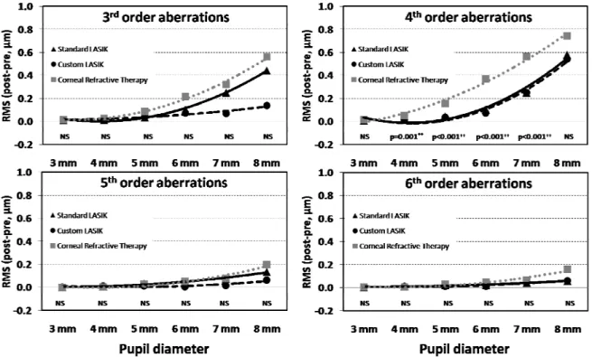
Figure 1 shows the differences of RMS values (after treatment minus baseline) for 3rd, 4th, 5th and 6th-order aberrations in the three clinical groups. Only in 4th-order aberrations did exist statistically significant differences in diameters from 4 mm to 7 mm.
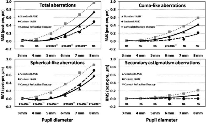
Figure 2 shows the differences of RMS values (after treatment minus baseline) for or total, spherical-like, coma-like and secondary astigmatism aberrations in the three clinical groups for different diameters from 3 mm to 8 mm. No statistically significant differences were present after the procedures in the secondary
astigmatism for any of the diameters analyzed in the three refractive techniques. Conversely, differences in the spherical-like aberrations were statistically significant for all diameters.
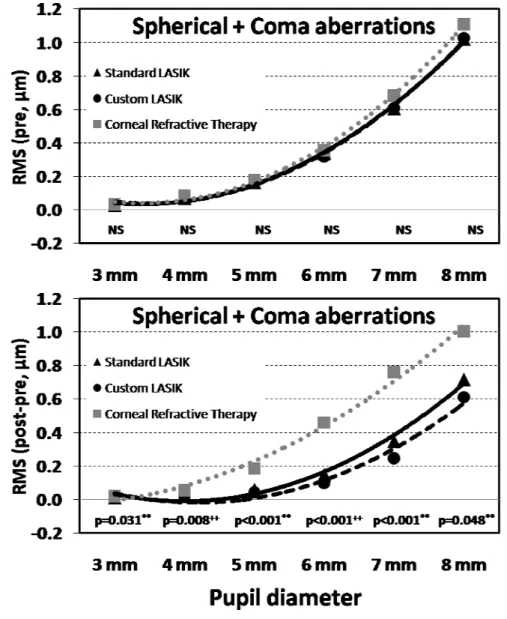
Figure 3 shows the differences of RMS values (after treatment minus baseline) for the combination of spherical-like and coma-like RMS. Although the analysis of isolated spherical-like aberration increases in the custom LASIK technique is greater than in standard LASIK, they are reversed when analyzed together with the coma-like aberration (P > .226) because coma-like aberrations was higher in custom LASIK.
DISCUSSION
Results from the present study showed that there is a significant increase in several aberrations among the three clinical groups and that this shift follows an exponential trend to increase as we enlarge the corneal area subjected to analysis (the induced change is up to 50x higher for 8 mm aperture diameter than for a 3 mm diameter). This is supported by different previous studies conducted by other authors after LASIK,13 and corneal refractive therapy29 or both treatments.11 However, to the best of our knowledge this is the first study combining two different surgical
treatments including custom LASIK and corneal refractive therapy. Also, this is the first study analyzing the aberration profiles with such a detail and for different diameters. Regarding to this analysis, we have observed that the three refractive techniques change aberrations similarly for 5th-order, 6th-order and Secondary astigmatism RMS with changes below 0.20 microns from 3 to 8 mm. Conversely, 3rd -order, 4th-order and Total RMS as well as spherical-like and coma-like RMS
increased significantly (up to 5x more than previously quoted aberrations) from 3 to 8 mm and in a different way for different refractive treatments. Overall, changes in HOA are not statistically different among techniques up to 4 mm becoming significantly different from 5 to 8 mm. This is in agreement also with previous studies that
analyzed the aforementioned techniques separately and not with such a detail as in the present work.17,18,30 Interestingly, RMS for spherical-like aberrations are much higher than the 4th order RMS. One would expect the 4th order RMS (which include Z40) to be equal if not higher than the spherical-like aberrations. A previous article has also demonstrated that for smaller areas of analysis (i.e., 3 mm), this effect is not found. However, when larger zones are analyzed (i.e. 6.5 mm), the RMS for
spherical-like aberrations are much higher than the 4th order RMS as shown by our study, which also includes other diameters of analysis, along with those reported by Moreno-Barriuso et al.1
These results are relevant because of the known influence of HOA on visual distortion, for example during night vision.31 Also, certain orders of aberration as spherical-like and coma-like aberrations have demonstrated to have a higher impact on image quality than others.16 In the present study these aberrations have
11
demonstrated to be significantly increased after all procedures. Present study showed that after corneal refractive treatments the area being analyzed in terms of optical quality of the anterior corneal surface plays an important role in the absolute values of aberration found as well as in the comparison among treatments,
particularly from spherical-like and coma-like aberrations. Considering that the optical zone dimensions might change slightly as the treatment progress in CRT treatment, it will be interesting to evaluate which is the impact of this on the optical quality of the eye.25 Lu et al. (2007) have analyzed longitudinally the time-course variation in HOA, spherical-like and coma-like aberrations during CRT treatment. They found an
increase in degradation of the optical quality of the eye up to the tenth day of treatment, with a trend towards stabilization thereafter up to the end of a month of treatment25. Other studies have also found an increase in the optical aberrations of the eye after orthokeratology in cross-sectional studies30,32 while Hiraoka et al. have demonstrated that these effects recover to baseline after a month once the treatment is interrupted.33
Another potential limitation related with stability of the aberration structure of the eye could be the fact that we assume stability of corneal parameters after 3rd month of treatment (for CRT and LASIK) but we have not considered this within the scope of the research work that produced this manuscript. According to the protocol followed, measurements for all patients were done (at baseline and after treatment) at the same time frame during the day to minimize potential diurnal variations. Any potential effect would be more relevant for CRT due to potential diurnal variations related with treatment regression, and this can be minimized by measuring every time at the same time. We assume that beyond the effect of circadian variations (edema, lid pressure,) that will recover during the morning hours, no significant effect is expected for the LASIK groups.
In the present study only anterior corneal surface aberrations, instead of total ocular aberrations (including internal aberrations), have been analyzed, what could be considered limiting in terms of understanding the whole eye optical quality. However, considering that all the procedures under evaluation affect the anterior corneal surface, with minor predicted impact in the posterior corneal surface and the
internal optics of the eye (mainly the crystalline lens), we believe that the results reported here reflect well the effects of these refractive procedures on the optical quality of the visual system as previous studies have reported.34-36 The study lacks to demonstrate the impact of the aberrations described on patients visual acuity or contrast sensitivity as well as their subjective complaints. Indeed, the study would benefit much from this approach to bring the results to a more clinical environment. However, considering the huge amount of information processed and presented here justifies its description separately in order to set the basis for future clinical studies that aim to demonstrate which could be the actual impact of present findings in patient’s subjective quality of vision. We expect that such results would agree with recently published work from Anera et al.11 The results found by those authors regarding worsen of contrast sensitivity function with CRT compared to LASIK are supported by our results that demonstrated the impact of CRT on HOA particularly when corneal areas larger than 6 mm of diameter are under analysis.
A closer observation of present results shows that corneal refractive therapy increases all aberrations orders at a higher rate than the surgical techniques, which is a clear disadvantage for this technique considering the outcomes solely in terms of the optical quality of the eye. This is explained by the dramatic change in corneal curvature at the margins of the optical zone created by as we showed in previous studies (Queiros et al 2009, accepted for publication). In that study it was observed how the transition zone between central treated and peripheral untreated cornea is smoother in both surgical techniques (custom LASIK and standard LASIK) compared with corneal refractive therapy. This is related to the redistribution of epithelial and stromal thickness37 with no tissue removal, compared with the ablation in the surgical techniques. As a consequence of this changes the asphericity of the cornea changes dramatically in corneal refractive therapy compared with custom LASIK and standard LASIK, and such changes have been related with degradation of the image quality by several authors after the aforementioned treatments.5,11,38,39 The previous facts are supported by the results of the present work in which the more significant differences between techniques, with corneal refractive therapy showing higher aberrations,
13
occurred between 4 to 7 mm, coincident with the transition zones analyzed previously for corneal curvature and asphericity by our group.
Considering both surgical treatments, interesting results also came out. In spite of absent of statistically significant differences between custom LASIK and standard LASIK what can be related with the high standard deviation compared to the average RMS values, and the limited sample size as shown in table 5, it is evident that the average behavior of both treatments differs in some way when we look at figures 1 and 2, with most of the aberrations analyzed showing lower increase in custom LASIK compared with standard LASIK. This is in agreement with previous studies that have found a significant increase in the visual performance after
customized treatments,40 and also when compared with standard LASIK41
convencional.42,43 However, this does not mean that the aberrations will be eliminated or maintained at the pretreatment levels, which seems to be quite difficult if not
impossible with current technology. Even if such complete control on the HOA with refractive treatments, this also raises the question if the compensation of aberrations is a desirable outcome which is not supported by some authors.16,44,45 The utility of a certain degree of aberrations is supported by the mechanism of increase in depth of focus they can provide and that might help to increase the tolerance to lower levels of defocus. Furthermore, Legras et al. using adaptative optics have shown that the correction of only 50% of coma-like and spherical-like aberration has a similar impact on the visual quality of the eye than full correction.46,47 The same authors reported negligible effects by correcting other aberrations more marginal aberrations such as trefoil, which is in agreement with Applegate results.16
Another limitation of the present study is the potential decentration between the treated areas which are targeted regarding the pupil center and the aberration analysis carried out with reference to the corneal center as analyzed by the corneal topographer. As reported by other authors,48 this differences can have an impact on comatic aberrations. This could be a potential explanation for the changes observed in comatic aberrations when those were not expected to exist considering that only well centered treatments were considered in this study. The other related issue is the different behavior of spherical-like aberration and coma-like in standard LASIK and
custom LASIK. While standard LASIK shows higher levels of coma-like, custom LASIK shows slightly higher values of spherical-like aberration compared to baseline as shown in figure 2. This could be related to slight decentrations of treatment of decentrations between treatment zone and topographically analyzed areas. These shifts in reference points could generate some kind of “transference” between spherical-like aberration and coma-like aberrations.49
In summary, corneal refractive therapy treatment impacts significantly the optical quality of the eye, particularly under pupil dilatation beyond 5 mm compared to the surgical treatments. For pupil size below 5 mm, results are not expected to be significantly different between surgical optics and corneal refractive therapy. This suggests that patients should be carefully selected for this treatment, with special considerations for those patients with higher refractive values as the increase in aberrations will be proportional to those values, and larger pupils under dim illumination. Also, lens designs should be improved in order to provide smoother transition between the central treated area and the peripheral untreated zone. Regarding the surgical techniques, although the present study has not been able to show differences among them, it is evident that they change the ocular aberrations in a different way. custom LASIK induces lower changes in RMS for most of the
aberrations considered and also shows more aberration remaining below the criteria for statistically significant change compared with standard LASIK and corneal
refractive therapy. Although the aims of customized refractive surgery to maintain the original level of aberrations of the eye seems not to be achievable with current
technology, there seems to be room to think that the improvements in visual
performance with customized treatments might be linked to a more moderate impact in corneal aberrations compared to standard procedures.50 In order to prove that, larger sample size might be considered in future studies.
Present study demonstrates that for apertures fewer than 4 mm, no significant differences in aberration induction are expected between LASIK and CRT. However, for larger apertures, significantly higher aberration is induced by CRT. For this
reason, it is highly important that clinicians give relevance to pupil size when performing CRT treatment
15
ACKNOWLEDGMENTS /DISCLOSURE
This work was supported by a grant from the Science and Technology Foundation (FCT) of Ministry of Science and Superior Education (MCES) (European Social Funding). The authors indicate no financial conflict of interest. Involved in design of the study (AQ, JG-M, JJ); conduct of the study (AQ, JG-M, JJ); analysis and
interpretation (AQ, CV-C, JG-M, JJ); writing the article (AQ, CV-C, JG-M, JJ, ARG); critical revision (AQ, CV-C, JG-M, JJ, ARG); final approval (AQ, CV-C, JG-M, JJ, ARG); data collection (AQ, CV-C, JG-M); management of patients and equipments (AQ, CV-C, JG-M, ARG); statistical expertise (AQ, JG-M, JJ) and literature review (AQ, JG-M). The study was approved by the School of Science Review Board (University of Minho, Braga, Portugal) and followed the tenets of the Declaration of Helsinki. Informed consent was obtained from all patients before all the interventions and they also gave their consent to treat their clinical data anonymously for research purposes.
Reference List
1. Moreno-Barriuso E, Lloves JM, Marcos S, Navarro R, Llorente L, Barbero S. Ocular aberrations before and after myopic corneal refractive surgery: LASIK-induced changes measured with laser ray tracing. Invest
Ophthalmol Vis Sci 2001;42(6):1396-1403.
2. Sorbara L, Fonn D, Simpson T, Lu F, Kort R. Reduction of myopia from corneal refractive therapy. Optom Vis Sci 2005;82(6):512-518.
3. Villa-Collar C, Gutierrez R, Jimenez JR, Gonzalez-Meijome JM. Night Vision Disturbances after Successful LASIK Surgery. Br J Ophthalmol 2007;91(8):1031-1037.
4. Holladay JT, Janes JA. Topographic changes in corneal asphericity and effective optical zone after laser in situ keratomileusis. J Cataract Refract Surg 2002;28(6):942-947.
5. Anera RG, Jimenez JR, Jimenez dB, Bermudez J, Hita E. Changes in corneal asphericity after laser in situ keratomileusis. J Cataract Refract Surg 2003;29(4):762-768.
6. Gonzalez-Meijome JM, Sanudo-Buitrago F, Lopez-Alemany A, Almeida JB, Parafita MA. Correlations between central and peripheral changes in anterior corneal topography after myopic LASIK and their implications in postsurgical contact lens fitting. Eye Contact Lens 2006;32(4):197-202.
7. Sridharan R, Swarbrick H. Corneal response to short-term orthokeratology lens wear. Optom Vis Sci 2003;80(3):200-206.
17
8. Gonzalez-Meijome JM, Jorge J, Queiros A, Almeida JB, Parafita MA. A comparison of the ARK-700A autokeratometer and Medmont E300 corneal topographer when measuring peripheral corneal curvature. Ophthalmic Physiol Opt 2004;24(5):391-398.
9. Gonzalez-Meijome JM, Villa-Collar C, Montes-Mico R, Gomes A.
Asphericity of the anterior human cornea with different corneal diameters. J Cataract Refract Surg 2007;33(3):465-473.
10. Applegate RA, Hilmantel G, Howland HC, Tu EY, Starck T, Zayac EJ.
Corneal first surface optical aberrations and visual performance. J Refract Surg 2000;16(5):507-514.
11. Anera RG, Villa-Collar C, Jimenez JR, Gutierrez R. Effect of LASIK and contact lens corneal refractive therapy on higher order aberrations and contrast sensitivity function. J Refract Surg 2009;25(3):277-284.
12. Montes-Mico R, Rodriguez-Galietero A, Alio JL, Cervino A. Contrast sensitivity after LASIK flap creation with a femtosecond laser and a mechanical microkeratome. J Refract Surg 2007;23(2):188-192.
13. DU CX, Shen Y, Wang Y. Comparison of high order aberration after
conventional and customized ablation in myopic LASIK in different eyes of the same patient. J Zhejiang Univ Sci B 2007;8(3):177-180.
14. Hiraoka T, Matsumoto Y, Okamoto F et al. Corneal higher-order aberrations induced by overnight orthokeratology. Am J Ophthalmol 2005;139(3):429-436.
15. Oshika T, Klyce SD, Applegate RA, Howland HC, El Danasoury MA. Comparison of corneal wavefront aberrations after photorefractive keratectomy and laser in situ keratomileusis. Am J Ophthalmol 1999;127(1):1-7.
16. Applegate RA, Sarver EJ, Khemsara V. Are all aberrations equal? J Refract Surg 2002;18(5):S556-S562.
17. Wang L, Koch DD. Ocular higher-order aberrations in individuals screened for refractive surgery. J Cataract Refract Surg 2003;29(10):1896-1903.
18. Wang Y, Zhao K, Jin Y, Niu Y, Zuo T. Changes of higher order aberration with various pupil sizes in the myopic eye. J Refract Surg 2003;19(2 Suppl):S270-S274.
19. Lopez-Gil N, Fernandez-Sanchez V, Legras R, Montes-Mico R, Lara F, Nguyen-Khoa JL. Accommodation-related changes in monochromatic aberrations of the human eye as a function of age. Invest Ophthalmol Vis Sci 2008;49(4):1736-1743.
20. He JC, Burns SA, Marcos S. Monochromatic aberrations in the accommodated human eye. Vision Res 2000;40(1):41-48.
21. Collins MJ, Buehren T, Iskander DR. Retinal image quality, reading and myopia. Vision Res 2006;46(1-2):196-215.
22. Llorente L, Barbero S, Cano D, Dorronsoro C, Marcos S. Myopic versus hyperopic eyes: axial length, corneal shape and optical aberrations. J Vis 2004;4(4):288-298.
19
23. Lu F, Simpson T, Sorbara L, Fonn D. Malleability of the ocular surface in response to mechanical stress induced by orthokeratology contact lenses. Cornea 2008;27(2):133-141.
24. Holladay JT, Dudeja DR, Chang J. Functional vision and corneal changes after laser in situ keratomileusis determined by contrast sensitivity, glare testing, and corneal topography. J Cataract Refract Surg 1999;25(5):663-669.
25. Lu F, Simpson T, Sorbara L, Fonn D. The relationship between the
treatment zone diameter and visual, optical and subjective performance in Corneal Refractive Therapy lens wearers. Ophthalmic Physiol Opt
2007;27(6):568-578.
26. Harper CL, Boulton ME, Bennett D et al. Diurnal variations in human corneal thickness. Br J Ophthalmol 1996;80(12):1068-1072.
27. Handa T, Mukuno K, Niida T, Uozato H, Tanaka S, Shimizu K. Diurnal variation of human corneal curvature in young adults. J Refract Surg 2002;18(1):58-62.
28. Gonzalez-Meijome JM, Villa-Collar C. Nomogram, corneal topography, and final prescription relations for Corneal Refractive Therapy. Optom Vis Sci 2007;84(1):59-64.
29. Hiraoka T, Okamoto C, Ishii Y, Kakita T, Oshika T. Contrast sensitivity function and ocular higher-order aberrations following overnight orthokeratology. Invest Ophthalmol Vis Sci 2007;48(2):550-556.
30. Joslin CE, Wu SM, McMahon TT, Shahidi M. Higher-order wavefront aberrations in corneal refractive therapy. Optom Vis Sci 2003;80(12):805-811.
31. Hong X, Thibos LN. Longitudinal evaluation of optical aberrations following laser in situ keratomileusis surgery. J Refract Surg 2000;16(5):S647-S650.
32. Berntsen DA, Mitchell GL, Barr JT. The effect of overnight contact lens corneal reshaping on refractive error-specific quality of life. Optom Vis Sci 2006;83(6):354-359.
33. Hiraoka T, Okamoto C, Ishii Y, Okamoto F, Oshika T. Recovery of corneal irregular astigmatism, ocular higher-order aberrations, and contrast sensitivity after discontinuation of overnight orthokeratology. Br J Ophthalmol 2009;93(2):203-208.
34. Barbero S, Marcos S, Merayo-Lloves J. Corneal and total optical aberrations in a unilateral aphakic patient. J Cataract Refract Surg 2002;28(9):1594-1600.
35. Artal P, Berrio E, Guirao A, Piers P. Contribution of the cornea and internal surfaces to the change of ocular aberrations with age. J Opt Soc Am A Opt Image Sci Vis 2002;19(1):137-143.
36. Lee JM, Lee DJ, Jung WJ, Park WC. Comparison between anterior corneal aberration and ocular aberration in laser refractive surgery. Korean J Ophthalmol 2008;22(3):164-168.
21
37. Alharbi A, Swarbrick HA. The effects of overnight orthokeratology lens wear on corneal thickness. Invest Ophthalmol Vis Sci 2003;44(6):2518-2523.
38. Jimenez JR, Anera RG, Jimenez dB. Equation for corneal asphericity after corneal refractive surgery. J Refract Surg 2003;19(1):65-69.
39. Jimenez JR, Anera RG, Diaz JA, Perez-Ocon F. Corneal asphericity after refractive surgery when the Munnerlyn formula is applied. J Opt Soc Am A Opt Image Sci Vis 2004;21(1):98-103.
40. Alio JL, Montes-Mico R. Wavefront-guided versus standard LASIK enhancement for residual refractive errors. Ophthalmology
2006;113(2):191-197.
41. Hament WJ, Nabar VA, Nuijts RM. Repeatability and validity of Zywave aberrometer measurements. J Cataract Refract Surg 2002;28(12):2135-2141.
42. Urbano AP, Nose W. [Visual quality after custom versus standard LASIK retreatment]. Arq Bras Oftalmol 2008;71(6):841-846.
43. Urbano AP, Nose W. [Refractional results of LASIK retreatment with wavefront-guided ablation versus standard ablation]. Arq Bras Oftalmol 2008;71(5):651-659.
44. Applegate RA, Marsack JD, Ramos R, Sarver EJ. Interaction between aberrations to improve or reduce visual performance. J Cataract Refract Surg 2003;29(8):1487-1495.
45. Applegate RA, Ballentine C, Gross H, Sarver EJ, Sarver CA. Visual acuity as a function of Zernike mode and level of root mean square error. Optom Vis Sci 2003;80(2):97-105.
46. Legras R, Rouger H. Calculations and Measurements of the Visual Benefit of Correcting the Higher-order Aberrations Using Adaptive Optics
Technology. J Optom 2008;1(1):22-29.
47. Legras R, Rouger H. Just-Noticeable Levels of Aberration Correction. J Optom 2008;1(2):71-77.
48. Lu F, Wu J, Qu J et al. Association between Offset of the Pupil Center from the Corneal Vertex and Wavefront Aberration. J Optom 2008;1(1):8-13.
49. Guirao A, Williams DR, Cox IG. Effect of rotation and translation on the expected benefit of an ideal method to correct the eye's higher-order aberrations. J Opt Soc Am A Opt Image Sci Vis 2001;18(5):1003-1015.
50. Villa C, Jimenez JR, Anera RG, Gutierrez R, Hita E. Visual performance after LASIK for a Q-optimized and a standard ablation algorithm. Appl Opt 2009;48(30):5741-5747.
23
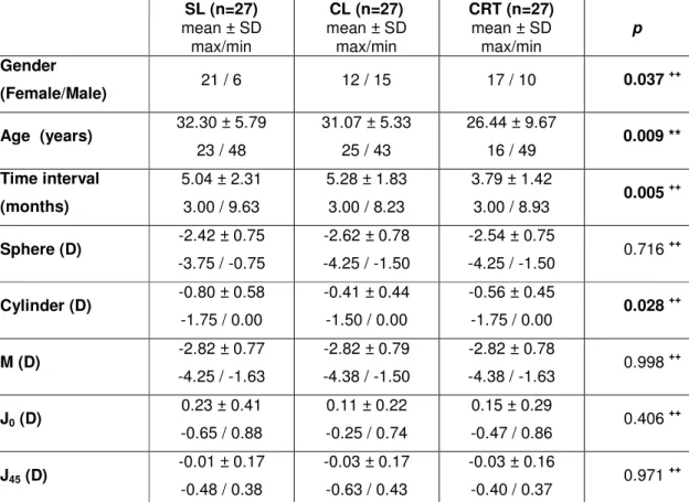
Table 1. Pretreatment demographic characteristics (mean±SD, maximum and minimum) for the population in each group: standard LASIK, custom LASIK and Corneal Refractive Therapy .
SL (n=27) mean ± SD max/min CL (n=27) mean ± SD max/min CRT (n=27) mean ± SD max/min p Gender (Female/Male) 21 / 6 12 / 15 17 / 10 0.037 ++ Age (years) 32.30 ± 5.79 23 / 48 31.07 ± 5.33 25 / 43 26.44 ± 9.67 16 / 49 0.009 ** Time interval (months) 5.04 ± 2.31 3.00 / 9.63 5.28 ± 1.83 3.00 / 8.23 3.79 ± 1.42 3.00 / 8.93 0.005 ++ Sphere (D) -2.42 ± 0.75 -3.75 / -0.75 -2.62 ± 0.78 -4.25 / -1.50 -2.54 ± 0.75 -4.25 / -1.50 0.716 ++ Cylinder (D) -0.80 ± 0.58 -1.75 / 0.00 -0.41 ± 0.44 -1.50 / 0.00 -0.56 ± 0.45 -1.75 / 0.00 0.028 ++ M (D) -2.82 ± 0.77 -4.25 / -1.63 -2.82 ± 0.79 -4.38 / -1.50 -2.82 ± 0.78 -4.38 / -1.63 0.998 ++ J0 (D) 0.23 ± 0.41 -0.65 / 0.88 0.11 ± 0.22 -0.25 / 0.74 0.15 ± 0.29 -0.47 / 0.86 0.406 ++ J45 (D) -0.01 ± 0.17 -0.48 / 0.38 -0.03 ± 0.17 -0.63 / 0.43 -0.03 ± 0.16 -0.40 / 0.37 0.971 ++
Statistically differences among groups highlighted in bold. ** ANOVA, ++ Kruskal-Wallis Test. SL - standard LASIK; CL - custom LASIK and CRT - Corneal Refractive Therapy, M, J0 and J45 is refractive components.
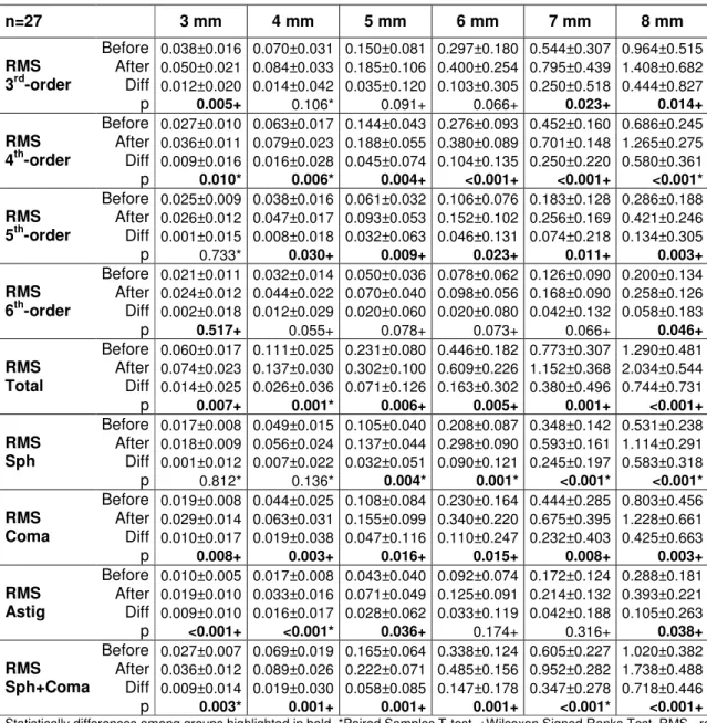
Table 2. Pretreatment, post treatment and difference (post-pre) values of RMS in the standard LASIK group (3rd to 6th-orders, total, spherical-like, coma-like, secondary astigmatism and spherical+coma aberrations) for different diameters (3 to 8 mm). Units for RMS are microns
n=27 3 mm 4 mm 5 mm 6 mm 7 mm 8 mm RMS 3rd-order Before 0.038±0.016 0.070±0.031 0.150±0.081 0.297±0.180 0.544±0.307 0.964±0.515 After 0.050±0.021 0.084±0.033 0.185±0.106 0.400±0.254 0.795±0.439 1.408±0.682 Diff 0.012±0.020 0.014±0.042 0.035±0.120 0.103±0.305 0.250±0.518 0.444±0.827 p 0.005+ 0.106* 0.091+ 0.066+ 0.023+ 0.014+ RMS 4th-order Before 0.027±0.010 0.063±0.017 0.144±0.043 0.276±0.093 0.452±0.160 0.686±0.245 After 0.036±0.011 0.079±0.023 0.188±0.055 0.380±0.089 0.701±0.148 1.265±0.275 Diff 0.009±0.016 0.016±0.028 0.045±0.074 0.104±0.135 0.250±0.220 0.580±0.361 p 0.010* 0.006* 0.004+ <0.001+ <0.001+ <0.001* RMS 5th-order Before 0.025±0.009 0.038±0.016 0.061±0.032 0.106±0.076 0.183±0.128 0.286±0.188 After 0.026±0.012 0.047±0.017 0.093±0.053 0.152±0.102 0.256±0.169 0.421±0.246 Diff 0.001±0.015 0.008±0.018 0.032±0.063 0.046±0.131 0.074±0.218 0.134±0.305 p 0.733* 0.030+ 0.009+ 0.023+ 0.011+ 0.003+ RMS 6th-order Before 0.021±0.011 0.032±0.014 0.050±0.036 0.078±0.062 0.126±0.090 0.200±0.134 After 0.024±0.012 0.044±0.022 0.070±0.040 0.098±0.056 0.168±0.090 0.258±0.126 Diff 0.002±0.018 0.012±0.029 0.020±0.060 0.020±0.080 0.042±0.132 0.058±0.183 p 0.517+ 0.055+ 0.078+ 0.073+ 0.066+ 0.046+ RMS Total Before 0.060±0.017 0.111±0.025 0.231±0.080 0.446±0.182 0.773±0.307 1.290±0.481 After 0.074±0.023 0.137±0.030 0.302±0.100 0.609±0.226 1.152±0.368 2.034±0.544 Diff 0.014±0.025 0.026±0.036 0.071±0.126 0.163±0.302 0.380±0.496 0.744±0.731 p 0.007+ 0.001* 0.006+ 0.005+ 0.001+ <0.001+ RMS Sph Before 0.017±0.008 0.049±0.015 0.105±0.040 0.208±0.087 0.348±0.142 0.531±0.238 After 0.018±0.009 0.056±0.024 0.137±0.044 0.298±0.090 0.593±0.161 1.114±0.291 Diff 0.001±0.012 0.007±0.022 0.032±0.051 0.090±0.121 0.245±0.197 0.583±0.318 p 0.812* 0.136* 0.004* 0.001* <0.001* <0.001* RMS Coma Before 0.019±0.008 0.044±0.025 0.108±0.084 0.230±0.164 0.444±0.285 0.803±0.456 After 0.029±0.014 0.063±0.031 0.155±0.099 0.340±0.220 0.675±0.395 1.228±0.661 Diff 0.010±0.017 0.019±0.038 0.047±0.116 0.110±0.247 0.232±0.403 0.425±0.663 p 0.008+ 0.003+ 0.016+ 0.015+ 0.008+ 0.003+ RMS Astig Before 0.010±0.005 0.017±0.008 0.043±0.040 0.092±0.074 0.172±0.124 0.288±0.181 After 0.019±0.010 0.033±0.016 0.071±0.049 0.125±0.091 0.214±0.132 0.393±0.221 Diff 0.009±0.010 0.016±0.017 0.028±0.062 0.033±0.119 0.042±0.188 0.105±0.263 p <0.001+ <0.001* 0.036+ 0.174+ 0.316+ 0.038+ RMS Sph+Coma Before 0.027±0.007 0.069±0.019 0.165±0.064 0.338±0.124 0.605±0.227 1.020±0.382 After 0.036±0.012 0.089±0.026 0.222±0.071 0.485±0.156 0.952±0.282 1.738±0.488 Diff 0.009±0.014 0.019±0.030 0.058±0.085 0.147±0.178 0.347±0.278 0.718±0.446 p 0.003* 0.001+ 0.001+ 0.001+ <0.001* <0.001+
Statistically differences among groups highlighted in bold. *Paired Samples T-test, +Wilcoxon Signed Ranks Test. RMS - root mean square, Total – total higher order aberrations, Sph - spherical-like aberrations, Coma - coma-like aberrations, Astig - secondary astigmatism aberrations and Sph+Coma – spherical aberrations +coma aberrations.
25
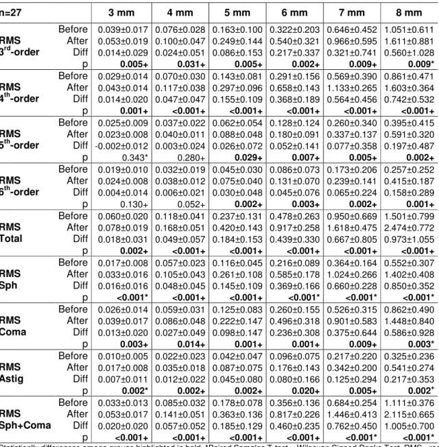
Table 3. Pretreatment, post treatment and difference (post-pre) values of RMS in the custom LASIK group (3rd to 6th-orders, total, spherical-like, coma-like, secondary astigmatism and spherical+coma aberrations) for different diameters (3 to 8 mm). Units for RMS are microns
n=27 eyes 3 mm 4 mm 5 mm 6 mm 7 mm 8 mm RMS 3rd-order Before 0.038±0.014 0.080±0.029 0.161±0.075 0.299±0.122 0.589±0.249 1.057±0.487 After 0.051±0.028 0.102±0.060 0.199±0.106 0.372±0.190 0.657±0.340 1.194±0.611 Diff 0.013±0.028 0.022±0.061 0.037±0.126 0.073±0.237 0.068±0.426 0.138±0.736 p 0.024+ 0.124+ 0.163+ 0.212+ 0.581+ 0.340* RMS 4th-order Before 0.026±0.009 0.060±0.015 0.130±0.060 0.255±0.071 0.435±0.125 0.721±0.243 After 0.038±0.015 0.073±0.028 0.165±0.077 0.328±0.134 0.684±0.190 1.260±0.322 Diff 0.012±0.018 0.013±0.030 0.035±0.096 0.073±0.155 0.249±0.226 0.540±0.421 p 0.002+ 0.032* 0.005+ 0.014+ <0.001+ <0.001+ RMS 5th-order Before 0.024±0.008 0.037±0.014 0.068±0.041 0.135±0.081 0.227±0.122 0.351±0.175 After 0.027±0.011 0.049±0.022 0.080±0.050 0.138±0.091 0.242±0.121 0.412±0.179 Diff 0.003±0.012 0.012±0.026 0.011±0.067 0.003±0.139 0.015±0.193 0.062±0.247 p 0.180* 0.037+ 0.239+ 0.923+ 0.597+ 0.163+ RMS 6th-order Before 0.018±0.008 0.029±0.011 0.051±0.033 0.091±0.064 0.143±0.092 0.210±0.107 After 0.023±0.007 0.042±0.025 0.060±0.041 0.102±0.073 0.187±0.124 0.270±0.133 Diff 0.005±0.010 0.012±0.028 0.009±0.056 0.011±0.108 0.044±0.178 0.060±0.185 p 0.013+ 0.046+ 0.442+ 0.866+ 0.230+ 0.149+ RMS Total Before 0.057±0.013 0.114±0.022 0.230±0.098 0.438±0.140 0.801±0.257 1.372±0.506 After 0.076±0.027 0.147±0.060 0.283±0.132 0.539±0.229 1.024±0.353 1.865±0.545 Diff 0.019±0.027 0.033±0.061 0.053±0.165 0.101±0.296 0.223±0.484 0.493±0.794 p 0.001+ 0.011+ 0.068+ 0.124+ 0.046+ 0.003* RMS Sph Before 0.015±0.007 0.046±0.013 0.090±0.041 0.160±0.073 0.264±0.148 0.425±0.236 After 0.018±0.011 0.046±0.024 0.116±0.058 0.267±0.093 0.609±0.201 1.142±0.362 Diff 0.003±0.013 0.000±0.029 0.027±0.063 0.108±0.113 0.344±0.185 0.717±0.332 p 0.194* 0.885+ 0.038* <0.001* <0.001* <0.001* RMS Coma Before 0.026±0.012 0.063±0.026 0.129±0.067 0.258±0.111 0.509±0.258 0.866±0.480 After 0.035±0.016 0.078±0.046 0.158±0.080 0.295±0.141 0.554±0.283 1.080±0.563 Diff 0.009±0.015 0.015±0.048 0.028±0.106 0.037±0.181 0.045±0.369 0.214±0.662 p 0.006+ 0.178+ 0.079+ 0.280+ 0.532+ 0.106* RMS Astig Before 0.012±0.006 0.022±0.010 0.050±0.045 0.112±0.093 0.208±0.157 0.372±0.245 After 0.018±0.008 0.034±0.024 0.061±0.050 0.099±0.069 0.201±0.098 0.377±0.180 Diff 0.006±0.009 0.011±0.026 0.011±0.074 -0.012±0.124 -0.006±0.188 0.005±0.302 p 0.001+ 0.032+ 0.280+ 0.581+ 0.866+ 0.938* RMS Sph+Coma Before 0.031±0.012 0.081±0.020 0.163±0.067 0.318±0.093 0.609±0.210 1.028±0.395 After 0.041±0.015 0.095±0.042 0.204±0.079 0.414±0.123 0.856±0.252 1.639±0.472 Diff 0.010±0.017 0.015±0.045 0.042±0.098 0.097±0.140 0.246±0.320 0.612±0.602 p 0.008+ 0.130+ 0.015+ 0.002+ 0.001+ <0.001+
Statistically differences among groups highlighted in bold. *Paired Samples T-test, +Wilcoxon Signed Ranks Test. RMS - root mean square, Total – total higher order aberrations, Sph - spherical-like aberrations, Coma - coma-like aberrations, Astig - secondary astigmatism aberrations and Sph+Coma – spherical aberrations +coma aberrations.
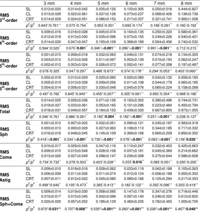
Table 4. Pretreatment, post treatment and difference (post-pre) values of RMS in the corneal refractive therapy group (3rd to 6th-orders, total, spherical-like, coma-like, secondary astigmatism and spherical+coma aberrations) for different diameters (3 to 8 mm). Units for RMS are microns
Statistically differences among groups highlighted in bold. *Paired Samples T-test, +Wilcoxon Signed Ranks Test. RMS - root mean square, Total – total higher order aberrations, Sph - spherical-like aberrations, Coma - coma-like aberrations, Astig - secondary astigmatism aberrations and Sph+Coma – spherical aberrations +coma aberrations.
n=27 3 mm 4 mm 5 mm 6 mm 7 mm 8 mm RMS 3rd-order Before 0.039±0.017 0.076±0.028 0.163±0.100 0.322±0.203 0.646±0.452 1.051±0.611 After 0.053±0.019 0.100±0.047 0.249±0.144 0.540±0.321 0.966±0.595 1.611±0.881 Diff 0.014±0.029 0.024±0.051 0.086±0.153 0.217±0.337 0.321±0.741 0.560±1.028 p 0.005+ 0.031+ 0.005+ 0.002+ 0.009+ 0.009* RMS 4th-order Before 0.029±0.014 0.070±0.030 0.143±0.081 0.291±0.156 0.569±0.390 0.861±0.471 After 0.043±0.014 0.117±0.038 0.297±0.096 0.658±0.143 1.133±0.265 1.603±0.364 Diff 0.014±0.020 0.047±0.047 0.155±0.109 0.368±0.189 0.564±0.456 0.742±0.532 p 0.001+ <0.001+ <0.001+ <0.001+ <0.001+ <0.001+ RMS 5th-order Before 0.025±0.009 0.037±0.022 0.062±0.054 0.128±0.124 0.260±0.340 0.395±0.415 After 0.023±0.008 0.040±0.011 0.088±0.048 0.180±0.091 0.337±0.137 0.591±0.320 Diff -0.002±0.012 0.003±0.024 0.026±0.072 0.052±0.141 0.077±0.358 0.197±0.487 p 0.343* 0.280+ 0.029+ 0.007+ 0.005+ 0.002+ RMS 6th-order Before 0.019±0.010 0.032±0.019 0.045±0.030 0.086±0.073 0.173±0.206 0.257±0.252 After 0.024±0.008 0.038±0.012 0.075±0.040 0.131±0.070 0.239±0.141 0.415±0.187 Diff 0.004±0.014 0.006±0.021 0.030±0.048 0.045±0.076 0.065±0.224 0.158±0.289 p 0.130+ 0.052+ 0.002+ 0.003+ 0.002+ 0.001+ RMS Total Before 0.060±0.020 0.118±0.041 0.237±0.131 0.478±0.263 0.950±0.669 1.501±0.799 After 0.078±0.019 0.168±0.051 0.420±0.143 0.917±0.258 1.618±0.475 2.474±0.772 Diff 0.018±0.031 0.049±0.057 0.184±0.153 0.439±0.330 0.667±0.805 0.973±1.055 p 0.002+ <0.001+ <0.001+ <0.001+ <0.001+ <0.001+ RMS Sph Before 0.017±0.008 0.057±0.023 0.116±0.045 0.216±0.089 0.364±0.164 0.552±0.307 After 0.033±0.016 0.105±0.043 0.261±0.108 0.585±0.178 1.024±0.266 1.402±0.408 Diff 0.016±0.016 0.048±0.045 0.145±0.109 0.369±0.166 0.660±0.228 0.850±0.352 p <0.001* <0.001+ <0.001+ <0.001* <0.001* <0.001* RMS Coma Before 0.026±0.014 0.059±0.031 0.125±0.083 0.260±0.155 0.526±0.315 0.862±0.490 After 0.039±0.017 0.086±0.048 0.222±0.147 0.496±0.318 0.901±0.583 1.448±0.840 Diff 0.013±0.020 0.027±0.049 0.098±0.147 0.236±0.308 0.375±0.644 0.586±0.928 p 0.003+ 0.014+ 0.001+ 0.001+ 0.009+ 0.003* RMS Astig Before 0.010±0.005 0.022±0.023 0.042±0.047 0.096±0.075 0.217±0.220 0.325±0.236 After 0.017±0.008 0.035±0.018 0.087±0.075 0.176±0.143 0.342±0.200 0.541±0.274 Diff 0.007±0.011 0.012±0.022 0.045±0.080 0.080±0.166 0.125±0.294 0.217±0.353 p 0.002* 0.002+ 0.002+ 0.020+ 0.005+ 0.002+ RMS Sph+Coma Before 0.033±0.013 0.085±0.032 0.178±0.078 0.356±0.136 0.684±0.254 1.111±0.376 After 0.053±0.017 0.141±0.051 0.363±0.136 0.817±0.226 1.446±0.413 2.115±0.665 Diff 0.020±0.020 0.057±0.052 0.185±0.129 0.460±0.235 0.762±0.450 1.005±0.700 p <0.001+ <0.001+ <0.001+ <0.001+ <0.001* <0.001*
27
Table 5. Values of differences in RMS (mean±SD) (3rd to 6th-orders, total, spherical-like, coma-spherical-like, secondary astigmatism and spherical+coma aberrations) and values of statistical significance between standard LASIK and custom LASIK (p£ ) and among the three techniques: standard LASIK, custom LASIK and Corneal Refractive Therapy (p¥). Units for RMS are microns
Statistically differences among groups highlighted in bold. *Independent sample T-test, +Mann-Whitney Test, ** ANOVA, ++Kruskal-Wallis Test. SL - standard LASIK; CL - custom LASIK and CRT - Corneal Refractive Therapy. RMS - root mean square, Total – total higher order aberrations, Sph - spherical-like aberrations, Coma - coma-like aberrations, Astig - secondary astigmatism aberrations and Sph+Coma – spherical aberrations +coma aberrations.
3 mm 4 mm 5 mm 6 mm 7 mm 8 mm RMS 3rd-order SL 0.012±0.020 0.014±0.042 0.035±0.120 0.103±0.305 0.250±0.518 0.444±0.827 CL 0.013±0.028 0.022±0.061 0.037±0.126 0.073±0.237 0.068±0.426 0.138±0.736 CRT 0.014±0.029 0.024±0.051 0.086±0.153 0.217±0.337 0.321±0.741 0.560±1.028 p£/p¥ 0.943+/0.761++ 0.573*/0.754** 0.953*/0.301** 0.692*/0.174** 0.168*/0.261** 0.160*/0.192** RMS 4th-order SL 0.009±0.016 0.016±0.028 0.045±0.074 0.104±0.135 0.250±0.220 0.580±0.361 CL 0.012±0.018 0.013±0.030 0.035±0.096 0.073±0.155 0.249±0.226 0.540±0.421 CRT 0.014±0.020 0.047±0.047 0.155±0.109 0.368±0.189 0.564±0.456 0.742±0.532 p£/p¥ 0.594+/0.520** 0.670* /0.001** 0.346+/<0.001++ 0.286+/<0.001++ 0.991*/<0.001++ 0.712*/0.215** RMS 5th-order SL 0.001±0.015 0.008±0.018 0.032±0.063 0.046±0.131 0.074±0.218 0.134±0.305 CL 0.003±0.012 0.012±0.026 0.011±0.067 0.003±0.139 0.015±0.193 0.062±0.247 CRT -0.002±0.012 0.003±0.024 0.026±0.072 0.052±0.141 0.077±0.358 0.197±0.487 p£/p¥ 0.578*/0.325** 0.547*/0.297** 0.488+/0.673++ 0.374+/0.178++ 0.294+/0.053++ 0.403+/0.060++ RMS 6th-order SL 0.002±0.018 0.012±0.029 0.020±0.060 0.020±0.080 0.042±0.132 0.058±0.183 CL 0.005±0.010 0.012±0.028 0.009±0.056 0.011±0.108 0.044±0.178 0.060±0.185 CRT 0.004±0.014 0.006±0.021 0.030±0.048 0.045±0.076 0.065±0.224 0.158±0.289 p£/p¥ 0.497*/0.782** 0.845+/0.940++ 0.455+/0.207++ 0.355+/0.061++ 0.950*/0.354++ 0.968*/0.186** RMS Total SL 0.014±0.025 0.026±0.036 0.071±0.126 0.163±0.302 0.380±0.496 0.744±0.731 CL 0.019±0.027 0.033±0.061 0.053±0.165 0.101±0.296 0.223±0.484 0.493±0.794 CRT 0.018±0.031 0.049±0.057 0.184±0.153 0.439±0.330 0.667±0.805 0.973±1.055 p£/p¥ 0.346+/0.761** 0.986+/0.261** 0.182+/0.004** 0.182+/<0.001** 0.251*/<0.001++ 0.238*/0.137** RMS Sph SL 0.001±0.012 0.007±0.022 0.032±0.051 0.090±0.121 0.245±0.197 0.583±0.318 CL 0.003±0.013 0.000±0.029 0.027±0.063 0.108±0.113 0.344±0.185 0.717±0.332 CRT 0.016±0.016 0.048±0.045 0.145±0.109 0.369±0.166 0.660±0.228 0.850±0.352 p£/p¥ 0.418*/<0.001** 0.334*/<0.001** 0.730*/<0.001++ 0.578*/<0.001** 0.063*/<0.001** 0.139*/0.026++ RMS Coma SL 0.010±0.017 0.029±0.045 0.047±0.116 0.110±0.247 0.232±0.403 0.425±0.663 CL 0.009±0.015 0.015±0.048 0.028±0.106 0.037±0.181 0.045±0.369 0.214±0.662 CRT 0.013±0.020 0.027±0.049 0.098±0.147 0.236±0.308 0.375±0.644 0.586±0.928 p£/p¥ 0.734*/0.732** 0.278*/0.503** 0.423+/0.228++ 0.223*/0.016** 0.085*/0.051** 0.250*/0.205** RMS Astig SL 0.009±0.010 0.018±0.018 0.028±0.062 0.033±0.119 0.042±0.188 0.105±0.263 CL 0.006±0.009 0.011±0.026 0.011±0.074 -0.012±0.124 -0.006±0.188 0.005±0.302 CRT 0.007±0.011 0.012±0.022 0.045±0.080 0.080±0.166 0.125±0.294 0.217±0.353 p£/p¥ 0.499+/0.640** 0.105+/0.473** 0.365*/0.413++ 0.182*/0.122++ 0.350*/0.096++ 0.203*/0.419++ RMS Sph+Coma SL 0.009±0.014 0.019±0.030 0.058±0.085 0.147±0.178 0.347±0.278 0.718±0.446 CL 0.010±0.017 0.015±0.045 0.042±0.098 0.097±0.140 0.246±0.320 0.612±0.602 CRT 0.020±0.020 0.057±0.052 0.185±0.129 0.460±0.235 0.762±0.450 1.005±0.700 p£/p¥ 0.870*/0.031** 0.707*/0.008++ 0.535*/<0.001** 0.260*/<0.001++ 0.226*/<0.001** 0.467*/0.048**
Figure Capture
Figure 1. Changes in values of Root Mean Square (RMS after treatments minus baseline) for the 3rd-order (Top left), 4th-order (Top right), 5th-order (Bottom left) and 6th-order (Bottom right) higher-order aberrations of the anterior corneal surface after standard LASIK, custom LASIK and Corneal Refractive Therapy. Significance values correspond to the comparison among results obtained for the three clinical groups. Lines represent the 2nd order polynomial fit.
**
29
Figure 2. Changes in values of Root Mean Square (RMS after treatments minus baseline) for the total (Top left), spherical-like (Bottom left), coma-like (Top right) and secondary astigmatism (Bottom right) higher-order aberrations of the anterior corneal surface after standard LASIK, custom LASIK and Corneal Refractive Therapy.
Significance values correspond to the comparison among results obtained for the three clinical groups. Lines represent the 2nd order polynomial fit.
**
Figure 3. Values of Root Mean Square (RMS) for the combination of spherical-like and coma-like aberrations at baseline (Top) and corresponding values of differences between post treatment and baseline values (Bottom) for standard LASIK, custom LASIK and Corneal Refractive Therapy . Significance values correspond to the comparison among results obtained for the three clinical groups. Lines represent the 2nd order polynomial fit.
**