Author
'
s Accepted Manuscript
Adjusting voltage criteria can unmask conducting channels in a patient with arrhythmogenic right ventricular cardiomyopathy and ventricular tachy-cardia
Prabhat Kumar MBBS, J Paul Mounsey BMBCh Ph. D., Eugene H Chung MD FHRS.
PII: S2214-0271(15)00057-3
DOI: http://dx.doi.org/10.1016/j.hrcr.2015.03.003 Reference: HRCR70
To appear in: Heart Rhythm Case Reports
Cite this article as: Prabhat Kumar MBBS, J Paul Mounsey BMBCh Ph.D., Eugene H Chung MD FHRS., Adjusting voltage criteria can unmask conducting channels in a patient with arrhythmogenic right ventricular cardiomyopathy and ventricular tachy-cardia,Heart Rhythm Case Reports, http://dx.doi.org/10.1016/j.hrcr.2015.03.003
This is a PDF file of an unedited manuscript that has been accepted for publication. As a service to our customers we are providing this early version of the manuscript. The manuscript will undergo copyediting, typesetting, and review of the resulting galley proof before it is published in its final citable form. Please note that during the production process errors may be discovered which could affect the content, and all legal disclaimers that apply to the journal pertain.
Adjusting voltage criteria can unmask conducting channels in a patient with
arrhythmogenic right ventricular cardiomyopathy and ventricular tachycardia
Prabhat Kumar MBBS, J Paul Mounsey BMBCh PhD and Eugene H Chung MD FHRS. Department of Medicine, Divisions of Cardiology and Cardiac Electrophysiology, University of North Carolina at Chapel Hill
Short Title: Voltage mapping in ARVC
Word count: 981, including headings and references
Address of Correspondence:
Eugene H Chung, MD, FHRS Associate Professor of Medicine
University of North Carolina at Chapel Hill
Division of Cardiology and Cardiac Electrophysiology 160 Dental Circle, CB 7075
Chapel Hill, NC 27599 Ph: (919) 966-4743
Email: [email protected]
Disclosures:
Prabhat Kumar: none; J Paul Mounsey: paid speaker and consultant to Boston
Keywords:
Arrhythmogenic right ventricular cardiomyopathy Ventricular tachycardia Epicardial Voltage 3D mapping CARTO-UNIVU Abbreviations:
Arrhythmogenic right ventricular cardiomyopathy (ARVC) Ventricular tachycardia (VT)
Introduction
Catheter ablation of ventricular tachycardia (VT) in patients with arrhythmogenic right ventricular cardiomyopathy (ARVC) is challenging. (1-4) Complex and predominantly epicardial substrate in the basal right ventricular free wall increases the risk of coronary injury during ablation. (5) The CARTO-UNIVU (Biosense Webster, Inc.) mapping system permits detailed voltage mapping as well as overlaying of the 3D anatomical map on the coronary angiogram. We present an epicardial ablation case in a patient with ARVC in which the CARTO-UNIVU system facilitated identification of the most critical targets for ablation and avoidance of coronary artery injury.
Case report
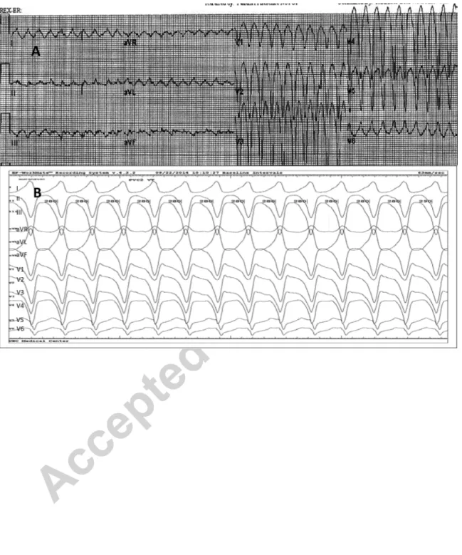
A 67 year-old male had an episode of hemodynamically stable monomorphic (VT) requiring cardioversion and he was diagnosed with ARVC at that time. The 12 lead ECG from that event is shown in Figure 1A. He underwent dual chamber ICD
implantation and was also initiated on sotalol. About two and half years later, he had an episode of VT with syncope while playing golf and received an ICD shock. He was subsequently referred for electrophysiology study and potential endocardial and epicardial catheter ablation.
During the procedure, VT was induced with double ventricular extrastimuli from the right ventricular apex. The VT was hemodynamically unstable (cycle length 270 msec) but did exactly match the clinical VT as recorded on the ECG from the first episode (Figure 1A). VT was terminated by synchronized DC shock after failed overdrive pacing.
The VT was seen to have a superior axis with positive QRS complexes in lateral leads and negative complexes in precordial leads. (Figure 1B)
An endocardial 3-D electroanatomic map during sinus rhythm was acquired using the Pentaray catheter and CARTO mapping system (Biosense Webster, Inc). The voltage map revealed a large scar (0.5 to 1.5 mV) in the inferior RV free wall. Pace-mapping produced QRS morphologies moderately similar (9/12 match) to the VT. Multiple
radiofrequency ablation lesions were placed with the Thermocool-SmartTouch ablation catheter (Biosense Webster, Inc) to transect the scar. Multiple areas of fractionated activity within the scarred area were also targeted. At this point, a slower (320 msec) and morphologically similar VT could be induced with double ventricular extrastimuli.
Pericardial access was obtained and the ablation catheter was advanced into the
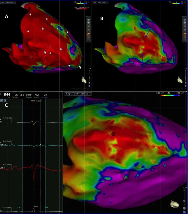
pericardial space via a 45cm Agilis (St Jude Medical) steerable sheath. A 3D epicardial map was acquired with voltage mapping and demonstrated a similar but much larger area of scar than the endocardial map (Figure 2A). Pace-mapping from the epicardium did not yield any good pace-maps. Coronary angiography did not identify any major epicardial coronary artery in that area and was displayed along with the 3D map during ablation with the CARTO-UNIVU protocol. This integration allowed us to place lesions in the epicardial locations without the need for repeated coronary angiography (Figure 3). Multiple lesions were placed in the broad scar but the VT was still easily inducible.
We then changed the voltage cut off in the epicardial map down to 0.05 mV- this
revealed discrete channels in the scar and multiple areas with late potentials (Figure 2B). Additional epicardial lesions placed along this region pf channels rendered the VT non-inducible even with triple ventricular extrastimuli. In three months follow up thus far, he has had no recurrent events.
Discussion
Our case highlights the roles of voltage mapping and CARTO-UNIVU for catheter ablation of ventricular tachycardia. Use of CARTO-UNIVU image integration permits overlaying the coronary angiographic images on the 3-D electroanatomic map and eliminates the need of repeated coronary angiography, especially when a large area of substrate modification is required on the epicardium.
Furthermore, our case demonstrates the challenges in defining the substrate from the epicardial surface. Unipolar voltage mapping from the endocardium may be used for evaluation of epicardial scar (6-8). But traditional cut-offs may not suffice for mapping from the epicardial surface, especially in the context of ARVC (1,3). In this patient, we attempted to recognize channels of relatively healthy tissue within the right ventricular free wall by selecting for voltage lower than 0.5 mV. Ablation of the channels unmasked by lowering the voltage cut-off was found to be an effective approach in ablating this patient’s VT.
Catheter ablation is rarely associated with coronary artery injury. The areas requiring ablation in patients with ARVC often involve the base of the right ventricle, beneath the tricuspid valve annulus, and the right ventricular outflow tract (1). These areas should be far from the right coronary artery, but coronary angiography is important to exclude unusual anatomy. Presentation of coronary injury can vary from immediate ECG changes to chest pain after discharge from the hospital (9). We believe the CARTO-UNIVU system offers an important tool for avoiding coronary injury, especially during epicardial ablation.
In conclusion, adjustment of the voltage cut-off during epicardial substrate mapping for scar related ventricular tachycardia might be helpful in defining the potential VT circuits by unmasking channels of relatively healthy tissue in the middle of a dense scar.
CARTO-UNIVU permits integration of coronary angiographic imaging with 3-D electroanatomic maps to guide safe epicardial catheter ablation for ventricular tachycardia.
REFERENCES
1. Arbelo E, Josephson ME: Ablation of ventricular arrhythmias in arrhythmogenic right ventricular dysplasia. J.Cardiovasc.Electrophysiol. 2010; 21:473-486.
2. Arruda M, Armaganijan L, Fahmy T, Di Biase L, Patel D, Natale A: Catheter ablation of ventricular tachycardia in arrhythmogenic right ventricular dysplasia.
J.Interv.Card.Electrophysiol. 2009; 25:129-133.
3. Bai R, Di Biase L, Shivkumar K, et al: Ablation of ventricular arrhythmias in arrhythmogenic right ventricular dysplasia/cardiomyopathy: arrhythmia-free survival after endo-epicardial substrate based mapping and ablation. Circ.Arrhythm
Electrophysiol. 2011; 4:478-485.
4. Satomi K, Kurita T, Suyama K, Noda T, Okamura H, Otomo K, Shimizu W, Aihara N, Kamakura S: Catheter ablation of stable and unstable ventricular tachycardias in patients with arrhythmogenic right ventricular dysplasia. J.Cardiovasc.Electrophysiol. 2006; 17:469-476.
5. Sacher F, Roberts-Thomson K, Maury P, et al: Epicardial ventricular tachycardia ablation a multicenter safety study. J.Am.Coll.Cardiol. 2010; 55:2366-2372.
6. Polin GM, Haqqani H, Tzou W, Hutchinson MD, Garcia FC, Callans DJ, Zado ES, Marchlinski FE: Endocardial unipolar voltage mapping to identify epicardial substrate in arrhythmogenic right ventricular cardiomyopathy/dysplasia. Heart Rhythm. 2011; 8:76-83.
7. Spears DA, Suszko AM, Dalvi R, Crean AM, Ivanov J, Nanthakumar K, Downar E, Chauhan VS: Relationship of bipolar and unipolar electrogram voltage to scar
transmurality and composition derived by magnetic resonance imaging in patients with nonischemic cardiomyopathy undergoing VT ablation. Heart Rhythm. 2012; 9:1837-1846.
8. Hutchinson MD, Gerstenfeld EP, Desjardins B, et al: Endocardial unipolar voltage mapping to detect epicardial ventricular tachycardia substrate in patients with
nonischemic left ventricular cardiomyopathy. Circ.Arrhythm Electrophysiol. 2011; 4:49-55.
9. Roberts-Thomson KC, Steven D, Seiler J, Inada K, Koplan BA, Tedrow UB, Epstein LM, Stevenson WG. Coronary artery injury due to catheter albation in adults:
HR Case Reports submission (HRCR-D-14-00135)
Adjusting voltage criteria can unmask conducting channels in a patient with arrhythmogenic right ventricular cardiomyopathy and ventricular tachycardia Key Teaching Points:
• Catheter ablation of ventricular tachycardia in patients with arhythmogenic right ventricular cardiomyopathy often requires a combined endocardial and epicardial approach
• Adjustment of the voltage criteria can unmask potential channels by discriminating subtle areas of healthy tissue
• CARTO-UNIVU integrates coronary angiography with 3D mapping to permit safer ablation on the epicardium
Figure Legends
Figure 1:
Electrocardiograms of clinical and induced ventricular tachycardia. Panel A shows clinical ventricular tachycardia at initial presentation and diagnosis 2 years before ablation: the left bundle branch morphology and negative QRS complexes in precordial leads suggested an apical inferior right ventricular exit. Panel B shows induced
ventricular tachycardia during catheter ablation procedure showing similar axes and directions of the QRS complexes; however, QRS was negative in all precordial leads with no transition likely due difference in lead location between the two recordings.
Figure 2:
Epicardial voltage map during sinus rhythm showing extensive scarring of the right ventricular free wall. Panel A shows voltage map with standard voltage threshold of 0.5 mV to 1.5 mV which shows uniform extensive scarring of the right ventricular free wall. Panel B unmasks channel of relatively higher voltage (blue arrows) after reducing the voltage threshold to 0.05 to 0.5 mV. Panel C shows unipolar and bipolar recordings from the epicardium with ablation catheter during sinus rhythm in the area of the channel (red tag) showing late potential.
Figure 3:
CARTO-UNIVU image showing electroanatomic voltage map in RAO projection with overlaid coronary angiogram.