Thyroid Disease in Pregnancy
LEO A. CARNEY, DO, Naval Hospital Pensacola Family Medicine Residency Program, Pensacola, Florida JEFF D. QUINLAN, MD, Uniformed Services University of the Health Sciences, Bethesda, Maryland JANET M. WEST, MD, Naval Hospital Pensacola Family Medicine Residency Program, Pensacola, Florida
T
hyroid disease is second only to diabetes mellitus as the most com-mon endocrinopathy that occurs in women during their reproduc-tive years. Symptoms of thyroid disease often mimic common symptoms of pregnancy, making it challenging to identify. Poorly controlled thyroid disease is associated with adverse outcomes during pregnancy, and treatment is an essential part of prenatal care to ensure maternal and fetal well-being.1-3 Thyroid Function Tests in Pregnancy To understand abnormal thyroid function in pregnancy, a review of normal physiologic changes is warranted (Table 1).4 Because of the estrogen-mediated increase in thyroid-binding globulin, the increased volume of distribution of thyroid hormone, and the pla-cental metabolism and transport of mater-nal thyroxine, there is a 20% to 40% increase in the thyroid hormone requirement as early as the fourth week of gestation.5During pregnancy, reference ranges for thyroid-stimulating hormone (TSH) are lower because of the cross-reactivity of the alpha subunit of human chorionic gonado-tropin with the TSH receptor.2,3 Changes in serum-binding protein levels can influ-ence measurements of free thyroxine (FT4) that rely on estimates rather than direct
measurements, resulting in inaccurate reported values.6 Physicians should know the limitations of locally available assay meth-ods. When preferred FT4 assay techniques are unavailable, a serum TSH level is a more accu-rate assessment of maternal thyroid status, and measurements of total thyroxine and the FT4 index can be used instead.3,6 Trimester- specific ranges for common serum thyroid studies are shown in Table 2.3,7
Screening
The Endocrine Society recommends screen-ing only pregnant women at high risk of thyroid disease using serum TSH measure-ment (Table 3).2,3 Although one study found that selectively screening women at high risk would miss 30% of those with overt or subclinical hypothyroidism,8 a random-ized controlled trial of 4,562 women did not show a reduction in adverse outcomes in those who were universally screened vs. case finding.9
Preconception Counseling
Women with hypothyroidism should be counseled about the importance of achiev-ing euthyroidism before conception because of the risk of decreased fertility and mis-carriage.1-3 They must also understand the importance of immediate monitoring Thyroid disease is the second most common endocrine disorder affecting women of reproductive age, and when untreated during pregnancy is associated with an increased risk of miscarriage, placental abruption, hypertensive disorders, and growth restriction. Current guidelines recommend targeted screening of women at high risk, includ-ing those with a history of thyroid disease, type 1 diabetes mellitus, or other autoimmune disease; current or past use of thyroid therapy; or a family history of autoimmune thyroid disease. Appropriate management results in improved outcomes, demonstrating the importance of proper diagnosis and treatment. In women with hypothyroidism, levo-thyroxine is titrated to achieve a goal serum thyroid-stimulating hormone level less than 2.5 mIU per L. The preferred treatment for hyperthyroidism is antithyroid medications, with a goal of maintaining a serum free thyroxine level in the upper one-third of the normal range. Postpartum thyroiditis is the most common form of postpartum thyroid dysfunction and may present as hyper- or hypothyroidism. Symptomatic treatment is recommended for the former; levothyroxine is indicated for the latter in women who are symptomatic, breastfeeding, or who wish to become preg-nant. (Am Fam Physician. 2014;89(4):273-278. Copyright © 2014 American Academy of Family Physicians.)
CME This clinical content
conforms to AAFP criteria for continuing medical education (CME). See CME Quiz Questions on page 251.
Author disclosure: No rel-evant financial affiliations. article that appeared in print.
at the onset of pregnancy, because by the first prenatal visit, many of these patients will already have an elevated TSH level consistent with uncontrolled hypothyroid-ism.5 Euthyroid women who are taking a stable dosage of levothyroxine should be counseled to notify their physi-cian and independently increase their medication by two additional doses per week after a missed menstrual cycle or positive home pregnancy test.3 In a study of 48 women treated for hypothyroidism with a normal prepreg-nancy serum TSH level, increasing levothyroxine by two doses per week prevented maternal TSH elevation above 2.5 mIU per L and above 5 mIU per L in 85% and 100% of patients, respectively, with only two patients requiring a subsequent dose reduction.5
Preconception counseling for women with known hyperthyroidism should include discussion of avail-able treatments and potential adverse effects, as well as the impact on future pregnancies. Standard treatments
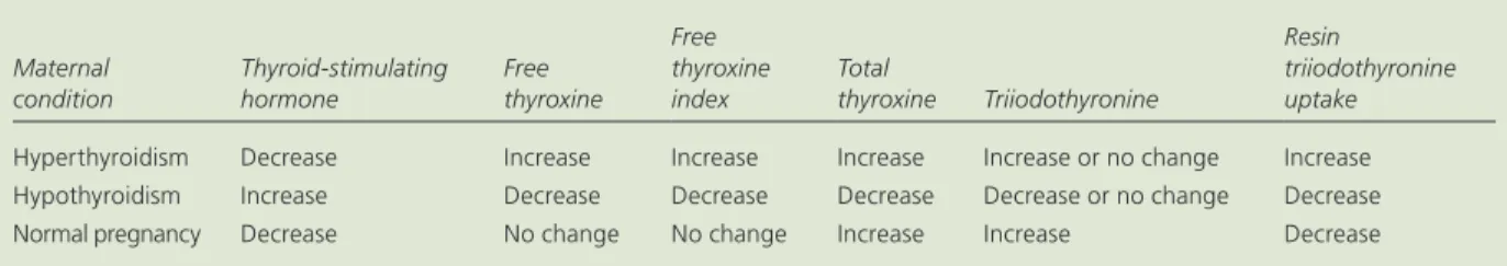
iodine ablation, and near-total thyroidectomy. Poten-tial adverse fetal effects of antithyroid medications include congenital abnormalities and neonatal hypo-thyroidism caused by transplacental transfer.2,3 Although radioactive iodine ablation is not associated with long-term consequences on gonadal function, fer-tility, or pregnancy outcomes, it is customary to wait six months after the therapeutic dose is administered before attempting conception.8 Radioactive ablation and surgery can increase the risk of neonatal goiter and hyperthyroidism because of the absence of maternal antithyroid medication, which crosses the placenta and counteracts the stimulatory effect of thyrotropin recep-tor antibodies on the fetal thyroid.2,3 The importance of achieving and maintaining euthyroidism before conception should be emphasized, because a significant increase in congenital malformations has been reported when hyperthyroidism is not controlled in the first tri-Table 1. Changes in Thyroid Function Test Results During Uncomplicated Pregnancy and in Pregnant Women with Thyroid Disease
Maternal condition Thyroid-stimulating hormone Free thyroxine Free thyroxine index Total thyroxine Triiodothyronine Resin triiodothyronine uptake
Hyperthyroidism Decrease Increase Increase Increase Increase or no change Increase Hypothyroidism Increase Decrease Decrease Decrease Decrease or no change Decrease Normal pregnancy Decrease No change No change Increase Increase Decrease
Adapted with permission from American College of Obstetrics and Gynecology. ACOG practice bulletin no. 37. Thyroid disease in pregnancy. Obstet
Gynecol. 2002;100(2):388.
Table 2. Trimester-Specific Reference Ranges for Common Thyroid Tests
Test Nonpregnant First trimester Second trimester Third trimester
Thyroid-stimulating hormone (mIU per L)
0.3 to 4.3 0.1 to 2.5 0.2 to 3.0 0.3 to 3.0 Thyroxine-binding globulin
(mg per dL)
1.3 to 3.0 1.8 to 3.2 2.8 to 4.0 2.6 to 4.2 Thyroxine, free (ng per dL) 0.8 to 1.7 0.8 to 1.2 0.6 to 1.0 0.5 to 0.8 Thyroxine, total (mcg per dL) 5.4 to 11.7 6.5 to 10.1 7.5 to 10.3 6.3 to 9.7 Triiodothyronine, free (pg per mL) 2.4 to 4.2 4.1 to 4.4 4.0 to 4.2 Not reported Triiodothyronine, total (ng per dL) 77 to 135 97 to 149 117 to 169 123 to 162
Hypothyroidism
The incidence of hypothyroidism during pregnancy is estimated to be 0.3% to 0.5% for overt hypothyroidism and 2% to 3% for subclinical hypothyroidism.11 Overt hypothyroidism is defined as thyroid hormone defi-ciency with low FT4 and elevated TSH levels, whereas subclinical hypothyroidism refers to asymptomatic indi-viduals with elevated TSH and normal FT4 levels.
Worldwide, the most common cause of hypothyroid-ism is iodine deficiency. In iodine-sufficient regions, the most common causes are autoimmune thyroid-itis and iatrogenic hypothyroidism after treatment for
hyperthyroidism.11 Symptoms such as fatigue, weight gain, decreased exercise capacity, and constipation are often confused with common symptoms of pregnancy; other symptoms such as hair loss, dry skin, and bradycar-dia may be evident only in more symptomatic persons.
Overt and subclinical hypothyroidism have been asso-ciated with adverse effects on pregnancy and fetal develop-ment (Table 4).1-3 These maternal conditions contribute to an increased risk of adverse neonatal outcomes, including preterm birth, low birth weight, and increased perinatal morbidity and mortality.12 Childhood neurodevelop-ment also seems to be contingent on thyroid hormone
regulation; impairment of neuropsychologic developmental indices and school learning abilities has been noted in children whose mothers had poorly controlled hypothyroid-ism during pregnancy.2,3,13
Levothyroxine is the mainstay of treatment for maternal hypothyroidism (Table 5).2,3,14-16 The increment of dose adjustment gener-ally is based on the degree of TSH elevation
(Table 6).17 Serum TSH should be measured
every four to six weeks until 20 weeks’ gesta-tion and until the patient is on a stable medi-cation dose; it should be measured again at
Table 4. Effects Associated with Thyroid Disease and Pregnancy
Condition Preconception Pregnancy Postpartum Medications
Hyperthyroidism, overt
Congenital malformations
Maternal: heart failure, placental abruption, preeclampsia, preterm delivery
Fetal: goiter, intrauterine growth restriction, small for gestational age, stillbirth, thyroid dysfunction
— Methimazole (Tapazole): aplasia cutis, choanal or esophageal atresia
Propylthiouracil: maternal liver failure
Hyperthyroidism, subclinical
— None — Not recommended
Hypothyroidism, overt
Decreased fertility, increased miscarriage
Anemia, fetal neurocognitive deficits, gestational hypertension, low birth weight, miscarriage, placental abruption, preeclampsia, preterm birth Maternal thyroid dysfunction, hemorrhage Levothyroxine: little to no effect on hypertensive disorders and abruption; reduces miscarriage and preterm birth, and improves fetal intellectual development Hypothyroidism,
subclinical
Effects similar to overt hypothyroidism, but less documentation exists
Information from references 1 through 3.
Table 3. Indications for Thyroid Testing in Pregnancy Current thyroid therapy
Family history of autoimmune thyroid disease
Goiter History of:
Autoimmune disorder High-dose neck radiation Information from references 2 and 3.
History of: (continued)
Postpartum thyroid dysfunction Previous delivery of infant with
thyroid disease
Therapy for hyperthyroidism Type 1 diabetes mellitus
24 to 28 weeks’ and 32 to 34 weeks’ gestation.2,3,17 Ante-natal testing is not recommended in women with well-controlled hypothyroidism, but it should be considered in patients with coexisting maternal or obstetric indica-tions. After delivery, levothyroxine should be decreased to the prepregnancy dosage over a four-week period, and further adjustment should be guided by TSH levels four to six weeks after delivery.2
Treatment seems to reduce the incidence of miscar-riage and preterm birth, and to improve fetal intellectual development; however, it has little impact on hyperten-sive disorders and placental abruption.1
Hyperthyroidism
Hyperthyroidism is less common than hypothyroid-ism, with an approximate incidence during pregnancy of 0.2%.11 Overt hyperthyroidism is defined as elevated FT4 and low TSH levels, whereas subclinical hyperthyroid-ism is defined as asymptomatic low TSH and normal FT4 levels. Clinical symptoms of hyperthyroidism include tachycardia, nervousness, tremor, sweating, heat intoler-ance, proximal muscle weakness, frequent bowel move-ments, decreased exercise tolerance, and hypertension.
Graves disease, which accounts for 95% of cases of hyperthyroidism, is an autoimmune disorder mediated by stimulatory antibodies against the TSH receptor. Other less common causes of hyperthyroidism include gestational trophoblastic disease, nodular goiter or soli-tary toxic adenoma, viral thyroiditis, and tumors of the pituitary gland or ovary. Transient hyperthyroidism may also be associated with hyperemesis gravidarum and ges-tational transient thyrotoxicity, most likely resulting from
on the thyroid.11 Although the radioactive iodine uptake scan used in the diagnosis of hyperthyroidism is contra-indicated during pregnancy, testing for the presence of antithyroid antibodies can be diagnostically useful.
The natural history of hyperthyroid disorders varies with the underlying etiology. Graves disease is typically characterized by an initial exacerbation of symptoms in the first trimester, and is thought to be caused by the ini-tial stimulatory effect of human chorionic gonadotropin on the thyroid. Symptoms usually improve during the second half of the pregnancy, only to worsen again in the postpartum period.11 Overt hyperthyroidism that is inadequately treated is associated with an increased risk of adverse maternal and neonatal outcomes (Table 4).1-3 However, one large prospective study of more than 25,000 pregnant women with subclinical hyperthyroid-ism showed no increase in adverse pregnancy outcomes; therefore, treatment is not recommended in these cases.18
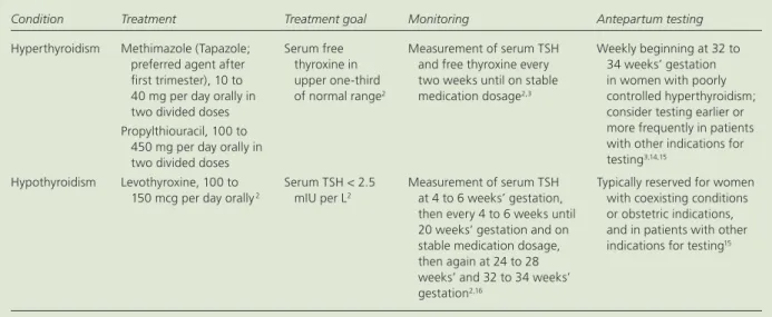
Overt hyperthyroidism during pregnancy is treated with methimazole (Tapazole) or propylthiouracil Table 5. Treatment of Thyroid Disease in Pregnancy
Condition Treatment Treatment goal Monitoring Antepartum testing
Hyperthyroidism Methimazole (Tapazole; preferred agent after first trimester), 10 to 40 mg per day orally in two divided doses Propylthiouracil, 100 to
450 mg per day orally in two divided doses
Serum free thyroxine in upper one-third of normal range2
Measurement of serum TSH and free thyroxine every two weeks until on stable medication dosage2,3
Weekly beginning at 32 to 34 weeks’ gestation in women with poorly controlled hyperthyroidism; consider testing earlier or more frequently in patients with other indications for testing3,14,15
Hypothyroidism Levothyroxine, 100 to 150 mcg per day orally 2
Serum TSH < 2.5 mIU per L2
Measurement of serum TSH at 4 to 6 weeks’ gestation, then every 4 to 6 weeks until 20 weeks’ gestation and on stable medication dosage, then again at 24 to 28 weeks’ and 32 to 34 weeks’ gestation2,16
Typically reserved for women with coexisting conditions or obstetric indications, and in patients with other indications for testing15
TSH = thyroid-stimulating hormone.
Information from references 2, 3, and 14 through 16.
Table 6. Adjustment of Levothyroxine Dosage Based on Thyroid-Stimulating Hormone Level
Thyroid-stimulating hormone level (mIU per L)
Levothyroxine dosage increase (mcg per day)
5 to < 10 25 to 50
10 to 20 50 to 75
> 20 75 to 100
(Table 5).2,3,14-16 Because the use of methimazole is asso-ciated with birth defects, including aplasia cutis and choanal or esophageal atresia,16,19 propylthiouracil is the preferred medication during the first trimester.3 How-ever, it is recommended that physicians consider switch-ing to methimazole after the first trimester because the risk of liver failure associated with propylthiouracil use is greater than the risk of congenital abnormalities.2,3
The main concern in women with hyperthyroidism is the potential effect on the fetus. Thyroid receptor anti-bodies should be measured by the end of the second trimester in women with active Graves disease, a his-tory of Graves disease treated with radioactive iodine or thyroidectomy, or a history of a previous infant with Graves disease.2,3,20 In women at high risk, including those receiving antithyroid medication and those with poorly controlled hyperthyroidism or high thyrotoxin receptor antibody levels, fetal ultrasonography should be performed monthly after 20 weeks’ gestation to detect evidence of fetal thyroid dysfunction (e.g., growth restriction, hydrops, goiter, cardiac failure).2,3,21 These women should also undergo antepartum testing at least weekly beginning at 32 to 34 weeks’ gestation (or earlier in particularly high-risk situations).22
Postpartum Thyroid Dysfunction
The most common cause of postpartum thyroid dys-function is postpartum thyroiditis, which affects 1.1% to 21.1% of women.23 Postpartum thyroiditis is defined as an abnormal TSH level within the first 12 months postpartum in the absence of a toxic thyroid nodule or thyrotoxin receptor antibodies.23 Clinical symptoms can
mimic the typical fatigue following delivery, as well as postpartum depression and Graves disease; a thorough assessment is required to differentiate these conditions. A radioactive iodine uptake scan can help distinguish postpartum thyroiditis from Graves disease, but is con-traindicated in breastfeeding women. Patients must limit close contact with others for a time after the study.
The clinical course of postpartum thyroiditis varies: approximately 25% of patients present with symptoms of hyperthyroidism, followed by hypothyroidism and then recovery; 43% present with symptoms of hypo-thyroidism; and 32% present with hyperthyroidism.3 Because the hyperthyroid phase of postpartum thyroid-itis is caused by autoimmune destruction of the thyroid, resulting in release of stored thyroid hormone, antithy-roid medications are not typically beneficial and treat-ment is generally symptomatic, using peripheral beta antagonists. Differentiation of the hyperthyroid phase of postpartum thyroiditis from Graves disease is important because Graves disease requires antithyroid therapy. In contrast, postpartum hypothyroidism should be treated with levothyroxine in women who are symptomatic or breastfeeding, or who wish to become pregnant, and may require lifetime supplementation.3,5
Women with a history of type 1 diabetes and women with thyroglobulin or thyroperoxidase autoantibod-ies are at increased risk of postpartum thyroiditis.14,15 Asymptomatic women should be screened at three and six months postpartum using serum TSH measurement.2 Additionally, women with a history of postpartum thy-roiditis are at increased risk of permanent hypothyroid-ism and should be screened annually thereafter.2,3,24
SORT: KEY RECOMMENDATIONS FOR PRACTICE
Clinical recommendation
Evidence
rating References
The optimal method to assess serum FT4 during pregnancy uses direct measurement techniques. Serum TSH
is a more accurate indicator of maternal thyroid status than alternative FT4 assay methods.
C 3, 6
Targeted screening for thyroid disease should be performed in pregnant women at high risk, including those with a history of thyroid disease, type 1 diabetes mellitus, or other autoimmune disease; current or past use of thyroid therapy; or a family history of autoimmune thyroid disease.
C 2, 3
Hypothyroidism during pregnancy should be treated with levothyroxine, with a serum TSH goal of less than 2.5 mIU per L.
A 1-3
Serum TSH should be measured in pregnant women who are being treated for hypothyroidism at four to six weeks’ gestation, then every four to six weeks until 20 weeks’ gestation and on a stable medication dosage, then again at 24 to 28 weeks’ and 32 to 34 weeks’ gestation.
C 2, 3
Propylthiouracil is the preferred agent for the treatment of hyperthyroidism during the first trimester of pregnancy and in women with methimazole (Tapazole) allergy and hyperthyroidism. Consideration should be given to switching to methimazole after the first trimester, and the dosage should be adjusted to maintain a serum FT4 level in the upper one-third of the normal range.
C 3
In pregnant women who are being treated for hyperthyroidism, serum TSH and FT4 should be measured
every two weeks until the patient is on a stable medication dosage.
C 2, 3
FT4 = free thyroxine; TSH = thyroid-stimulating hormone.
A = consistent, good-quality patient-oriented evidence; B = inconsistent or limited-quality patient-oriented evidence; C = consensus, disease-oriented evidence, usual practice, expert opinion, or case series. For information about the SORT evidence rating system, go to http://www.aafp.org/afpsort.
Data Sources: Essential Evidence Plus was searched using PubMed, and OVID was searched. Key words were thyroid disease and pregnancy. Article selection was limited to human studies, original research, system-atic reviews, and current clinical practice guidelines. Search date: August 22, 2013.
The views expressed in this article are those of the authors and do not necessarily reflect the official policy or position of the U.S. Navy Medical Corps, the U.S. Navy, or the U.S. Department of Defense.
The Authors
LEO A. CARNEY, DO, is a faculty member at the Naval Hospital Pensacola (Fla.) Family Medicine Residency Program and an assistant professor in the Department of Family Medicine at the Uniformed Services University of the Health Sciences, Bethesda, Md.
JEFF D. QUINLAN, MD, is the Navy Family Medicine Specialty Leader and an assistant professor in the Department of Family Medicine at the Uni-formed Services University of the Health Sciences.
JANET M. WEST, MD, is a faculty member at the Naval Hospital Pensacola Family Medicine Residency Program and the U.S. Army School of Aviation Medicine Occupational Medicine Residency Program.
Address correspondence to Leo A. Carney, DO, Naval Hospital Pen-sacola, 6000 W. Hwy 98, PenPen-sacola, FL 32512 (e-mail: leo.carney@med. navy.mil). Reprints are not available from the authors.
REFERENCES
1. Reid SM, Middleton P, Cossich MC, Crowther CA. Interventions for clini-cal and subcliniclini-cal hypothyroidism in pregnancy. Cochrane Database Syst Rev. 2010;(7):CD007752.
2. De Groot L, Abalovich M, Alexander EK, et al. Management of thy-roid dysfunction during pregnancy and postpartum: an Endocrine Society clinical practice guideline. J Clin Endocrinol Metab. 2012; 97(8):2543-2565.
3. Stagnaro-Green A, Abalovich M, Alexander E, et al.; American Thy-roid Association Taskforce on ThyThy-roid Disease During Pregnancy and Postpartum. Guidelines of the American Thyroid Association for the diagnosis and management of thyroid disease during pregnancy and postpartum. Thyroid. 2011;21(10):1081-1125.
4. American College of Obstetrics and Gynecology. ACOG prac-tice bulletin no. 37. Thyroid disease in pregnancy. Obstet Gynecol.
2002;100(2):387-396.
5. Yassa L, Marqusee E, Fawcett R, Alexander EK. Thyroid hormone early adjustment in pregnancy (the THERAPY) trial. J Clin Endocrinol Metab. 2010;95(7):3234-3241.
6. Lee RH, Spencer CA, Mestman JH, et al. Free T4 immunoassays are flawed during pregnancy. Am J Obstet Gynecol. 2009;200(3):260. e1-260.e6.
7. Abbassi-Ghanavati M, Greer LG, Cunningham FG. Pregnancy and laboratory studies: a reference table for clinicians [published correc-tion appears in Obstet Gynecol. 2010;15(2 pt 1):387]. Obstet Gynecol. 2009;114(6):1326-1331.
8. Vaidya B, Anthony S, Bilous M, et al. Detection of thyroid dysfunction in early pregnancy: universal screening or targeted high-risk case finding?
J Clin Endocrinol Metab. 2007;92(1):203-207.
9. Negro R, Schwartz A, Gismondi R, Tinelli A, Mangieri T, Stagnaro- Green A. Universal screening versus case finding for detection and treatment of thyroid hormonal dysfunction during pregnancy. J Clin
Endocrinol Metab. 2010;95(4):1699-1707.
10. Momotani N, Ito K, Hamada N, Ban Y, Nishikawa Y, Mimura T. Mater-nal hyperthyroidism and congenital malformation in the offspring. Clin Endocrinol (Oxf). 1984;20(6):695-700.
11. Neale DM, Cootauco AC, Burrow G. Thyroid disease in pregnancy. Clin Perinatol. 2007;34(4):543-557, v-vi.
12. Stagnaro-Green A. Overt hyperthyroidism and hypothyroidism during pregnancy. Clin Obstet Gynecol. 2011;54(3):478-487.
13. Rovet JF. Neurodevelopmental consequences of maternal hypothyroid-ism during pregnancy. In: Program and abstracts from the 76th annual meeting of the American Thyroid Association; September 30 – Octo-ber 3, 2004; Vancouver, British Columbia. Thyroid. 2004;14(9):710. Abstract 88.
14. Männistö T, Vääräsmäki M, Pouta A, et al. Thyroid dysfunction and autoantibodies during pregnancy as predictive factors of pregnancy complications and maternal morbidity in later life. J Clin Endocrinol
Metab. 2010;95(3):1084-1094.
15. Mamede da Costa S, Sieiro Netto L, Coeli CM, Buescu A, Vaisman M. Value of combined clinical information and thyroid peroxidase antibod-ies in pregnancy for the prediction of postpartum thyroid dysfunction.
Am J Reprod Immunol. 2007;58(4):344-349.
16. Di Gianantonio E, Schaefer C, Mastroiacovo PP, et al. Adverse effects of prenatal methimazole exposure. Teratology. 2001;64(5):262-266. 17. Mandel SJ. Hypothyroidism and chronic autoimmune thyroiditis in the
pregnant state: maternal aspects. Best Pract Res Clin Endocrinol Metab. 2004;18(2):213-224.
18. Casey BM, Dashe JS, Wells CE, McIntire DD, Leveno KJ, Cunningham FG. Subclinical hyperthyroidism and pregnancy outcomes. Obstet Gynecol. 2006;107(2 pt 1):337-341.
19. Wing DA, Millar LK, Koonings PP, Montoro MN, Mestman JH. A com-parison of propylthiouracil versus methimazole in the treatment of hyperthyroidism in pregnancy. Am J Obstet Gynecol. 1994;170(1 pt 1): 90-95.
20. Mitsuda N, Tamaki H, Amino N, Hosono T, Miyai K, Tanizawa O. Risk factors for developmental disorders in infants born to women with Graves disease. Obstet Gynecol. 1992;80(3 pt 1):359-364.
21. Luton D, Le Gac I, Vuillard E, et al. Management of Graves’ disease during pregnancy: the key role of fetal thyroid gland monitoring. J Clin
Endocrinol Metab. 2005;90(11):6093-6098.
22. ACOG practice bulletin. Antepartum fetal surveillance. Number 9, Octo-ber 1999 (replaces Technical Bulletin NumOcto-ber 188, January 1994). Clini-cal practice management guidelines for obstetrician-gynecologists. Int J
Gynaecol Obstet. 2000;68(2):175-185.
23. Muller AF, Drexhage HA, Berghout A. Postpartum thyroiditis and autoimmune thyroiditis in women of childbearing age: recent insights and consequences for antenatal and postnatal care. Endocr Rev. 2001;22(5):605-630.
24. Azizi F. The occurrence of permanent thyroid failure in patients with sub-clinical postpartum thyroiditis. Eur J Endocrinol. 2005;153(3):367-371.