The following full text is a publisher's version.
For additional information about this publication click this link.
http://repository.ubn.ru.nl/handle/2066/127112
Please be advised that this information was generated on 2017-03-10 and may be subject to
change.
non-small cell lung cancer,
a critical evaluation
Proefschrift
ter verkrijging van de graad van doctor aan de Radboud Universiteit Nijmegen
op gezag van de rector magnificus prof. mr. S.C.J.J. Kortmann volgens besluit van het college van decanen
in het openbaar te verdedigen op maandag 26 mei 2014 om 14.30 uur precies door Ad Verhagen geboren op 8 november 1961 te Nijmegen
a critical evaluation
Publication of this thesis was supported by: Maquet Nederland, St. Jude Medical, Sorin Group Nederland N.V., Edwards Lifesciences, Krijnen Medical Innovations, Covidien Nederland, Takeda Nederland, Johnson & Johnson Medical, Stöpler Instrumenten & Apparaten.
A.F.T.M. Verhagen
Aspects of staging in non-small cell lung cancer, a critical evaluation.
Thesis Radboud University Nijmegen Medical Centre
ISBN
978-94-6259-097-7
Cover
Pantheon, Rome
Cover and lay-out design
Promotie In Zicht, Arnhem, The Netherlands
Ipskamp Drukkers, Enschede, The Netherlands
Copyright
© A.F.T.M. Verhagen 2014
All rights reserved. No part of this thesis may be reproduced or transmitted, in any form or by any means, without written permission of the author.
Copromotor:
Dr. H.F.M. van der Heijden
Manuscriptcommissie:
Prof. dr. P.N.R. Dekhuijzen (voorzitter) Prof. dr. H.J.M. Groen (UMCG) Dr. A. Brutel de la Rivière
Introduction.
Development of a lung cancer staging system.
TNM classification. TNM in lung cancer staging.
The current, 7th, edition of the staging system. Future developments. Staging methods. Clinical evaluation. Imaging. Endoscopy. Surgical staging. Pathologic assessment.
Aim of the thesis.
11 12 12 12 13 14 15 15 15 16 18 20 21
Chapter 2 FDG-PET in staging lung cancer. How does it change the algorythm?
Lung Cancer 2004;44(2):175-181.
27
Chapter 3 Mediastinal staging in daily practice: endosonography, followed by cervical mediastinoscopy. Do we really need both?
Interact Cardiovasc Thorac Surg 2013;17(5):823-828.
41
Chapter 4 Completeness of lung cancer surgery: is mediastinal dissection common practice?
Eur J Cardiothorac Surg 2012;41:834-838.
55
Chapter 5 The clinical value of lymphatic micrometastases in patients with non-small cell lung cancer.
J Thorac Oncol 2010;5:1201-1205.
51
Chapter 6 General discussion. 83
Chapter 7 Summary. Samenvatting. 93 103 Acknowledgement. 111 List of publications. 117 Curriculum Vitae. 123
General introduction
and outline of the thesis.
Introduction.
Lung cancer accounts for a huge disease burden worldwide: it is the leading cause of cancer in the entire population, representing 12.7% of all newly diagnosed malignant diseases and responsible for 18.2% of all cancer related deaths (1).
Also in The Netherlands lung cancer is frequently diagnosed: in 2011, in almost 12000 patients a lung carcinoma was detected. It was more frequently diagnosed in men (7,100 pts., being 13.5% of all cancers diagnosed in men) than in women (4,600 pts., being 9.5% of all cancers diagnosed in women), although the incidence among women is still slowly increasing (2).
Treatment of patients with lung cancer is based on both local therapeutic modalities, like surgery or radiotherapy, and several forms of systemic therapy, sometimes in combination, as well as providing best supportive care.
Despite improvements in diagnostic procedures and therapeutic regimens, the overall prognosis of patients with lung cancer remains poor (3). Nevertheless, there is a clear prognostic difference between patient subgroups, depending on the extent of the disease, which is based on clinical aspects, like tumour size and the presence or absence of lymphatic and/or haematogenous metastases (4). Besides these anatomic parameters, the histological type, specific tissue characteristics (5) and both molecular- and genomic characteristics, are also important prognostic factors.
Staging in lung cancer comprises multiple diagnostic procedures to determine the extent of the disease at the time of presentation, as well as during and after treatment. Accurate staging is a multidisciplinary responsibility of all physicians involved in the diagnosis and treatment of patients with lung cancer. This is essential to select the appropriate therapy for an individual patient, but also to have an indication of the prognosis and to compare results of treatment between distinct patient groups (6).
The staging algorithm is not static, but evolving over time, and is dependent on available techniques and performance.
When these techniques reveal no sign or symptom of distant metastasis, the mediastinal lymph nodes are crucial in the staging process and will determine the contribution and sequence of treatment modalities.
The aim of this thesis is to contribute to the evolution of the staging process in patients with non-small cell lung cancer, by a critical evaluation of four different aspects, with special focus on the mediastinum.
Additionally, a new system for regional lymph node classification was proposed, since local differences appeared in classifying lymph nodes, as a result of two different systems that were in use: the lymph node map developed by Naruke and adopted by the AJCC (16), and a slightly different scheme approved by the American Thoracic Society (17). Both systems were unified into a new, so called “Mountain-Dresler American Thoracic Society (MD-ATS)” lymph node map (18).
The revisions in this latest staging system were based on analysis of 5,319 patients, primarily treated in a single hospital, the University of Texas M.D. Anderson Cancer Center in the USA. Most of these patients were treated surgically.
Serious doubts about the representativeness of these data with regard to medically treated patients, as well as patients treated in other hospitals in other parts of the world, led to the initiative of the International Association for the Study of Lung Cancer (IASLC), in 1998, to develop an international lung cancer database, from a large number of patients, in all stages of the disease, from all over the world. Analysis would have to lead to validated revisions of the staging system (19).
The current, 7th, edition of the staging system.
As a result of this initiative, data from 68,463 patients with non-small cell lung cancer and an additional 13,032 patients with small cell lung cancer, originating from 20 countries in four continents, were analysed. The analysis led to reclassification of the “T” and the “M” components with regard to lung cancer, in the current 7th edition of the TNM classification of malignant tumours (20). The “N” component remained unchanged, although the concept of nodal zones was introduced. Seven different zones were defined, 1 cervical-, 4 mediastinal- and 2 intrapulmonary zones, each consisting of closely related nodal stations (21).
Because of the reclassification of the T and M components, changes were also proposed for the stage grouping of lung cancer. Based on these proposals, for each stage in the current TNM classification there is a clear relation between stage and survival, as well as a significant separation of survival curves between stages (22).
According to the TNM principles, in the 1980’s a distinction was made between a classification based on clinical findings only, defined as cTNM, and a TNM classification also based on postoperative pathologic examination, pTNM. According to the current stage groupings, the survival rates of the pathological stages exceed the survival rates of the corresponding clinical stages at every stage.
In conjunction with the 7th edition of the TNM staging system, a new lymph node map was again proposed. Despite the development of the MD-ATS lymph node map, the former Naruke map was still used by Asian, especially Japanese, physicians. This hampered the interpretation of treatment results in the lung cancer database. To overcome the discrepancies and to facilitate future analysis, a new IASLC lymph node map has become part of the current staging system, providing precise anatomic
Development of a lung cancer staging system.
TNM classification.
As early as the 18th century the concept of staging was introduced by a Scottish surgeon, John Hunter, who divided tumours into “movable” and “not only movable” as a criterion for resection (7). But it was only in the 1940’s that a systematic method to describe the stage of cancer was introduced by Pierre Denoix, a French oncologic surgeon. He presented a “uniform technique for clinical classification”, which was based on the anatomic extent of a carcinoma and defined by three parameters: a “T”, concerning the extent of the primary tumour, an “N”, recording the presence or absence of lymph node metastases and an “M”, describing distant metastases. This method was adopted by the Union Internationale Contre le Cancer (UICC), an organisation founded in 1933 by cancer researchers worldwide to share their knowledge, now called the Union for International Cancer Control (UICC), based in Geneva. In the 1950’s this organisation established a Committee on Clinical Stage Classification and Applied Statistics to “extend the general technique of classification to cancer at all sites” (8). In the 1960’s they published nine brochures, proposing a classification for 23 tumour sites. The first edition of a comprehensive TNM classification of malignant tumours was published in 1968. Since then new editions have been published, containing extensions and changes to previous versions, but by users in different parts of the world local variations in the rules of classification were introduced (9). Despite this, worldwide agreement on cancer staging was reached in the 1980’s and secured by the publication in 1997 of the 5th edition of the “TNM classification of malignant tumours” by the UICC (10), simultaneously with- and identical to- the “Cancer Staging Manual” by the American Joint Committee on Cancer (AJCC) (11).
Subsequent to the TNM classification, the concept of stage grouping was proposed, actually by the AJCC, to facilitate analysis and improve transparency. Patient cohorts with different TNM subsets, but nevertheless a comparable survival, were grouped together in the same stage group, while survival rates for each stage group had to be distinctive (9).
TNM in lung cancer staging.
With regard to lung cancer, a first staging system according to the TNM principles, was proposed by Clifton F. Mountain in the 1970’s, but in 1986 he published the “new international staging system” (12). This was adopted by both the UICC and the AJCC and published in the 4th edition of both staging manuals (13,14). Unfortunately, in this system the end results of TNM subsets within stage groups diverged. Furthermore, greater specificity with respect to the end results, of especially the TNM subsets in stage I, II and IIIa, was needed. As a result, a revised version of the staging system, based on a larger patient cohort, was published by Mountain in 1997 (15).
Staging methods.
Clinical evaluation.
Determining the anatomical extent of lung cancer starts by the interpretation of symptoms and signs of a patient. Common complaints like coughing and haemoptysis are not specific, but pain referring to the skeleton, or neurologic disorders may be a first sign of distant metastases. In the same way, pain of the chest wall can result from a tumour invading the pleura or beyond, and fever may be a sign of post-obstruction atelectasis or pneumonia, once the diagnosis of a tumour is established. Furthermore, physical examination may reveal enlarged supra-clavicular lymph nodes, suspicious of metastatic involvement. Nevertheless, additional investigation will be necessary to confirm the diagnosis and demonstrate the extent of the disease.
Laboratory tests are, in general, of little help with regard to the detection of a lung carcinoma or identification of metastases. Only in the case of extensive bone metastases an elevated serum calcium concentration will be found. Also results from liver function tests are seldom abnormal, unless numerous metastases are present. However, anaemia at presentation is a poor prognostic factor and seems to correlate with metastatic disease (27).
Imaging.
Once a lung carcinoma is considered, chest radiography is routinely performed to detect a tumour, determine the site and, roughly, extent of the process. Nevertheless in about 20 % of patients with lung cancer, a tumour is not seen on chest X-ray, in the majority due to superimposed structures or additional alterations, for example as a result of pneumonia (28). Also the size, density and ill-defined margins of a lesion are important factors that contribute to detection error (29).
A diagnostic Computed Tomographic (CT) scan of the chest should therefore be performed when a carcinoma is suspected, not only to establish a tumour , but also to gather information about the size of the process, the anatomic location within the lung, the relationship with surrounding structures and to screen for additional nodules or parenchyma disorders, as well as pleural collections.
Furthermore, enlargement of both hilar- and mediastinal lymph nodes has to be assessed on the mediastinal setting of a CT scan. In general, lymph nodes with a short axis of more than 1 cm. are considered enlarged, being suggestive of metastatic involvement.
In conjunction with imaging of the chest, a CT scan of the upper abdomen provides information about the liver and adrenal glands, both being sites of preference for metastatic disease. Overall, a CT scan provides essential information to stage a lung carcinoma, both with regard to the T, N and M parameters.
definitions for all lymph node stations and including the grouping of separate stations into seven zones (23).
The redefined anatomic boundaries of lymph node stations can be easily applied to clinical staging, among others by CT scanning. Nevertheless, the redefinition of boundaries between intrapulmonary- and mediastinal lymph node stations, and the distinction between left- and right sided stations, will have an impact on clinical practice, both in determining the correct stage and optimal therapy. For example, the lower border of the paratracheal- and the upper border of the hilar lymph nodes are now defined by the caudal margin of the azygos vein. As a result, hilar lymph nodes have become easily accessible for invasive mediastinal staging by mediastinoscopy. Furthermore, the border between left- and right sided lymph nodes has moved from the midline to the left margin of the trachea, whereas the area of the subcarinal lymph nodes has been extended caudally.
The current staging system only encloses anatomical parameters, although descriptors exist to indicate histopathological grading (G), lymphatic- (L) or venous invasion (V) and descriptors to indicate special circumstances, like classification of multiple primary tumours (m), staging following multimodality therapy (y), recurrent disease (r), or at autopsy (a) (19). Moreover, for the first time, the 7th edition of the staging system is also applicable to small cell lung cancer and carcinoid tumours. Nevertheless, in patients with non-small cell lung cancer, histology appeared to have only limited influence on prognosis and thus was not included as a staging parameter (24).
Future developments.
Analysis of the IASLC database revealed additional non-anatomic parameters, like performance status, age and gender, as significant prognostic factors. These are not taken into account yet, but may be included in the next version of the staging system, although they do not refer to the extent of the process (25). Also functional- or metabolic data, which have become available only after the inclusion period of the current database, as a result of the widespread use of 18FluoroDeoxyGlucose-Positron Emission Tomographic (FDG-PET) scanning in the work-up of patients with lung cancer, will probably have an impact on the next staging system.
Finally, the amount of data, based on molecular- and genomic characteristics of the primary tumour, are rapidly increasing and, once validated with regard to prognosis, are expected to play an important role in future staging systems. The next, 8th , version of the UICC lung cancer staging manual, for which patient data are prospectively collected between 2009 and 2012, is intended to be published in 2016 (26).
the procedure may be increased by navigation bronchoscopy or radial endobronchial ultrasound-guided lung biopsy, but these methods are not yet widely used (37).
In addition to establishing a diagnosis and determining the type of resection, recently endoscopic techniques have emerged in hilar and mediastinal lymph node staging and have become an important method in determining the N status as part of the staging procedure.
Initially Endoscopic Ultrasound guided-Fine Needle Aspiration (EUS-FNA) was introduced, being a method to puncture and aspirate mediastinal lymph nodes by an oesophageal endoscope, using ultrasound to detect lymph nodes and guide a hollow needle (38). In addition to assessment of the mediastinal lymph nodes, the left-, but also right adrenal gland are accessible by this method, making it possible to prove a metastatic lesion in a suspicious adrenal gland on FDG-PET and/or CT scan (39,40).
Because the oesophagus is running down in the mediastinum behind, but also somewhat on the left side of the trachea, this technique is less suitable for the assessment of lymph nodes on the right side of the trachea. Visibility by ultrasound is hampered by the interfering air. Nevertheless, lymph nodes in the lower mediastinum, in the pulmonary ligament and paraoesophageally, which cannot be reached by me-diastinoscopy, are accessible on both sides. Therefore this technique seems primarily complementary to surgical staging of the mediastinum by cervical mediastinoscopy.
However, also Endobronchial Ultrasound guided-Transbronchial Needle Aspiration (EBUS-TBNA) has become available, making use of a bronchoscope to detect and aspirate lymph nodes under real-time guidance by ultrasound. By this technique the same mediastinal lymph nodes can be reached as by mediastinoscopy, moreover also hilar- and even interlobar lymph nodes are accessible (41,42).
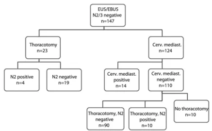
A major advantage of both techniques is their minimally invasive character, making it possible to perform them under conscious sedation, without anaesthesia, in an outpatient clinical setting. However, pathologic assessment of the yield of both procedures is only possible by cytology instead of histology, and there is some doubt about the completeness of the mediastinal examination. Furthermore, the sample obtained by needle aspiration is non-diagnostic in a significant number of cases (43). Consequently, the use of Rapid On-Site Evaluation (ROSE) of the aspirate is advised to increase accuracy, by repeating the needle passes and aspiration of a lymph node, until a representative sample is obtained (44). Despite this, endosonography, being the combination of EBUS-TBNA and/or EUS-FNA, has become the first line diagnostic procedure in a growing number of hospitals, when invasive mediastinal lymph node staging is indicated. Whether endosonography can completely replace mediastinos-copy in primary lung cancer staging is still a matter of debate (45). To assess the additional value of cervical mediastinoscopy in routine clinical practice, in patients with suspected or proven non-small cell lung cancer, after a negative result of endosonography, was the goal of the study in chapter 3.
Even with a CT scan, a tumour may be missed due to the size and characteristics of a nodule, or failure to distinguish a nodule from an adjacent structure or atelectasis (30).
Moreover, with regard to mediastinal lymph node staging, the accuracy of CT scan is low, since size is the main criterion for malignancy. In mediastinal lymph nodes with a size of 10 to 15 mm, the prevalence of metastatic involvement is still 30%, increasing to 67% in lymph nodes over 15 mm (31). In a meta-analysis of 43 studies, concerning 7,368 patients, the pooled sensitivity and specificity of CT scan for mediastinal lymph node metastases was 55% and 81% respectively, with a negative predictive value of 83% (32). These numbers are insufficient for an optimal assessment and thus, historically, a mediastinoscopy had to be performed to improve the reliability of mediastinal staging.
Since the mid 1990’s FDG-PET scanning has emerged as a valuable staging tool, which is based on the increased glucose uptake of malignant cells, in this way providing metabolic information. Addition of the metabolic information of FDG-PET to the anatomic information provided by CT scan has improved the reliability of the staging process, both with regard to the detection of distant metastases and with regard to the mediastinal lymph nodes.
In extrathoracic staging up to 30 % unexpected metastases are found by the use of FDG-PET, in addition to conventional imaging by CT scan (33). Furthermore, FDG-PET proved to be more effective in detecting bone metastases than MRI or bone scintigraphy (34).
With regard to the mediastinal lymph nodes, the addition of FDG-PET improved the accuracy of non-invasive staging (35), probably diminishing the need for a medi-astinoscopy. Nevertheless, the poor spatial resolution of FDG-PET is a disadvantage, which is only partially solved by the fusion of FDG-PET and CT in a single scan, a so-called integrated PET-CT scan (36).
To assess the additional value of FDG-PET in detecting extrathoracic metastases and to compare the accuracy of FDG-PET in mediastinal lymph node staging to cervical mediastinoscopy, was the aim of the study in chapter 2.
Endoscopy.
To confirm the diagnosis of lung cancer, suspected on chest radiography, CT- and/or FDG-PET scan, bronchoscopy is indicated, possibly providing tissue for histological assessment or the yield of bronchial washing and brushing for cytology. Moreover, in case of a centrally located tumour, the endobronchial extent of the process can be assessed. This may help to define the T status and gives insight to the potential type of resection.
The sensitivity of bronchoscopy for the confirmation of lung cancer in central lesions is high, 88%, but diminishes in peripherally located tumours to 63% and 34%, depending on their size (over- and under 2 cm in diameter, respectively). The yield of
endosonography, in patients suspected to have mediastinal lymph node involvement, mediastinoscopy is still indicated to reduce the false-negative rate (see chapter 3).
This is in agreement with the recent update of the European guidelines on pre-operative staging: apart from the addition of FDG-PET as an imaging modality, endosonography, if available, is suggested the primary staging tool for tissue confirmation, both in cases of only hilar suspicion, a central tumour, or a tumour over 3 cm in diameter, and in cases of positive mediastinal lymph nodes on imaging techniques. However, especially in these latter patients, confirmation of a negative result by mediastinoscopy is recommended (58).
In early stage lung cancer, without mediastinal lymph node involvement by clinical staging, surgical resection is regarded as the standard of care. The goal of surgery is to perform a complete resection, providing the best outcome for an individual patient. The type of resection is mainly dependent on the T stage of the tumour. In case of a T1 or T2 tumour (20), confined to a single lobe, in general a lobectomy is the treatment of choice (59). However, in case of a central location, with tumour invading the pulmonary artery or main bronchus, a sleeve resection or pneumonectomy may be needed. In case of a T3 tumour with invasion of the chest wall or other surrounding tissue, or a T4 tumour, supposed to be resectable, an “en-bloc” resection of the originating lobe or lung, together with the invaded structure, leading to negative resection margins, should be performed. Yet, a prerequisite for a complete resection is confirmation of the clinical stage during surgery, since unexpected findings may be encountered for each of the stage descriptors. Therefore the pre-operative staging process has to be continued during surgery, assessing both the primary tumour and remaining lung parenchyma, to determine the type of resection. Moreover, special attention has to be paid to the intrapulmonary- and mediastinal lymph nodes, since unforeseen lymphatic dissemination may be present despite a thorough pre-operative work-up (60). Nodal metastases themselves may influence the type of parenchyma resection, for example in case of extra-nodal growth. Yet, in addition to the pulmonary resection, a systematic nodal dissection should primarily be performed to accurately determine the pathologic stage and, ideally, consists of a meticulous excision of both lobar-, interlobar-, hilar- and all ipsilateral mediastinal lymph node stations.
To determine completeness of resection, a definition has been proposed by the IASLC and has been adopted by the UICC (61). This definition not only includes a free resection margin at the vascular-, bronchial- and parenchyma margins or pleural surface, but also minimal requirements with regard to lymph node dissection. Moreover, the European Society of Thoracic Surgeons (ESTS) guidelines for intraoperative lymph node staging have been published (62). According to these guidelines a systematic nodal dissection is recommended and should consist, on the right side, of an en-bloc resection of the upper- and lower paratracheal lymph nodes,
Surgical staging.
In the absence of distant metastases, pre-operative surgical staging of the mediastinum by mediastinoscopy, has proven to be a valuable method to select patients who might benefit most from an intended curative resection (46).
The technique was described by Carlens, in 1959, as a procedure “for inspection and tissue biopsy in the superior mediastinum” (47). Since then the procedure has appeared to be accurate, with a low complication rate and is still considered the gold standard in mediastinal lymph node staging (48). To ensure a high reliability of mediastinoscopy, minimal requirements have been defined (49), although in daily practice they are not always met (50,51). The yield of the procedure is dependent on the thoroughness of the mediastinal exploration and seems related to the experience of the surgeon.
Although mediastinoscopy was initially used to select appropriate candidates for surgical treatment, based on the absence of mediastinal lymph node metastases, its role has changed over time. Since two randomized trials, both published in 1994, demonstrated a survival benefit of chemotherapy prior to surgery, over surgery alone, in patients with resectabel non-small cell lung cancer and ipsilateral mediastinal lymph node metastases, cervical mediastinoscopy became important, not only to demonstrate, but also to discriminate between ipsilateral and contra-lateral mediastinal lymph node involvement (52,53).
Following this combination treatment of systemic and local therapy, the question raised whether the same local control with regard to the primary tumour site could be achieved by radiotherapy instead of surgery. In two landmark trials, designed to define the role of surgery in patients with ipsilateral mediastinal lymph node metastases, overall survival curves of treatment arms with- and without surgery were not significantly different (54,55), resulting in chemo-radiotherapy as the standard of care. However, subgroup analysis showed that in patients with a good response to induction therapy, leading to clearance or so-called down staging of the mediastinal lymph nodes, the surgical arm led to a favourable outcome. As a result of these studies among others (56), the concept of re-staging was introduced. Restaging, or reassessment of the mediastinal lymph nodes to prove down staging after chemo- and/or radiotherapy, can theoretically be performed by the same modalities as primary staging: by imaging, endosonography and/or (repeat) mediastinoscopy. The reliability of both imaging techniques and endosonography in restaging appears to be low. Although a repeat mediastinoscopy is technically possible, this method too has a lower yield compared to an initial procedure (57). Therefore, the highest accuracy in both initial staging and restaging may be achieved by the use of endosonography first, to prove ipsilateral lymph node involvement, followed by the use of a primary mediastinoscopy to evaluate the result of induction therapy in patients with locally advanced lung cancer. However, in cases of an unexpected negative result of
assessment is critical. By serial sectioning and the use of immunohistochemistry or molecular techniques as real-time polymerase chain reaction, micrometastases may be detected in up to 30% of histologically negative lymph nodes (65). Nevertheless, the clinical impact of this “occult” lymph node involvement remains unclear (66).
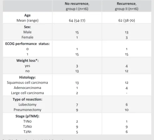
To assess the clinical impact of lymphatic micrometastases, by determining whether recurrent disease was associated with the presence of lymphatic isolated tumour cells and/or micrometastases at the time of the lung resection, was the goal of the study in chapter 5.
Aim of the thesis.
Determining the optimal treatment for each individual patient with a non-small cell lung cancer, is the main goal of staging. Over the last decades, not only therapeutic regimens have changed and are still changing, but also new techniques and insights into lung cancer staging have become available.
The aim of this thesis is to scrutinize the role and performance of four techniques used in mediastinal lymph node staging:
Firstly, FDG-PET. Over the last 15 years, this technique has become widely available and adopted, but what is the accuracy of FDG-PET with regard to the mediastinal lymph nodes? Does it replace invasive mediastinal staging? To what extent?
Secondly, endosonography. This technique has emerged as an attractive first line method for invasive mediastinal lymph node staging. But is there still a place for cervical mediastinoscopy, if endosonography appears to be negative?
Thirdly, as part of an intended curative resection, a systematic lymph node dissection is recommended to obtain optimal loco-regional staging. However, what is routine performance during lung cancer surgery?
Finally, pathologic assessment of the mediastinal lymph nodes, both taken pre-operatively and during surgery, is critical in treatment planning. What is the clinical impact of occult lymph node involvement, detected by immunohistochemistry, in addition to conventional examination techniques?
Each of these questions is relevant, as they are encountered in daily practice when treating patients with non-small cell lung cancer.
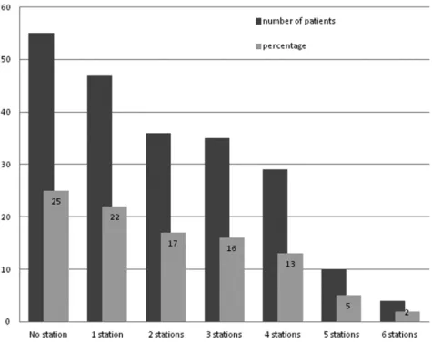
any visible lymph nodes in front of the superior vena cava and behind the trachea, as well as lymph nodes in the subcarinal space, next to the oesophagus and in the pulmonary ligament. On the left side, dissection of the sub- and para-aortic lymph nodes, the lower paratracheal lymph nodes and also the lymph nodes in the subcarinal space, next to the oesophagus and in the pulmonary ligament should be performed. Besides these mediastinal lymph nodes, the hilar, interlobar and pulmonary lymph nodes should be dissected. However, based on two large surveys concerning surgical care in the United States, it is questionable whether these guidelines have been adopted by the surgical community (51,63). To assess the extent of mediastinal lymph node dissection, routinely performed during lung cancer surgery, and hereby the completeness of resection according to the guidelines of the ESTS, was the goal of the study in chapter 4.
Pathologic assessment.
Pathology plays an essential role at multiple phases in the staging algorithm. In patients with a suspicious lesion on chest X-ray, confirmation of a primary lung carcinoma by examination of the yield of bronchoscopy is a first step. Furthermore, confirmation of distant metastasis in case of clinical suspicion on tumour dissemination is necessary. In the absence of distant metastasis the focus of clinical staging is oriented towards the mediastinum. Depending on the indication for pre-operative mediastinal lymph node staging, cytological assessment of the yield of endosonography will be performed, in some cases followed by histological examination of lymph node biopsies taken by mediastinoscopy.
During surgery confirmation of malignancy may still have to be performed and additional frozen section analysis to assess the resection margin may be indicated, especially in central tumours, if an extended resection is feasible (64).
Finally, the pTNM stage is determined by pathological examination of the surgical specimen. Based on the size of the tumour, associated atelectasis or pneumonitis, invasion of pleura, chest wall, bronchus, vessels, nerves or surrounding organs, as well as the presence of co-existing tumour nodules, the T stage is recorded. In the current staging system, the M stage can only rarely be determined from the surgical specimen (only in case of pleural or pericardial tumour nodules), however, the N stage is critical to define completeness of resection in addition to assessment of the hilar- and pleural- or parenchyma resection margin.
Examination of lymph nodes, both taken during mediastinoscopy and during surgery, is traditionally performed by assessment of haematoxylin and eosin stained slides, taken at two levels of formalin fixed and paraffin embedded lymph nodes.
However, by this method micrometastases and isolated tumour cells may be overlooked, potentially leading to understaging. Since lymph node involvement is a strong prognostic factor and key to the application of (neo-)adjuvant therapy, accurate
25. Sculier JP, Chansky K, Crowley JJ, Van Meerbeeck J, et al. The impact of additional prognostic factors on survival and their relationship with the anatomical extent of disease expressed by the 6th edition of the
TNM Classification of Malignant Tumours and the proposals for the 7th edition. J Thorac Oncol
2008;3:457-466.
26. Giroux DJ, Rami-Porta R, Chansky K, Crowley JJ, et al. The IASLC lung cancer staging project. Data elements for the prospective project. J Thorac Oncol 2009;4:679-683.
27. Spiro SG, Gould MK and Colice GL. Initial evaluation of the patient with lung cancer: symptoms, signs, laboratory tests, and paraneoplastic syndromes: ACCP evidence-based clinical practice guidelines (2nd edition). Chest 2007;132(3S):149S-160S.
28. Quekel LG, Goei R, Kessels AG and van Engelshoven JM. The limited detection of lung cancer on chest X-rays. Ned Tijdschr Geneesk 2003;147:1048-1056.
29. White CS, Flukinger T, Jeudy J and Chen JJ. Use of a computer-aided detection System to detect missed lung cancer at chest radiography. Radiology 2009;252:273-281.
30. Fardanesh M and White C. Missed lung cancer on chest radiography and computed tomography. Semin Ultrasound CT MRI 2012;33:280-287.
31. De Langen AJ, Raijmakers P, Riphagen I, Paul MA, et al. The size of mediastinal lymph nodes and its relation with metastatic involvement: a meta-analysis. Eur J Cardiothorac Surg 2006;29:26-29. 32. Silvestri GA, Gonzalez AV, Jantz MA, Margolis ML, et al. Methods for staging non-small cell lung cancer.
Diagnosis and Management of Lung Cancer, 3rd ed: American College of Chest Physicians Evidence-Based
Clinical Practice Guidelines. Chest 2013;143(5)(Suppl):211-250.
33. Fischer B, Lassen U, Mortensen J, Larsen S, et al. Preoperative staging of lung cancer with combined PET-CT. N Engl J Med 2009;361:32-39.
34. Qu X, Huang X, Yan W, Wu L, et al. A meta-analysis of 18FDG-PET-CT, 18FDG-PET, MRI and bone scintigraphy
for diagnosis of bone metastases in patients with lung cancer. Eur J Radiol 2012;81:1007-1015. 35. Pieterman RM, van Putten JW, Meuzelaar JJ, Mooyaart EL, et al. Pre-operative staging of non-small-cell
lung cancer with positron-emission tomography. N Engl J Med 2000;343(4):254-261.
36. Lardinois D, Weder W, Hany T, Kamel EM, et al. Staging of non-small-cell lung cancer with integrated Positron-Emission Tomography and Computed Tomography. N Engl J Med 2003;348:2500-2507. 37. Rivera MP, Mehta AC and Wahidi MM. Establishing the diagnosis of lung cancer: diagnosis and
management of lung cancer, 3rd ed: American College of Chest Physicians evidence-based clinical
practice guidelines. Chest 2013;143(5)(Suppl):142-165.
38. Annema JT, Versteegh MI, Veseliç M, Voigt P, et al. Endoscopic ultrasound-guided fine-needle aspiration in the diagnosis and staging of lung cancer and its impact on surgical staging. J Clinic Oncol 2005;23:8357-8361.
39. Schuurbiers OJC, Tournoy KG, Schoppers HJ, Dijkman BG, et al. EUS-FNA for the detection of left adrenal metastasis in patient with lung cancer. Lung Cancer 2011;73:310-315.
40. Eloubeidi MA, Black KR, Tamhane A, Eltoum IA, et al. A large single-center experience of EUS-guided FNA of the left and right adrenal glands: diagnostic utility and impact on patient management. Gastrointest Endosc 2010;71:745-53.
41. Yasufuku K, Chiyo M, Sekine Y, Chhajed PN, et al. Real-time endobronchial ultrasound-guided transbronchial needle aspiration of mediastinal and hilar lymph nodes. Chest 2004;126:122-128. 42. Ernst A, Eberhardt R, Krasnik M and Herth FJ. Efficacy of endobronchial ultrasound-guided transbronchial
needle aspiration of hilar lymph nodes for diagnosing and staging cancer. J Thorac Oncol 2009;8:947-950.
43. Whitson BA, Groth SS, Odell DD, Briones EP, et al. True negative predictive value of endobronchial ultrasound in lung cancer: are we being conservative enough? Ann Thorac Surg 2013;95:1689-1694. 44. Nakajima T, Yasufuku K, Saegusa F, Fujiwara T, et al. Rapid on-site cytologic evaluation during
endobronchial ultrasound-guided transbronchial needle aspiration for nodal staging in patients with lung cancer. Ann Thorac Surg 2013;95:1695-1699.
45. Tournoy KG, Keller SM and Annema JT. Mediastinal staging of lung cancer: novel concepts. Lancet Oncol 2012;5:221-229.
References:
1. Ferlay J, Shin HR, Bray F, Forman D, et al. Estimates of worldwide burden of cancer in 2008: Globocan 2008. Int J Cancer 2010;127:2893-2917.
2. Nederlandse Kankerregistratie, beheerd door IKNL©, 2013. (Dutch National Cancer Registry).
3. Van der Drift MA, Karim-Kos HE, Siesling S, Groen HJM, et al. Progress in standard of care therapy and modest survival benefits in the treatment of NSCLC patients in the Netherlands in the last 20 years. J Thorac Oncol 2012;7:291-298.
4. Goldstraw P, Crowley J, Chansky K, Giroux DJ, et al. The IASLC lung cancer staging project: proposals for the revision of the TNM stage groupings in the forthcoming (seventh) edition of the TNM classification of malignant tumours. J Thorac Oncol 2007;2:706-714.
5. Tomoyuki Hishida, Junji Yoshida, Ryo Maeda, Genichiro Ishii et al. Prognostic impact of intratumoural microvascular invasion and microlymphatic permeation on node-negative non-small-cell lung cancer: which indicator is the stronger prognostic factor? Eur J Cardiothoracic Surg 2013;43(4):772-777. 6. Gospodarowicz MK, Miller D, Groome PA, Greene FL, et al. The process for continuous improvement of
the TNM classification. Cancer 2004;100;1-5.
7. John Hunter, Frederick James. Lectures on the principles of surgery. Haswell, Barrington and Haswell, Philadelphia, 1839.
8. Greene FL and Sobin LH. The TNM System: Our language for cancer care. J Surg Oncol 2002;80:119-120. 9. Sobin LH. TNM. Principles, history and relation to other prognostic factors. Cancer 2001;91:1589-1592. 10. Sobin LH and Wittekind C. UICC: TNM classification of malignant tumours, 5th ed. Wiley-Liss, Inc., New
York, 1997.
11. Fleming ID, Cooper JS, Henson DE, Hutter RVP et al. American Joint Committee on Cancer: Cancer staging manual, 5th ed. Lippincott, Philadelphia, 1997.
12. Mountain CF. A new international system for staging lung cancer. Chest 1986;89:225s-233s.
13. Hermanek P and Sobin LH. UICC: TNM classification of malignant tumours, 4th ed. Springer Verlag,
Berlin, 1987.
14. Beahrs OH, Hensen DE, Hutter RVP, et al. American Joint Committee on Cancer (AJCC): Manual for Staging of Cancer, 4th ed. Lippincott, Philadelphia, 1992.
15. Mountain CF. Revisions in the international system for staging lung cancer. Chest 1997;111:1710-1717. 16. Naruke T, Suemasu K and Ishikawa S. Lymph node mapping and curability at various levels of metastasis
in resected lung cancer. J Thorac Cardiovasc Surg 1978;76(6):832-839.
17. Glazer GM, Gross BH, Quint LE, et al. Normal mediastinal lymph nodes: number and size according to American Thoracic Society mapping. AJR Am J Roentgenol 1985;144(2):261-265.
18. Mountain CF and Dresler CM. Regional lymph node classification for lung cancer staging. Chest 1997;111:1718-1723.
19. Goldstraw P. IASLC: Staging Manual in Thoracic Oncology. Editorial Rx Press, Orange Park,FL,USA,2009. 20. L sobin, M Gospodarowicz and Wittekind C. UICC: TNM classification of Malignant Tumours, 7th ed.
Wi-ley-Blackwell, New York, 2009.
21. Rusch VW, Crowley J, Giroux DJ, Goldstraw P, et al. The IASLC lung cancer staging project: proposals for the revision of the N descriptors in the forthcoming seventh edition of the TNM classification for lung cancer. J Thorac Oncol 2007;2:603-612.
22. Goldstraw P, Crowley J, Chansky K, Giroux DJ, et al. The IASLC lung cancer staging project: proposals for the revision of the TNM stage groupings in the forthcoming (seventh) edition of the TNM classification of malignant tumours. J Thorac Oncol 2007;2:706-714.
23. Rusch VW, Asamura H, Watanabe H, Giroux DJ, et al. The IASLC lung cancer staging project: a proposal for a new international lymph node map in the forthcoming seventh edition of the TNM classification for lung cancer. J Thorac Oncol 2009;4:568-577.
24. Chansky K, Sculier JP, Crowley JJ, Giroux D, et al. The International Association for the Study of Lung Cancer staging project. Prognostic factors and pathologic TNM stage in surgically managed non-small cell lung cancer. J Thorac Oncol 2009;4:792-801.
46. Pearson FG, DeLarue NC, Ilves R, Todd TR, et al. Significance of positive superior mediastinal nodes identified at mediastinoscopy in patients with resectable cancer of the lung. J Thorac Cardiovasc Surg 1982;83:1-11.
47. Carlens E. Mediastinoscopy: a method for inspection and tissue biopsy in the superior mediastinum. Dis Chest 1959;36:343-352.
48. Lemaire A, Nikolic I, Petersen T, Haney JC, et al. Nine-year single center experience with cervical mediastinoscopy: complications and false negative rate. Ann Thorac Surg 2006;82:1185-1190. 49. De Leyn P, Lardinois D, van Schil PE, Rami-Porta R, et al. ESTS guidelines for preoperative lymph node
staging for non-small cell lung cancer. Eur J Cardiothorac Surg 2007;32:1-8.
50. Van Albada ME, Eldering MJ, Post WJ, Klinkenberg TJ, et al. The biopsying of at least 5 mediastinal lymph node stations for presurgical staging in patients with a non-small-cell lung carcinoma. Ned Tijdschr Geneeskd 2004;148:281-286.
51. Little AG, Rusch VW, Bonner JA, Gaspar LE, et al. Patterns of surgical care of lung cancer patients. Ann Thorac Surg 2005;80:2051-2056.
52. Roth JA, Fossella F, Komaki R, Ryan MB, et al. A randomized trial comparing perioperative chemotherapy and surgery with surgery alone in resectable stage IIIA non-small-cell lung cancer. J Natl Cancer Inst 1994;86:673-680.
53. Rosell R, Gomez-Codina J, Camps C, Maestre J, et al. A randomized trial comparing pre-operative chemotherapy plus surgery with surgery alone in patients with non-small-cell lung cancer. N Engl J Med 1994;330:153-158.
54. Van Meerbeeck JP, Kramer GW, van Schil PE, Legrand C, et al. Randomized controlled trial of resection versus radiotherapy after induction chemotherapy in stage IIIA-N2 non-small-cell lung cancer. J Natl Cancer Inst 2007;99:442-450.
55. Albain KS, Swann RS, Rusch VW, Turrisi AT 3rd, et al. Radiotherapy plus chemotherapy with or without
surgical resection for stage III non-small-cell lung cancer: a phase III randomised controlled trial. Lancet 2009;374:379-386.
56. Betticher DC, Hsu Schmitz SF, Tötsch M, Hansen E, et al. Mediastinal lymph node clearance after docetaxel-cisplatin neoadjuvant chemotherapy is prognostic of survival in patients with stage IIIA pN2 non-small-cell lung cancer: a multicenter phase II trial. J Clin Oncol 2003;21:1752-1759.
57. De Cabanyes Candela S and Detterbeck FC. A systematic review of restaging after induction therapy for stage IIIa lung cancer. Prediction of pathologic stage. J Thorac Oncol 2010;5:389-398.
58. De Leyn P, Dooms C, Kuzdzal J, Lardinois D, et al. Revised ESTS guidelines for preoperative mediastinal lymph node staging for non-small cell lung cancer. www.ests.org.
59. Ginsberg RJ and Rubinstein LV. Randomized trial of lobectomy versus limited resection for T1 N0 non-small cell lung cancer. Lung Cancer Study Group. Ann Thorac Surg 1995;60:615-622.
60. Graham ANJ, Chan KJM, Pastorino U and Goldstraw P. Systematic nodal dissection in the intrathoracic staging of patients with non-small cell lung cancer. J Thorac Cardiovasc Surg 1999;117:246-251. 61. Rami-Porta R, Wittekind C and Goldstraw P. Complete resection in lung cancer surgery: proposed
definition. Lung Cancer 2005;49:25-33.
62. Lardinois D, De Leyn P, van Schil P, Rami-Porta R, et al. ESTS guidelines for intraoperative lymph node staging in non-small cell lung cancer. Eur J Cardiothoracic Surg 2006;30:787-792.
63. Boffa DJ, Allen MS, Grab JD, Gaissert HA, et al. Data from the Society of Thoracic Surgeons General Thoracic Surgery database: The surgical management of primary lung tumors. J Thoracic Cardiovasc Surg 2008;135:247-254.
64. Maygarden SJ, Detterbeck FC and Funkhouser WK. Bronchial margins in lung cancer resection specimens: utility of frozen section and gross evaluation. Mod Pathol 2004;17:1080-1086.
65. Coello MC, Luketich JD, Litle VR and Godfrey TE. Prognostic significance of micrometastasis in Non-Small-Cell Lung Cancer. Clin Lung Cancer 2004;5:214-225.
66. Marchevsky AM, Gupta R, Kusuanco D, Mirocha J et al. The presence of isolated tumor cells and micrometastases in the intrathoracic lymph nodes of patients with lung cancer is not associated with decreased survival. Hum Pathol 2010;41:1536-1543.
A.F.T. Verhagen, G.P. Bootsma, V.C.G. Tjan-Heijnen, G.J. van der Wilt, A.L. Cox, M.H.J. Brouwer, F.H.M. Corstens, W.J.G. Oyen.
Lung Cancer 2004 May;44(2):175-81.
FDG-PET in staging lung cancer.
How does it change the algorythm?
2
Introduction
To determine the appropriate therapy in patients with lung cancer, accurate staging is mandatory. Both detection of extrathoracic metastases (ETM) and, when there is no sign of distant metastases, assessment of mediastinal lymph node involvement are essential.
For detection of extrathoracic metastases a wide variety of diagnostic procedures is available, such as CT-scan, bone scintigraphy and MRI.
In mediastinal lymph node staging (MLS) the reliability of CT scan is low [1]. Therefore, in the work-up for a thoracotomy a cervical mediastinoscopy is still considered the gold standard, providing a high negative- and positive predictive value and a low morbidity and mortality rate [2,3]. However, it remains an invasive procedure and not every surgeon feels comfortable performing it.
In the last decade increasing evidence on the utility of positron emission tomography (PET) using fluor-18-fluorodesoxyglucose (FDG) to detect both extra- thoracic metastases and mediastinal lymph node involvement has become available. In contrast to the anatomic information provided by CT scan, FDG-PET capitalizes on the increased glucose uptake of malignant cells, thus providing complimentary diagnostic information. The positioning of FDG-PET in the diagnostic work-up of lung cancer patients depends on its reliability to detect both extrathoracic- and lymph node metastases.
Several reports [4,5], using FDG-PET in MLS, suggest that the need for mediastinos-copy in patients potentially eligible for a curative resection is reduced.
Particularly the high negative predictive value of a mediastinal FDG-PET would make a mediastinoscopy redundant.
The aim of this study was to assess the added value of FDG-PET in detecting extrathoracic metastases and to compare the reliability of FDG-PET in MLS to that of mediastinoscopy in patients with non-small cell lung cancer.
Patients and methods
Patients.
From July 2000 until March 2001 we studied 72 consecutive patients (60 man, 12 women; mean age 62 years, range 18-87 years) with suspected or proven primary non-small cell lung cancer at the University Medical Center Nijmegen. The final histological diagnosis was squamous cell carcinoma (n=35), adenocarcinoma (n=19), undifferentiated non-small cell carcinoma (n=16), and carcinoid (n=2, 1 typical, 1 atypical).
All patients underwent full conventional clinical staging, consisting of history taking and physical examination, laboratory investigations, X-chest, bronchoscopy,
Summary
Background: In patients with lung cancer FDG-PET may be used both to detect
extrathoracic metastases (ETM) and for mediastinal lymph node staging (MLS), potentially reducing the need for mediastinoscopy.
We assessed the added value of FDG-PET in detecting ETM and focused on the reliability of FDG-PET and mediastinoscopy for MLS.
Patients and methods: In 72 consecutive patients with non-small cell lung cancer, the
impact of adding FDG-PET to full conventional clinical staging was prospectively analyzed. The predictive value of FDG-PET findings and tumor location for pathologic mediastinal lymph node status were assessed in a logistic regression analysis.
Results: Unexpected extrathoracic metastases were detected by FDG-PET in 15% of
patients. In MLS overall negative- and positive predictive values were 71% and 83% for FDG-PET and 92% and 100% for mediastinoscopy. However, the negative predictive value of FDG-PET was only 17% in case of FDG-PET positive N1 nodes and/or a centrally located primary tumor, whereas it was 96% in case of FDG-PET negative N1 nodes and a non-centrally located primary tumor.
Conclusion: By incorporating FDG-PET in clinical staging 15% of patients with lung
cancer are upstaged due to unexpected extrathoracic metastases. In case of a negative mediastinal FDG-PET, mediastinoscopy can only be omitted in the presence of a non-centrally located primary tumor and without FDG-PET positive N1 nodes.
2
transmission images of the area between proximal femora and the base of the skull were acquired (10 minutes per bedposition). When only an emission study was recorded, the images were not corrected for attenuation and reconstructed using filtered backprojection (Butterworth filter with a cut-off frequency of 0.4 Nyquist). When an emission and transmission study were recorded, the images were corrected for attenuation and reconstructed using the Ordered-Subsets Expectation Maximization (OSEM) algorithm. Reconstructed images were displayed in coronal, transverse and sagittal planes. All images were read by two experienced nuclear medicine physicians. Standard uptake values were not calculated.
When FDG-PET suggested supraclavicular lymph node involvement or extra- thoracic metastases, confirmation was obtained by needle biopsy or correlative imaging by X-ray or MRI.
FDG-PET was considered positive for mediastinal involvement when lesions were detected in any mediastinal lymph node station, not separating ipsilateral (N2) from contralateral (N3) lymph nodes. The decision to perform mediastinoscopy was not influenced by the mediastinal status on FDG-PET.
All patients without lymph node involvement at mediastinoscopy underwent thoracotomy during which mediastinal lymph node sampling was performed. An average of 3.2 lymph node stations was explored.
To assess the influence of the location of the primary tumor on the result of mediastinal FDG-PET evaluation, all tumors were classified as centrally-, intermediately- or peripherally located. A central tumor was located into the inner 1/3 of the lung parenchyma (adjacent to the mediastinum) on a transverse image on the CT-scan. An intermediate or peripheral tumor was located in outer 2/3 of the parenchyma.
Statistical analysis.
The reliability of FDG-PET in MLS was assessed using mediastinoscopy and/or thoracotomy as the gold standard. Sensitivity, specificity, negative- and positive predictive values were calculated by cross tabulation. Posterior probabilities of mediastinal lymphnode involvement were calculated in case of positive and negative test results.
The value of N1 status and N2 status according to FDG-PET, and location of the primary tumor (central, intermediate or peripheral) in predicting pathologic mediastinal lymph node status was explored in a logistic regression analysis.
Difference in proportions of a false negative FDG-PET for MLS between patients with positive hilar nodes and/or a central location of the primary tumor and patients with negative hilar nodes and an intermediate or peripheral location was assessed using Fisher’s exact test.
A P value of less than 0.05 was considered to indicate statistical significance. Statistical analysis was carried out with SPSS software.
CT-scan of the chest and upper abdomen, and, only in case of symptoms, bone scintigraphy and/or CT-scan of the brain (Fig. 1). When there was no evidence of distant metastases, cervical mediastinoscopy (n=50) and, in case of a carcinoma in the left upper lobe, parasternal mediastinoscopy (n=14) was performed in every patient, except for those patients with a peripheral T1 lesion without hilar or mediastinal lymph nodes larger than 1cm. in shortest axis on CT-scan (n=6). At cervical mediastinoscopy multiple biopsies were taken of at least lymph node station 4R, 4L and 7 [6]. An average number of 3.4 lymph node stations was biopsied. Patients were staged according to the revised International System for Staging Lung Cancer [7].
FDG-PET.
In case conventional screening was negative for supraclavicular lymph node involvement or distant metastases, patients underwent FDG-PET scanning in the week before mediastinoscopy. A dedicated, rotating half-ring PET-scanner (ECAT-ART, Siemens/CTI, Knoxville, Tn, USA) was used for data acquisition. Prior to FDG-injection, patients were fasting for at least 6 hours. Intake of sugar-free liquids was permitted. Immediately prior to the procedure, patients were hydrated with 500 ml of water. One hour after intravenous injection of 200-220 MBq FDG (Mallinckrodt Medical, Petten, The Netherlands) and 20 mg furosemide, emission images or emission and
2
Of 18 patients with FDG-PET positive lesions in the mediastinum, mediastinoscopy was positive in 14, but negative in 4 patients. Thoracotomy proved a positive paratracheal lymph node in one of these latter 4 patients (Fig. 2).
Based on the findings at mediastinoscopy and thoracotomy, the overall sensitivity and specificity of FDG-PET with regard to the mediastinum were 58% and 90%. The negative predictive value was 71%, the positive predictive value 83%. The likelihood ratio for mediastinal lymph node involvement in case of a “FDG-PET positive” mediastinum was 5.77, with a posterior probability of mediastinal lymph node metastases of 0.83. The likelihood ratio in case of a “FDG-PET negative” mediastinum was 0.47, with a posterior probability of lymph node metastases of 0.29.
Based on findings at thoracotomy, both sensitivity and negative predictive value of mediastinoscopy in 48 patients were 92%. Specificity and positive predictive value were 100% by definition. When mediastinoscopy was negative, the likelihood ratio for mediastinal lymph node metastases was 0.08. With a prior probability of mediastinal lymph node metastases of 0.5, the posterior probability was 0.07. In case of a positive mediastinoscopy the posterior probability was by definition 1.0.
False-negative FDG-PET.
Of the 11 patients with a false-negative result of FDG-PET for mediastinal involvement, 6 patients showed positive hilar (N1) nodes on PET scan (Fig. 3). Review of the
Results
Findings by FDG-PET.
Of the 72 patients extrathoracic metastases were detected in 5 patients by conventional diagnostic methods and one patient was considered irresectable due to tumor invasion into the vertebral column on CT-scan (T4). Therefore, no FDG-PET was performed in these patients.
In the remaining 66 patients FDG-PET revealed previously unexpected metastases in 10 patients (15%): in 8 patients extrathoracic metastases (M1), in 2 patients supra-clavicular lymph node involvement (N3). In another patient an FDG-accumulating lesion at the base of the tongue proved to be a second primary carcinoma. In 4 additional patients FDG-positive lesions suggestive of metastasis could not be confirmed by conventional methods and were therefore ignored.
Of 56 patients without any sign of tumor dissemination beyond the mediastinum, FDG-PET was positive in the mediastinum in 18 patients and negative in 38 patients.
In 13 patients both FDG-positive mediastinal ànd hilar (N1) lymph nodes were found, in 5 patients only FDG-positive mediastinal lymph nodes.
Only positive hilar lymph nodes (N1) were found in 8 out of 56 patients.
Findings by mediastinoscopy.
48 out of the 56 patients without tumor dissemination beyond the mediastinum underwent cervical- and when indicated also parasternal mediastinoscopy (n=14), which was positive for mediastinal lymph node metastases in 22 patients and negative in 26 patients. In 2 of these mediastinoscopy-negative patients unexpected N2 disease was found during thoracotomy.
In 2 additional patients with a carcinoma in the left upper lobe only cervical mediastinoscopy was performed, which turned out to be negative. Parasternal mediastinoscopy was omitted because of previous coronary bypass surgery. At thoracotomy in both patients lymph nodes in the aorto-pulmonary window proved to be positive, whereas subcarinal and paratracheal nodes were negative.
Six patients with a peripheral tumor clinically staged T1N0M0 directly underwent thoracotomy, without mediastinoscopy. FDG-PET did not show any pathologic uptake except in the primary tumor. Their pathologic stage also proved to be T1N0M0.
Comparison of FDG-PET to mediatinoscopy.
Of 38 patients with a “FDG-PET negative” mediastinum, mediastinoscopy was positive in 8 patients, while at thoracotomy tumor-positive N2 nodes were found in 3 additional patients. Histologically these 11 patients with a false-negative FDG-PET had squamous cell carcinoma (n=8), adenocarcinoma (n=2) and an atypical carcinoid (n=1).
2
according to PET, but also N1 status (p=0.001) and central location (p=0.04) had an independent predictive value with regard to the pathologic N2 stage. When the group of 38 patients with a negative mediastinum on FDG-PET was divided into a group of 12 patients with positive hilar nodes and/or a central location and a group of 26 patients with negative hilar nodes and an intermediate or peripheral location, mediastinal FDG-PET was false-negative in 10 out of 12 patients in the first group, which was significantly different from only 1 false-negative result out of 26 in the second group (Fisher’s exact test, p<0.001) (Table 1).
Sensitivity and specificity of FDG-PET in the first group of patients, with FDG-positive hilar nodes and/or a central location, were 57% and 50%. The negative predictive value was only 17%, the positive predictive value 87%.
In the second group of patients, with FDG-negative hilar nodes and a non-central location, sensitivity and specificity of FDG-PET were 67% and 96%, with a negative predictive value of 96% and a positive predictive value of 67%.
Discussion
In our study FDG-PET scan improved clinical staging of lung cancer patients by detecting unexpected extrathoracic metastases in 15% of patients without any evidence of metastases after conventional staging, thus avoiding futile mediastinos-copy and eventually thoracotomy. This is in accordance with previous publications [8,9]. Routine use of FDG-PET in staging lung cancer may thus lead to a further reduction of patients presenting with metastases within a few months after a “curative” treatment. Of note, conventional staging performed according to the guidelines of the American Thoracic Society and the European Respiratory Society pathology report showed micrometastases in mediastinal (N2) nodes in all 6 of them.
Four other patients had a central location of the primary tumor (adjacent to the mediastinum) on CT scan (Fig. 4). In a logistic regression model not only N2 status
Figure 3. FDG-positive N1 nodes, but negative N2 nodes.
At histology, micrometastases were found in N2 nodes.
Figure 4. Central primary NSCLC, adjacent to the mediastinum,
obscuring histologically proven N2 metastases.
CORONAL TRANSAXIAL SAGITTAL
CORONAL TRANSAXIAL SAGITTAL
Table 1. Histological mediastinal lymph node status in patients with a negative
mediastinal FDG-PET scan (n=38).
Histological positive mediastinal lymph nodes Histological negative mediastinal lymph nodes Total
FDG-PET negative N1 nodes
and non-central tumor 1 25 26 FDG-PET positive N1 nodes
and/or central tumor 10 2 12
2
known pitfall. In this patient group FDG-PET should be used for guidance during mediastinoscopy with a potential increase in sensitivity. Moreover, mediastinoscopy may also be indicated to make a distinction between stage IIIa and stage IIIb.
Conclusion
By incorporating FDG-PET in the preoperative work-up of all patients with potentially resectable non-small cell lung cancer, 15% of patients are upstaged due to detection of unexpected extrathoracic metastases, thus avoiding futile mediastinoscopy and eventually thoracotomy.
With strict criteria, FDG-PET reduces the number of mandatory mediastinoscopy procedures by 46% without an increase in unexpected N2 involvement at thoracotomy. However, mediastinoscopy should not routinely be omitted in patients with apparently a negative mediastinal FDG-PET, but with a centrally located primary tumor and/or FDG-positive hilar lymph nodes: the primary tumor may obscure positive mediastinal lymph nodes and positive hilar lymph nodes are predictive for mediastinal micrometastases.
[10] revealed metastases in 7% (5 of 72) of patients. This is less than expected, but it may reflect the referral pattern to our center, since approximately half of our population consisted of patients who were considered to be surgical candidates by the referring hospital.
Conventionally, when there is no evidence of distant metastases, accurate MLS is the next step in clinical staging, since mediastinal lymph node involvement significantly reduces the benefit of surgery as a single therapy [11,12].
Whether or not FDG-PET should be used as a routine procedure in MLS, replacing mediastinoscopy, is still a matter of debate [13]. On the one hand, a particularly high negative predictive value of mediastinal FDG-PET (up to 97%) is reported [4,5,14]. This led to the recommendation to omit mediastinoscopy in case of a negative mediastinal FDG-PET [15,16].
On the other hand, two important points need to be addressed. First, failure to distinguish involved lymph nodes from activity in the primary tumor on FDG-PET has been reported earlier [8]. Consequently, negative interpretation of mediastinal FDG-PET in patients with centrally located tumors may lead to understaging, because of the primary tumor obscuring the mediastinal lymph nodes [17,18,19]. This is in agreement with our results. Second, we observed that 6 out of 8 patients with FDG-positive N1 nodes but FDG-negative N2 nodes, had microscopic involvement in N2 nodes, ranging in size from a few cells to a few millimeters. Such small volume disease is beyond detectability of any currently available imaging technique. Thus, FDG-PET positive hilar lymph nodes are predictive for microscopic involvement of mediastinal lymph nodes.
The present study convincingly shows that the negative predictive value was only 17% in case of FDG-PET positive N1 nodes and/or a centrally located primary tumor, whereas it was 96% in case of FDG-PET negative N1 nodes and a non-centrally located primary tumor. These conditions clearly identify the limitations of FDG-PET for MLS and strongly support the continued use of mediastinoscopy in the first group of patients. Conversely, the true-negative result of mediastinal FDG-PET in 25 out of 26 patients with a non-centrally located primary tumor and without any FDG-PET positive lymph nodes, makes FDG-PET very reliable in this specific patient group, resulting in a significant reduction in the need for mediastinoscopy, in our series of 46% (26 out of 56 patients undergoing MLS). In the future, fusion of the CT scan with FDG-PET into one study may be helpful to improve the diagnostic accuracy of these imaging procedures.
Although the reliability of a positive mediastinal (N2 or N3) FDG-PET was high in our series (15 histologic positive patients out of 18 FDG-PET positive), we feel that mediastinoscopy should still be performed in these patients to confirm nodal metastases histologically. Otherwise, a potentially curative operation may be withheld from patients due to FDG-uptake in inflammatory lymph nodes, being a
2
References
1. Dillemans B, Deneffe G, Verschakelen J, Decramer M. Value of computed tomography and mediastinos-copy in preoperative evaluation of mediastinal nodes in non-small cell lung cancer. A study of 569 patients. Eur J Cardio-thorac Surg 1994; 8:37-42.
2. Hammoud ZT, Anderson RC, Meyers BF, et al. The current role of mediastinoscopy in the evaluation of thoracic disease. J Thoracic Cardiovasc Surg 1999; 118:894-9.
3. Gdeedo A, van Schil P, Corthouts B, et al. Prospective evaluation of computed tomography and medias-tinoscopy in mediastinal lymph node staging. Eur Respir J 1997; 10:1547-51.
4. Vansteenkiste JF, Stroobants SG, De Leyn PR, et al. Lymph node staging in non-small-cell lung cancer with FDG-PET scan: a prospective study on 690 lymph node stations from 68 patients. J Clin Oncol 1998; 16:2142-9.
5. Pieterman RM, van Putten JW, Meuzelaar JJ, et al. Preoperative staging of non-small-cell lung cancer with Positron-Emission Tomography. N Engl J Med 2000; 343:254-61.
6. Mountain CF, Dresler CM. Regional lymph node classification for lung cancer staging. Chest 1997; 111:1718-23.
7. Mountain CF: Revisions in the International System for Staging Lung Cancer. Chest 1997; 111:1710-7. 8. Valk PE, Pounds TR, Hopkins DM, et al. Staging non-small cell lung cancer by whole-body positron
emission tomographic imaging. Ann Thorac Surg 1995; 60:1573-82.
9. Weder W, Schmid RA, Bruchhaus H, et al. Detection of extrathoracic metastases by positron emission tomography in lung cancer. Ann Thorac Surg 1998; 66:886-93.
10. American Thoracic Society / European Respiratory Society. Pretreatment Evaluation of Non-Small-cell Lung Cancer. Am J Respir Crit Care Med 1997; 156:320-32.
11. Roth JA, Fossella F, Komaki R, et al. A randomized trial comparing perioperative chemotherapy and surgery with surgery alone in resectable stage IIIA non-small-cell lung cancer. J Natl Cancer Inst 1994; 86:673-80.
12. Rosell R, Gomez-Codina J, Camps C, et al. A randomized trial comparing preoperative chemotherapy plus surgery with surgery alone in patients with non-small-cell lung cancer. N Engl J Med 1994; 330:153-8.
13. Kernstine KH, McLaughlin KA, Menda Y, et al. Can FDG-PET reduce the need for mediastinoscopy in potentially resectable nonsmall cell lung cancer? Ann Thorac Surg 2002; 73:394-402.
14. Gupta NC, Tamim WJ, Graeber GM, et al. Mediastinal lymph node sampling following positron emission tomography with fluorodeoxyglucose imaging in lung cancer staging. Chest 2001; 120:521-27. 15. Jett JR. How to optimize staging in early non-small cell lung cancer. Lung Cancer 2002; 38:S13-S16. 16. Kramer H, Groen HJM. Current Concepts in the Mediastinal Lymph Node Staging of Nonsmall Cell Lung
Cancer. Ann Surg 2003; 238:180-8.
17. van Tinteren H, Hoekstra OS, Smit EF, et al. Effectiveness of positron emission tomography in the preoperative assessment of patients with suspected non-small-cell lung cancer: the PLUS multicentre randomised trial. Lancet 2002; 359:1388-92.
18. Graeter TP, Hellwig D, Hoffmann K, et al. Mediastinal Lymph Node Staging in Suspected Lung Cancer: Comparison of Positron Emission Tomography With F-18-Fluorodeoxyglucose and Mediastinoscopy. Ann Thorac Surg 2003; 75:231-6.
19. Cerfolio RJ, Ojha B, Bryant AS, et al. The Role of FDG-PET Scan in Staging Patients With Nonsmall Cell Carcinoma. Ann Thorac Surg 2003; 76:861-6.
20. Lardinois D, Weder W, Hany TF, et al. Staging of non-small-cell lung cancer with integrated posi-tron-emission tomography and computed tomography. N Engl J Med 2003;9; 348:2500-7.
Ad F. Verhagen, Olga C.J. Schuurbiers, Monika G. Looijen-Salamon,
Stefan M. van der Heide, Henry A. van Swieten and Erik H.F.M. van der Heijden.
Interact Cardiovasc Thorac Surg 2013;17:823-828.