A Stable, Sensitive Fluorescence-based Method for
Measuring Cyclic GMP
Janet Daijo
1, Anne T. Ferguson
1and David Morton
2*1
Molecular Devices Corporation, Sunnyvale, CA, USA.
2Oregon Health and Sciences University, Portland, OR
Email: [email protected]
*To whom correspondence should be addressed
Application Note
INTRODUCTION 3´, 5´ cyclic guanosine monophosphate (cGMP) is a secondary messenger in signal transduction pathways that is produced by the enzyme guanylyl cyclase (GC). Cyclic GMP signaling is important in many physiological responses including relaxation of smooth muscles, penile erection, kidney function and inflammatory responses1, 2, 3. Also, phototransduction in both vertebrate and invertebrate involve signaling pathways that regulate guanylyl cyclase activity4, 5.
In this report, we demonstrate the use of a new cGMP assay (CatchPoint™ cGMP
fluorescent assay) to measure GC activity in two model systems. The first model system involves activation of GC by a polypeptide hormone, atrial natriuretic peptide (ANP)6, 7. This hormone is found in many species of vertebrates and is released in response to elevated blood pressure. ANP reduces blood pressure by stimulating the excretion of sodium and water from the kidneys and relaxing vascular smooth muscle. We study the activity of ANP in the rat lung fibroblast cell line, RFL-6.
In the second model system, we study the activity of a novel GC, MsGC-
β
3,localized in the central nervous system of the tobacco hornworm, Manduca sexta 8–10. Unlike other soluble GCs that require heterodimer formation and activation by NO, MsGC-
β
3 is unique in that it has high basal activity when expressed alone in COS-7 cells and shows only modest stimulation in the presence of nitric oxide9.The cGMP assay that is used to measure cGMP in these two model systems is a competitive immunoassay that is based on the same principles as the CatchPoint cAMP fluorescent assay kit from Molecular Devices (cat #R 8044, R 8053, see also MaxLine Application Note 46, downloadable from the MaxLine product family literature page of Molecular Devices’ web site, www.moleculardevices.com/pages /literature.html). This means that as the amount of cGMP in the sample increases, the fluorescent signal output decreases. In this report, we demonstrate high sensitivity, reliability and convenience of the assay.
MATERIALS 1 Cells. RFL-6 rat lung fibroblast cells (ATCC cat #CCL-192) were grown in Ham's F-12 media supplemented with 20% fetal bovine serum, glutamine (2 mM), penicillin (50 U/mL) and streptomycin (10 mg/mL). COS-7 monkey kidney cells (ATCC cat #CRL-1651) were grown in DMEM supplemented with 10% FCS.
2 Reagents. CatchPoint cyclic GMP fluorescent assay kit, including lyophilized cGMP calibrator, rabbit-anti-cGMP antibody, HRP-cGMP conjugate and Stoplight Red™ substrate (Molecular Devices, cat #R8074, 96-well assay kit Tel: 1-800-636-5577), sodium bicarbonate (Sigma, cat #S5761), human ANP (Sigma cat #A-1663), bovine serum albumin, BSA 30% (Sigma cat #A9576), phosphodiesterase inhibitor, 3-isobutyl-1-methylxanthine or IBMX (Sigma, cat #I7018), EDTA-free Complete protease inhibitors (Roche Applied Science,
4 Medium and buffers. Ham's F-12 medium (Irvine Scientific, cat #9058, Tel: 1-800-577-6097), DMEM (Irvine Scientific, cat #9031), fetal bovine serum (Irvine Scientific, cat #3000), glutamine pen-strep solution (Irvine Scientific,
cat #9316), Krebs-Ringer bicarbonate buffer (KRGB buffer) (Sigma,
cat #K4002), M-PER mammalian cell extraction reagent (Pierce, cat #78501, Tel: 1-800-874-3723) and phosphate buffered saline, PBS (Invitrogen™ Life Technologies, cat #10010-023). Cell Lysis Buffer (pH 7.3), cGMP Assay Buffer (pH 5.8) and 10X Wash Solution are all supplied with the kit.
Reagent Preparation
1 KRGB Buffer. Sigma provides detailed instructions for preparing this buffer in the product information sheet.
2 800 mM IBMX. Dissolve 100 mg of IBMX in 563 µL of DMSO. Store at -20 °C. IBMX blocks the breakdown of cGMP by cGMP-phosphodiesterases.
3 Pre-stimulation buffer. Add 9.4 µL of 800 mM IBMX to 10 mL of KRGB buffer. Vortex vigorously to ensure that IMBX is completely dissolved. This buffer should be prepared fresh on the day of the experiment.
4 cGMP calibrator. Reconstitute one vial of lyophilized calibrator in 5 mL of PBS. This results in 30 µM cGMP calibrator concentration. Mix well to ensure dissolution of all contents. Store on ice or at 4 °C. Further dilutions are made with cGMP assay buffer as shown in Table 1.
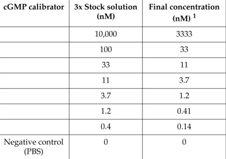
Table 1: Doses of cGMP calibrator used to create the standard dose response curve. 1Final concentration in a 120 µL assay volume.
cGMP calibrator 3x Stock solution (nM) Final concentration (nM) 1 10,000 3333 100 33 33 11 11 3.7 3.7 1.2 1.2 0.41 0.4 0.14 Negative control (PBS) 0 0
5 60 µM ANP. Dissolve 100 µg ANP in 541 µL of sterile PBS supplemented with 0.1% BSA. Aliquot and store at -20 °C. Further dilutions are prepared in PBS containing 0.1% BSA as shown in Table 2.
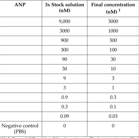
Table 2: Doses of ANP used for cell-based experiments. 1Final concentration in a 600 µL assay volume.
6 Reconstituted rabbit anti-cGMP antibody and HRP-cGMP conjugate. Add 10 mL of cGMP Assay Buffer to each of the two vials, mix well to dissolve contents, store on ice or at 4 °C for up to two weeks.
7 Stoplight Red substrate solution. This solution should be prepared fresh before use and added within one hour after preparation. Dilute 150 µL of 100X stock Stoplight Red substrate into 15 mL of substrate buffer, and then add 17 µL of 3% hydrogen peroxide. Keep the solution protected from light. METHODS Detailed instructions for use of the CatchPoint cyclic-GMP assay kit (Molecular
Devices cat #R8074) with the cGMP calibrator can be found in the product insert that accompanies the kit or on our web site:
www.moleculardevices.com/pages/reagents.html Cell preparation for RFL-6 cell studies
ANP 3x Stock solution
(nM) Final concentration (nM) 1 9,000 3000 3000 1000 900 300 300 100 90 30 30 10 9 3 3 1 0.9 0.3 0.3 0.1 0.09 0.03 Negative control (PBS) 0 0
Step 3 In order to lyse the cells, 200 µL of Cell Lysis Buffer were added to each well, and the microplate was agitated for 10 minute at room tempera-ture using a plate shaker.
Step 4 Using a multichannel pipettor, a 40 µL volume was removed from each well and transferred to separate wells of the 96-well plate. Each well of the 12-well plate was used for quadruplicates in the 96-well assay microplate.
Cell preparation for COS-7 cell studies
Step 1 COS-7 cells were transiently transfected with either the control
pcDNA3.1 plasmid (Invitrogen) or MsGC-
β
3 coding sequence clonedinto pcDNA3.1 as described previously8.
Step 2 Three days after transfection, the cells were harvested and homoge-nized in M-PER extraction reagent containing EDTA-free Complete pro-tease inhibitors.
Step 3 COS-7 cell extracts were incubated for 30 minutes in a buffer containing
50 mM MOPS-KOH (pH 7.5), 60 mM KCl, 8 mM NaCl, 4 mM MgCl2 or
4 mM MnCl2, 10 mM each of the cGMP phosphodiesterase inhibitors
dipyridamole and zaprinast and 1 mM GTP.
Step 4 The reaction was stopped with 0.2 M zinc acetate, and excess GTP was precipitated with 0.2 M Na2CO3. The samples were centrifuged and the supernatant was diluted with CatchPoint Assay Buffer (1:40 dilution). Step 5 Using a multichannel pipettor, a 40 µL volume was removed from each
well and transferred to separate wells of the 96-well plate, with each sample assayed in duplicate. Samples were assayed using the TK Catch-Point assay and in parallel using a colorimetric assay11.
Assay procedure for CatchPoint cGMP assay
Step 1 Forty microliters of reconstituted rabbit anti-cGMP antibody were added to all of the wells. The microplate was gently agitated on a plate shaker for 5 minutes to ensure proper mixing.
Step 2 Forty microliters of reconstituted HRP-cGMP conjugate were added to all wells of the microplate and gently mixed. The plate was then incu-bated for 2 hours at room temperature.
Step 3 When incubation was complete, the assay microplate was washed four times with 300 µL wash buffer per wash.
Step 4 One hundred microliters of Stoplight Red substrate solution were added to every well as quickly as possible. The microplate was covered and protected from light.
Step 5 Measurements were taken after 60 minutes by reading the fluorescence intensity at the settings described in the CatchPoint cGMP product
insert. Fluorescence was measured using either Analyst™ AD,
RESULTS Assay Performance
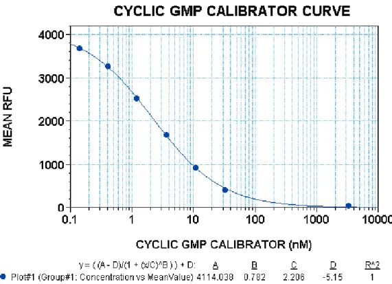
Cyclic GMP calibrator
First assay performance was examined using the cGMP calibrator. Each concentra-tion of cGMP calibrator was performed in quadruplicates. Results that are
repre-sentative of more than three experiments are shown in Figure 1. The range of EC50
values obtained using all three fluorescence microplate readers was between 1.7 and 2.8 nM. The range of signal-to-background values was calculated using the zero calibrator and 3333 nM calibrator concentrations. The values obtained for the Analyst were between 300 and 340; the values obtained for the Gemini XS were between 420 and 430; and the values for FlexStation were between 400 and 480.
Figure 1: Dose response curve for cGMP calibrator using the FlexStation. Data was obtained 60 minutes after addition of Stoplight Red substrate. Each point on the graph represents an average of four replicate samples. The error bars denote standard deviation from the mean. The R2 value is 1.0 for a 4-parameter curve fit. For this experiment, the resulting EC50 value for cGMP calibrator is 2.2 nM. RFU denotes relative fluorescence units.
The Z´ factor was determined using sixteen replicates of the zero calibrator and 3333 nM calibrator concentrations. The range of Z´ factor values obtained using all three fluorescence microplate readers was between 0.89 and 0.95.
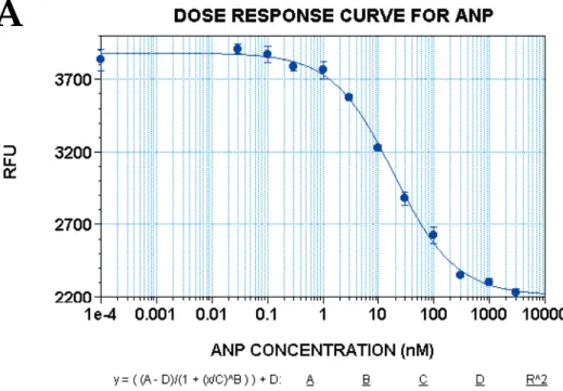
Figure 2: Dose response curve for ANP-treated RFL-6 cells. Each point on the dose response curve represents the average of four replicate samples for one concentration of the activator. The plate was incubated with Stoplight Red substrate for 60 minutes before reading the data set. The dose response shown in (A) represents data obtained using Gemini XS and SoftMax® Pro and (B) represents data obtained using Analyst AD using Graph-Pad Prism® software. The EC50 values obtained from repeated experiments for both instruments were between 11 and 19 nM.
The rat lung fibroblast cell line RFL-6 was used to study activation of GC by the
peptide hormone, ANP. As shown in Figure 2, the EC50 values obtained from
repeated experiments using either the Gemini XS or Analyst AD were between 11 and 19 nM. The signal-to-background values ranged from 400 to 450 using Gemini XS, 300 to 350 using Analyst and 425 to 500 using FlexStation. The Z´ factors ranged from 0.5 to 0.73 for all of the instruments used.
300 100 0 0.01 0.1 1 10 100 103 104 200 cGMP (fmol/well)
Atrial natriuretic peptide (nM) Dose response curve for
atrial natriuretic peptide
A
Comparison to a colorimetric assay
Next, we were interested in determining how the CatchPoint cGMP assay com-pared to other commercially available cGMP assays. We comcom-pared the results of the calibrator curve using the CatchPoint assay to a colorimetric assay using
reagents from American Qualex (San Clemente, CA)11. The procedure for the
col-orimetric assay requires an overnight incubation of primary antibody, HRP-cGMP conjugate (competitor) and cGMP-containing samples. In contrast, the CatchPoint kit requires only a two-hour incubation. Note that the two assays yield the same
EC50 value (~250 fmol) when used at their optimal incubation times (see
Figure 3).
Figure 3: Comparison of EC50 values obtained at optimal incubation times for each assay. The time to complete the colorimetric assay (EIA, solid squares) was approximately 18 hours, and the time for the CatchPoint assay (dashed line) was three hours. Both assays yielded approximately the same EC50 value for the calibrator.
However, when a two-hour incubation time was used with the colorimetric assay, the EC50 value was significantly compromised (see Table 3). In addition, the signal-to-noise ratio for the CatchPoint assay was 22 times greater than the colorimetric assay.
Kit manufacturer EC50 value Signal-to-noise ratio
American Qualex 948 4.9 Molecular Devices 230 110 1.0 0.8 0.2 0.0 -1 0 1 2 3 4 0.4 0.6 B/B 0 log (cGMP) (fmol/well) Standard curves EIA vs. CatchPoint
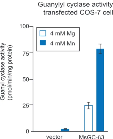
enzyme, Mg2+ and Mn2+. Guanylyl cyclases have been shown to have different levels of activity in the presence of Mn and Mg13. Specifically, one other NO-insensitive GC showed higher levels of activity in the presence of Mn2+ than Mg2+
(13). The results are shown in Figure 4.
Figure 4: Guanylyl cyclase activity in GC-overexpressing COS-7 cells. Cells were transfected with vector (nega-tive control) or MsGC-
β
3, which is a GC variant that has high basal activity. The divalent cations magnesium and manganese are co-factors required for MsGC-β
3 activity.CONCLUSIONS In this report, we show that the CatchPoint cGMP assay is a sensitive assay with both higher signal-to-background ratios (22:1 direct comparison) and Z-factor values when using purified cGMP (calibrator) and cell lysates containing cGMP. We used two different cell systems to measure cGMP. First, we measured the levels of peptide hormone-inducible cGMP, and then we measured the levels of cGMP produced by transiently transfected cells expressing a unique GC. Both systems demonstrated that the assay was reliable and highly sensitive.
When we compared the CatchPoint assay to a colorimetric assay, we found that the two assays produced very similar results when each was used under their optimal conditions. However, the time to process the CatchPoint assay was three hours compared to the 18 hours required for the colorimetric assay. Additionally, the colorimetric assay has a mandatory termination step (addition of acetic acid) and the signal is not stable for long periods of time. Also, if the reaction is not terminated within a certain period of time, all samples, including the controls, will become saturated and the values meaningless. In contrast, the signal
produced from the Stoplight Red substrate used in the CatchPoint assay is stable for up to 24 hours. Of note, if we compared the colorimetric assay using a two-hour incubation instead of the suggested overnight incubation, the EC50 value for the calibrator increased by over four-fold. These two features, no termination step and faster processing time, provide the convenience that high throughput
laboratories require.
REFERENCES 1 Wedel, B. and Garbers, D. 2001. The guanylyl cyclase family at Y2K. Annu. MsGC-β3
4 mM Mn 4 mM Mg
Guanyl cyclase activity (pmol/min/mg protein) 100 75 50 25 0 vector
Guanylyl cyclase activity in transfected COS-7 cells
2 Mateo, O. A. and de Artinano, A. A. 2000. Nitric oxide reactivity and mecha-nisms involved in its biological effects. Pharmacol. Res. 42: 421–427.
3 Patel, M. J., Wypij, D. M., Dudley, A., Rimele, T. J. and Wiseman, J. S. 1995. Secretion of cyclic GMP by cultured epithelial and fibroblast cell lines in response to nitric oxide. J. Pharm. Exp. Therap. 273: 16–25.
4 Dizhoor, A. M. and Hurley, J. B. 1999. Regulation of photoreceptor membrane
guanylyl cyclases by guanylyl cyclase activator proteins. Methods 10: 521–531.
5 Pugh, E. N., Duda, T., Sitaramayya, A. and Sharma, R. K. 1997. Photoreceptor
guanylate cyclases: a review. Biosci. Rep. 17: 429–473.
6 Vesely, D. L. 2002. Atrial natriuretic peptide prohormone gene expression:
hormones and diseases that upregulate its expression. IUBMB Life 53: 153–
159.
7 Azizo, V. A. and Muradova, S. R. 2001. Atrial natriuretic peptide and
cardio-vascular system. Anadolu Kardiyol.Derg. 2001. 1: 297–300.
8 Morton, D. B. 1996. Neuropeptide-stimulated cyclic guanosine
monophos-phate immunoreactivity in the neurosecretory terminals of a neurohemal organ. J. Neurobiol. 29: 341–353.
9 Nighorn, A. Byrnes, K. A., and Morton, D. B. 1999. Identification and
charac-terization of a novel β subunit of soluble guanylyl cyclase that is active in the absence of a second subunit and is relatively insensitive to nitric oxide. J. Biol. Chem. 274: 2525–2531.
10Morton, D. B. and Hudson, M. L. 2002. Cyclic GMP regulation and function
in insects. Advances in Insect Physiol. In press.
11 Kingan, T. G., Gray, W. Zitnan, D. and Adams, M. E. 1997. Regulation of
ecdysis-triggering hormone release by eclosion hormone. J. Exp. Biol.
200: 3245–3256.
12Tremblay, J., Gerzer, R. and Hamlet, P. 1988. Cyclic GMP in cell function. Adv. Second Messenger Phosphoprotein Res. 22: 319–383.
13Simpson, P. J., Nighorn, A. and Morton, D. B. 1999. Identification of a novel
guanylyl cyclase that is related to receptor guanylyl cyclases, but lacks