Neurocognitive mechanisms of
anxiety: an integrative account
Sonia J. Bishop
1,21Behavioural and Clinical Neuroscience Institute, Department of Experimental Psychology, University of Cambridge, Downing
Street, Cambridge, CB2 3EB, UK
2
Medical Research Council Cognition and Brain Sciences Unit, 15 Chaucer Road, Cambridge, CB2 2EF, UK
Anxiety can be hugely disruptive to everyday life. Anxious individuals show increased attentional capture by poten-tial signs of danger, and interpret expressions, comments and events in a negative manner. These cognitive biases have been widely explored in human anxiety research. By contrast, animal models have focused upon the mechan-isms underlying acquisition and extinction of conditioned fear, guiding exposure-based therapies for anxiety dis-orders. Recent neuroimaging studies of conditioned fear, attention to threat and interpretation of emotionally ambiguous stimuli indicate common amygdala–prefron-tal circuitry underlying these processes, and suggest that the balance of activity within this circuitry is altered in anxiety, creating a bias towards threat-related responses. This provides a focus for future translational research, and targeted pharmacological and cognitive interventions. Introduction
It has been argued that fear mechanisms evolved to enable us to shift our focus from the task at hand at the first suggestion of potential danger; for example, interrupting foraging at the glimpse of a potential predator behind a tree. However, in a short evolutionary timescale our world has changed dramatically. The mass media brings news of natural disasters, potential pandemics, terrorist atrocities and violent crime straight into our homes. Perhaps it is not surprising that nearly one in four of us will experience a clinical level of anxiety within our lifetimes[1]. Previously, it might have been adaptive to attend to any potential source of danger and to interpret each ambiguous event as threat-related, but this is no longer the case. Arguably, a more interesting question is why only some individuals experience the excessive fear, worry and disruption to everyday function that characterizes clinical anxiety. This article focuses on the neurocognitive mechanisms impli-cated in anxiety; the literature on the genetics and neuro-chemistry of anxiety having been reviewed elsewhere[2,3]. Fear is viewed as a biologically adaptive physiological and behavioral response to the actual or anticipated occur-rence of an explicit threatening stimulus. Anxiety crucially involves uncertainty as to the expectancy of threat[4], is triggered by less explicit or more generalized cues [5], and is characterized by a more diffuse state of distress, with symptoms of hyperarousal and worry. The human
Glossary
Backward masking:brief presentation of a given stimulus is followed by a visual mask that limits processing of the stimulus and can prevent volunteers from being able to identify it or even detect its presence. Dot probe:a task in which participants are briefly presented with two words or two faces on either side of the screen. These stimuli are replaced by a probe (e.g. a pair of dots), which is presented in the spatial location previously occupied by one of the faces or words. Participants are asked to either simply detect the occurrence of the probe or to make a decision about its shape or orientation. On key trials, one of the two faces or words is threat-related and the other is neutral in valence. Anxious individuals are faster to respond to probes that occur in the position previously occupied by the threat-related stimulus than to probes that occur in the position previously occupied by the neutral stimulus. This difference in response times is held to provide an index of attentional capture by threat.
Emotional Stroop:a variant of the standard Stroop task in which participants are asked to name the ink color of words, which them-selves are the names of colors. In this variant, the key variable is the emotional valence of the words presented, with anxious participants being slower to indicate the color of threat-related words. Genetic polymorphism:a genetic variation that is seen in at least 1% of a given population. Each polymorphism produces two or more differ-ent alleles or versions of the DNA sequence at the locus of the poly-morphism. Some genetic polymorphisms lead to changes in gene expression. These are described as functional genetic polymorphisms. Genomic imaging:a term coined to refer to studies that use neuroima-ging techniques to examine the impact of functional genetic poly-morphisms upon neural activity during cognitive or emotional processing.
Pavlovian fear conditioning:repeated pairing of a neutrally valenced conditioned stimulus (CS), such as a tone or a light, with an uncondi-tioned aversively valenced stimulus (US), such as a footshock, results in the CS alone elicitingconditioned fear responses (CRs), such as freezing, increased startle reflexes and behavioral response suppression. Subsequent repeated presentation of the CS alone, in the absence of the US, leads to extinction of conditioned fear responses.Extinction recallrefers to the retention of extinction after a period of time and is thought to depend upon presentation of the CS in the context in which extinction took place.Reinstatementof the conditioned fear response can occur following re-exposure to the US. Renewal of the conditioned fear response can occur if the CS is presented in a different context to that used for extinction, especially if this is the context in which CS–US pairings were initially established. Perceptual load:the demand or load placed upon perceptual proces-sing is held to become higher when the number of different-identity items that need to be perceived is increased and/or, for the same number of items, when perceptual identification is made more demanding on attention[59].
State and trait anxiety:state anxiety refers to current levels of anxiety and trait anxiety refers to the disposition to experience anxiety across multiple time points. In the human literature these are measured by means of self-report questionnaires that primarily assess symptoms of hyperarousal and worry.
Corresponding author:Bishop, S.J. (sb445@cam.ac.uk). Available online xxxxxx.
cognitive anxiety literature has provided compelling evidence that anxious individuals show increased atten-tional capture by cues signaling danger and are more likely to interpret emotionally ambiguous stimuli in a threat-related manner. It has been suggested that these cognitive biases are implicated in the maintenance, and possibly even the etiology, of anxiety[6,7]. The neural substrate of these processes, however, is not easily amenable to inves-tigation with animal models. This contrasts with associ-ative fear mechanisms, where basic neuroscience studies of Pavlovian fear conditioning (see Glossary) have exten-sively investigated the neural mechanisms mediating the acquisition and extinction of learned or conditioned fear. Although research into factors influencing extinction has been influential in informing exposure therapy for anxiety disorders, there has been little integration of this work with the literature on attentional and interpretative biases in anxiety.
Several recent findings have highlighted the possible interaction of associative and attentional processes in deter-mining the response to threat-related stimuli, while also suggesting conceptual links between associative and inter-pretative processes[8–10]. Crucially, the advent of neuro-imaging has provided a route for examining the neural substrate of these processes in humans. Thus, we can inves-tigate whether the neurocognitive mechanisms underlying attention to, and interpretation of, potentially threat-related stimuli are threat-related to those identified by the animal literature as underlying conditioned fear. The emergence of affective cognitive neuroscience has seen a surge in neuroi-maging studies in this area. Findings from these studies support the contention that amydala-prefrontal circuitry is centrally involved in enabling both representations of stimulus emotional salience and top-down control mechan-isms to influence associative, attentional and interpretative processes. Initial evidence suggests disruption of this cir-cuitry in anxiety, with deficient recruitment of prefrontal control mechanisms and amygdaloid hyper-responsivity to threat potentially leading to alterations in associative, attentional and interpretative processes that sustain a threat-related processing bias in anxious individuals. Mechanisms involved in the processing of threat and their disruption in anxiety
Selective attention to threat
Patients suffering from anxiety disorders have been reported to show a bias in selective attention towards threat-related stimuli [11,12]. Similar findings have been observed for individuals with high levels of trait anxiety. Here, however, the results are less robust, and it has been suggested that a combination of high trait and high state anxiety might be required for threat-related attentional biases to be observed in non-clinical populations[13].
Several paradigms, including the emotional Stroop and dot probe tasks, have been used to establish the presence of anxiety-related biases in selective attention. Anxiety is associated with slower reaction times and increased error rates in conditions requiring a response to an emotionally neutral stimulus or stimulus attribute (e.g. word color) presented simultaneously with task-irrelevant threat-related information. Conscious awareness of threat-threat-related
distractors is not necessary for attentional capture, anxious individuals orienting to the position previously occupied by briefly presented backward-masked threat-related stimuli despite being unable to identify these stimuli or even to detect their occurrence[13].
Drawing on these findings, cognitive models of anxiety have extended biased competition models of attention[14]
to argue that selective attention to threat is determined by the relative signal strength from a pre-attentive threat evaluation mechanism versus that from top-down control mechanisms[12]. Anxiety is held to increase the output from the threat evaluation mechanism, biasing attentional competition in a threat-related direction, even when con-scious awareness of the threat-related stimulus is absent.
Interpretation of emotionally ambiguous stimuli
Anxious individuals also judge future negative life events to be more likely to occur and are more prone to choose negative (or less positive) interpretations of emotionally ambiguous stimuli than non-anxious volunteers [12]. Negative interpretative biases of emotionally ambiguous stimuli have been reported across studies using verbal stimuli (e.g. threat-neutral homophones such as die-dye)
[11], facial expressions[15]and complex social vignettes
[16]. Both clinically anxious populations and high trait anxious individuals have been found to show threat-related interpretative biases, although, in the latter case, manipulations are often used to elevate state anxiety prior to task performance, leaving open the possibility that a combination of high trait and high state anxiety might increase the likelihood of interpretative biases[17,18]. It has been argued that these interpretative biases can be accounted for by the same mechanisms as those held to explain threat-related attentional biases. Specifically, competition is held to occur between alternate interpret-ations of emotionally ambiguous stimuli (e.g die versus dye), the outcome of this competition being influenced by threat evaluation and top-down mechanisms, with anxiety strengthening the activation of threat-related representa-tions by augmenting the output from the proposed threat-evaluation mechanism and so making the selection of threat-related interpretations more likely[12].
Fear conditioning
Processes involved Stimuli with acquired or conditioned
threat value can capture attention, and provoke physio-logical and behavioral fear responses, in a similar manner to intrinsically threat-related stimuli[8,9]. In patients with anxiety disorders, stimuli that for many of us are neutral or only mildly aversive (the sight of a spider, a car back-firing, or the perception of one’s heart beating while giving a talk) can give rise to extreme hyperarousal, vigilance, emotional distress and attempts to escape from or avoid the anxiety-provoking object or situation. How do these disproportionate fear responses develop? Consideration of this issue has led Pavlovian fear conditioning to become widely used as a theoretical framework for the pathogenesis and treatment of anxiety disorders[19]. In conditioned fear, a conditioned stimulus (CS) generates a conditioned fear response (CR) as a result of its association with an intrinsically aversive unconditioned stimulus (US). Repeated presentation of
the CS in the absence of the US can lead to extinction of the CR, with a change in context potentially resulting in its renewal.
Animal studies The extensive basic neuroscience literature on Pavlovian fear conditioning has been subject to several recent reviews[20–22]and will only be dealt with briefly here.
The amygdala is thought to be important in the acquisition and expression of conditioned fear, with lesion-ing or pharmacological disruption of the basolateral amyg-dala interfering with these aspects of fear conditioning
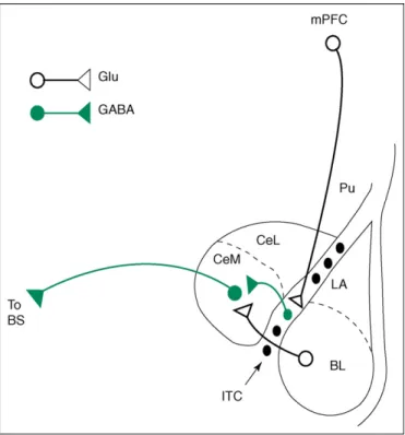
[20]. It is increasingly recognized that extinction of con-ditioned fear does not entail the original CS–US associ-ation being eradicated, but rather it being overshadowed by a stronger association between the CS and the non-occurrence of the US. This process is thought to depend upon prefrontal mechanisms, lesioning of medial prefron-tal cortical (mPFC) areas in particular having been shown to disrupt extinction[21,22]. Recently, it has been argued that these frontal mechanisms are especially cru-cial to the recall of fear extinction and active inhibition of previously conditioned fear responses [21–23]. This is thought to involve prefrontal downregulation of amygdala output, with mPFC neurons exciting GABAergic neurons within the basolateral complex of the amygdala or the nearby intercalated cells (ITC), and these, in turn, inhibit-ing output from the central nucleus of the amygdala
[21,22,24,25](Figure 1).
In addition, the hippocampus is thought to have a central role in the contextual modulation of the acquisition of conditioned fear and of its renewal or reinstatement following extinction [26,27]. It should be noted that although there is strong evidence that the hippocampus enables information about contexts in which a CS is safe versus dangerous (predictive of an US) to influence CR generation, some accounts posit a much more extensive role for the hippocampus in anxiety-related behavioral inhibition[28].
Human studies Fear conditioning studies in humans have
replicated many of the findings from the animal literature. Both experimental and clinical studies have shown that contextual influences on conditioned fear extinction are observable in humans [29,30], with context-renewal ef-fects being demonstrated in exposure therapy for anxiety disorders[29]. There is also evidence that clinically anxi-ous patient groups show stronger acquisition and retention of novel conditioned fear associations than low anxious controls. However, it remains unclear as to whether anxi-ety is primarily characterized by stronger acquisition, weaker extinction and/or greater generalization of cond-itioned fear[19].
Are there common neurocognitive mechanisms underlying the attentional, interpretative and associative processing of threat?
It has been argued that fear extinction results in the CS becoming, in effect, an emotionally ambiguous stimulus linked to representations of both threat (from acquisition) and safety (from extinction) [10]. In extinction recall,
prefrontal inhibition of amygdala output is held to support activation of the latter representation over the former[21], providing a clear parallel to the proposed interaction of control mechanisms and threat evaluation mechanisms in attentional regulation of threat and the interpretation of emotionally ambiguous stimuli [12]. This prompts the question of whether an interplay between amygdala and prefrontal mechanisms influences not only associative but also attentional and interpretative processes, with anxiety biasing this interplay by upregulation of the amygdala response to potentially threat-related cues and/or down-regulation of prefrontal control mechanisms. To some extent, this question can be addressed by consideration of recent findings from the affective cognitive neuroscience literature where neuroimaging techniques, such as func-tional magnetic resonance imaging (fMRI), have enabled investigation, in humans, of the neural mechanisms under-lying fear conditioning, regulation of attention to threat distractors and the interpretation of stimuli of uncertain threat value.
Neuroimaging studies of fear conditioning
Human neuroimaging studies of fear conditioning have broadly confirmed findings from the basic neuroscience literature. Amygdala activity is observed during the acqui-sition of conditioned fear [31–35], with the magnitude of the amygdala response to the CS being strongest during the early stages of acquisition [31–33] and correlating Figure 1. Illustration of one of the proposed mechanisms for prefrontal downregulation of amygdala output (as studied in the rat). Infralimbic (IL) neurons in the medial prefrontal cortex (mPFC) are proposed to inhibit central medial nucleus (CeM) projection neurons via GABAergic intercalated (ITC) cells, thereby reducing CeM responses to inputs from the basolateral nucleus of the amygdala (BL) or cortex (adapted from [25]). An alternate proposal involving medial prefrontal cortical (mPFC) inhibition of CeM output via excitation of BL GABAergic interneurons[24]is not shown here. LA, lateral nucleus of the amygdala; CeL, central lateral nucleus of the amygdala; BS, brain stem; Pu, putamen; GLU, excitatory glutamatergic projections; GABA, inhibitory GABAergic projections.
positively with physiological measures of arousal, such as skin conductance responses (SCRs) [36]. Several studies have also reported enhanced CS-related amygdala activity during extinction and/or extinction recall[31,34,35,37]. In addition, extinction recall is associated with increased CS related activity in the medial prefrontal cortex (mPFC) and hippocampus[35–37], with there being evidence for strong functional connectivity between activity in these regions and the amygdala during this stage[35].
fMRI studies of fear conditioning are subject to a number of limitations. First, the spatial resolution is not good enough to make definite statements about the acti-vation of specific amygdala nuclei. Second, positive con-nectivity between regions can reflect either excitatory or inhibitory connections and is agnostic as to the direction of projections. That said, it has been argued that results indicating coactivation of the mPFC and amygdala during extinction recall could reflect prefrontal projections acting upon inhibitory GABAergic neurons within the basolateral nucleus or ITC of the amygdala, in line with proposals from the basic neuroscience literature[35]. Coactivation of the hippocampus, meanwhile, is thought to relate to contex-tual modulation of extinction recall[35,37].
Despite the centrality of fear conditioning paradigms to animal models of anxiety, none of the neuroimaging stu-dies reviewed have examined the impact of trait or state anxiety upon amygdala and prefrontal activation during the acquisition and extinction of conditioned fear. How-ever, within the clinical literature, adults with post trau-matic stress disorder (PTSD) have been reported to show amygdala hyper-responsivity during conditioned fear acquisition and frontal hyporesponsivity during extinction
[38]. This provides some initial support for the suggestion that anxiety might be linked to an altered balance of amygdala–prefrontal activity.
Neuroimaging studies of selective attention to threat Several models have suggested that anxiety biases attention towards threat-related stimuli by augmenting the output from an amygdala-centered pre-attentive threat evaluation mechanism [12,39]. Within the neuroimaging literature, there has been a heated debate as to whether the amygdala does indeed show a pre-attentive response to threat-related stimuli. Studies using manipulations of spatial and object-based attention have reported amygdala activation to unattended threat-related stimuli [40,41]. However, contrary findings have led others to argue that this is only observed when the perceptual processing demands of the primary task are low, allowing attentional spillover to nominally unattended threat distractors[42,43]. Consideration of the modulatory effects of anxiety raises the possibility that the discrepancies in findings could also reflect variability in the composition of volunteer samples, participants’ anxiety levels typically going unreported. One study that addressed this directly reported that both low and high anxious individuals showed an increased amygdala response to attended threat-related stimuli (fearful faces), but only high anxious volunteers showed an increased amygdala response to unattended threat-related stimuli[44]. At first glance, this seems consistent with modulation by anxiety of the output of a pre-attentive
threat evaluation mechanism [12,39]. However, more recent results indicate that anxiety only modulates the amygdala response to threat distractors under conditions of low perceptual load, suggesting that this influence might occur subsequent to an initial stage of perceptual compe-tition[45](Box 1).
The focus upon the amygdala in neuroimaging studies of selective attention to threat has led the potential impact of individual differences in recruitment of prefrontal control mechanisms to be overlooked. Biased competition models of selective attention suggest that, under conditions of atten-tional competition, frontal control mechanisms are engaged to support the processing of task-related stimuli [14]. Although the influence of such control mechanisms has been recognized by cognitive models of anxiety, they are not traditionally seen as the locus of anxiety-related individual differences [12]. However, there is evidence for working memory and inhibitory control deficits in anxiety[46,47]. Furthermore, recent neuroimaging studies have reported that anxious individuals show weaker recruitment of pre-frontal control mechanisms in response to attentional com-petition from threat-related distractors than low anxious volunteers [45,48,49]. For example, when expectancy of threat distractors is manipulated, high anxious individuals show both reduced activity in a rostral anterior cingulate region implicated in detecting processing conflict from emotional stimuli and a reduced response to increased expectancy of threat distractors in lateral prefrontal regions implicated in augmenting attentional control[48](Figure 2). Discussion of the implications of the results reviewed here for neurocognitive models of anxiety-related biases in se-lective attention can be found inBox 1. In broad terms, these findings provide evidence for anxiety-related frontal hypo-responsivity, as well as amygdala hyper-hypo-responsivity, during the regulation of attention to threat-related stimuli, paral-leling the findings previously reported for the acquisition and extinction of fear conditioning in adults with PTSD[38]. Neuroimaging studies of threat interpretation
Recently, several neuroimaging studies have examined the neural mechanisms underlying the interpretation of poten-tially threat-related stimuli. It has been shown that the amygdala response to neutral facial expressions, perceived by some as mildly negative, increases as a function of anxiety [50]. Expressions of surprise are also relatively ambiguous, signaling the occurrence of either a positive or a negative unexpected event, and sharing certain features with expressions of fear. The selective amygdala response to these expressions also varies between individuals, cor-relating with the extent to which surprise expressions are perceived as negatively valenced[51]. These findings are consistent with the suggestion that the magnitude of the amygdala response to emotionally ambiguous sti-muli influences the competitive success of threat-related interpretations. However, the correlational nature of these findings prevents any strong causal interpretations from being drawn.
Top-down contextual influences and conscious attempts at reinterpretation have been suggested to influence the interpretation of potentially threat-related stimuli, with the PFC playing a central role. Evidence for this proposal
Box 1.Modeling the effects of anxiety upon the neural mechanisms supporting visual selective attention Consideration of biased competition models of selective attention[14]
and related cognitive models of anxiety[12]suggests that the outcome of attentional competition between affectively neutral task-related stimuli and task-irrelevant threat-related stimuli is potentially deter-mined by the relative strength of modulatory signals from the PFC (supporting task-related processing) and the amygdala (supporting the processing of threat-related stimuli). Re-entrant connections from both these regions enable modulation of activity within visual processing pathways (Figure I). Additionally, a direct thalamo–amygdala pathway has been suggested to enable non-re-entrant amygdala influences upon attentional competition, although this is still a matter of some debate[60].
In the context of this basic framework, anxiety could lead to threat-related biases in selective attention by amplifying the amygdala threat signal and/or by reducing the prefrontal control signal. Initial findings suggest that anxiety is associated with both an augmented amygdala response to threat distractors and reduced recruitment of frontal control mechanisms[44,48,49](Figure IIa).
Recently, it has been argued that there are two stages of attentional competition, with early perceptual competition preventing distractors from being processed further when the perceptual load of the primary task is high, and with active recruitment of control mechanisms being required to prevent salient distractors from competing for further processing resources when perceptual load is low[59]. In line with this account, it has been reported that the amygdala response to threat distractors is abolished under high perceptual load[43], with individual differences in the prefrontal and amygdala response to threat distractors primarily being observed under low perceptual load [45]. The latter study found elevated state anxiety to be associated with increased amygdala activity and high trait anxiety with weakened recruitment of prefrontal control mechanisms in response to threat-related distractors under conditions of low perceptual load. These results are consistent with findings linking high trait anxiety to impoverished attentional control [61], and with findings indicating that recruitment of prefrontal mechanisms to aid in the appraisal and regulation of aversive stimuli is reduced when cognitive resources are otherwise depleted[62], as has been argued to be the case in anxiety [46]. The apparent dissociation in the effects of trait and state anxiety upon prefrontal and amygdala function, although requiring replica-tion, is supportive of findings in non-human primates[63]. A revised
neurocognitive model of anxiety-related attentional biases that incorporates these additional findings is shown inFigure IIb.
Figure I. Anatomical substrate for modulatory influences of the amygdala (red arrows) and PFC (green arrows) upon the ventral visual processing stream (solid black arrows). Dashed black arrows illustrate projections emanating from the ventral visual processing stream. The amygdala receives strong input from inferior temporal cortex (area TE), returning projections to all levels of the visual pathway from primary visual cortex (V1) through to temporal cortical regions TEO and TE. There are also reciprocal connections between TE and PFC with lateral prefrontal cortical (lPFC) regions having strong modulatory influences upon activity in TE and weaker, potentially indirect, modulatory influences upon earlier visual regions (not shown). Frontal projections to the amygdala are strongest from the orbital frontal cortex (OFC), medial PFC (mPFC) and anterior insula (AI) (frontal cortical subdivisions are not illustrated here). These projections mainly target the basolateral nucleus and are predominantly reciprocated by amygdaloprefrontal projections from the same regions. Within the frontal cortex, there are strong connections between lPFC, OFC, AI and mPFC, the latter including the paracingulate gyrus and dorsal, rostral and subgenual regions of the anterior cingulate cortex. The strong interconnectivity within frontal regions provides a mechanism for cohesive regulation of both cognitive and affective processing. Pathways are illustrated on a lateral view of a monkey brain. The diagram is schematic and not intended to illustrate anatomical specificity of origin and terminal sites.
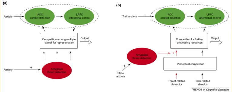
Figure II. Two versions of a neurocognitive model of anxiety-related biased in selective attention.(a)Allocation of attentional resources to threat distractors is influenced both by the strength of a threat-detection signal from the amygdala and the strength of a top-down control signal supporting task-related processing. The latter is thought to emanate from the lateral prefrontal cortex (LPFC), with a rostral anterior cingulate cortical (ACC) region signaling the presence of attentional competition from threat distractors[48]. Anxiety is held to modulate the magnitude of both the amygdala and prefrontal signals, being associated with amygdala hyper-responsivity and frontal hyporesponsivity.(b)An extended version of the model in(a)incorporates recent findings suggesting that: (i) attentional competition involves both early competition for perceptual resources and later competition for further processing, with anxiety modulating processing subsequent to the initial stage of perceptual competition; and (ii) amygdala responsivity is primarily modulated by state anxiety whereas prefrontal recruitment is primarily influenced by individual differences in trait anxiety[45].
comes from studies showing that prefrontal regions are activated by attempts to interpret negative stimuli in a less threat-related manner [52–54]. In addition, success in decreasing negative affect through stimulus re-interpret-ation has been reported to be correlated with increases in prefrontal activity and decreases in amygdala activity
[53,54]. Similarly, amygdala activity associated with nega-tive interpretations of surprise expressions decreases as a function of prefrontal activity [51] and in response to contextual cues that disambiguate expressions of surprise in a positive direction [55].
These findings suggest that individual differences in prefrontal recruitment might well influence success in reinterpreting the threat value of a given stimulus. In line with this, individuals reporting high levels of rumination, a characteristic of generalized anxiety, show poorer recruit-ment of medial prefrontal regions when attempting to reinterpret negative stimuli in a less threat-related
manner [56]. So here, as in the neuroimaging literature on fear conditioning and selective attention to threat, there is some initial suggestive evidence of anxiety-related affec-tive styles being associated with decreased recruitment of prefrontal control mechanisms, as well as increased amyg-dala activity during implicit or explicit evaluation of poten-tially threat-related stimuli. These results complement findings from symptom provocation studies of anxiety – which also report frontal hyporesponsivity and amygdala hyper-responsivity to threat-related stimuli (Box 2) – but, in addition, link disruption of this frontal-amygdala cir-cuitry to cognitive processes that have been implicated in the maintenance of anxiety[6,7].
Towards a central role for amygdala–prefrontal interactions in anxiety
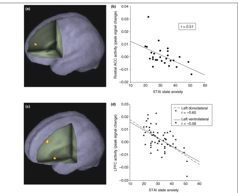
Heightened acquisition and/or diminished extinction of learned fear, enhanced selective attention to threat, and Figure 2. Results from a study examining the modulatory effects of anxiety upon prefrontal recruitment during regulation of attention to threat-related distractors.
(a)Elevated rostral anterior cingulate (ACC) activity is observed in response to unexpected attentional competition from threat-related distractors; peak activation: 2 50 18 (x y z), Z = 3.44, p<0.02 corrected.(b)Across conditions, high anxious individuals show a reduced rostral ACC response [graphed for the peak voxel from (a)].(c)Lateral prefrontal cortical regions responding to increased expectancy of threat distractors as an inverse function of anxiety; peak activations – left dorsolateral PFC: 34 36 32 (x y z), Z = 3.29, p<0.05 corrected; left ventrolateral PFC: 36 16 6 (x y z), Z = 3.18, p<0.05 corrected.(d)Recruitment of both the left dorsolateral (broken line) and left ventrolateral (unbroken line) regions decreases as anxiety levels increase [graphed for the peak voxels from (c)]. Note, in this study, state and trait anxiety were highly correlated and no attempt was made to dissociate their effects. STAI, State-Trait Anxiety Inventory[75]. Reproduced, with permission, from[48].
negative biases in the interpretation of emotionally ambiguous stimuli have all been held to characterize anxiety and to potentially play a role in its maintenance and even possibly its etiology[6,7,12,19,21]. Despite this, there has been little crosstalk between basic neuroscience studies of conditioned fear, and human cognitive studies of attentional and interpretative biases in anxiety. The emer-gent affective cognitive neuroscience literature will hope-fully help to change this. Neuroimaging studies of fear conditioning, selective attention to threat, and interpret-ation of potentially threat-related stimuli indicate a com-mon neural circuitry crucial to each of these domains of cognitive-affective function. Both the amygdala and PFC are central to this circuitry. Heightened amygdala activation is associated with stronger conditioned fear responses during initial periods of fear acquisition, and with negative biases in the interpretation of emotionally ambiguous stimuli [36,51]. In addition, initial findings indicate that anxious individuals show an increased amyg-dala response to threat distractors[44,45], and during the
acquisition of conditioned fear[38]. Meanwhile, prefrontal cortical activity is associated with attempts to regulate the outcome of attentional, interpretive and associative pro-cesses triggered by the occurrence of potentially threat-related cues[34,35,37,45,48,52–54]. This activity is reduced in anxiety[38,45,48,49,56].Figure 3illustrates the common-ality in the lateral and medial prefrontal regions activated across the different paradigms, highlighting those showing a reduced response in high anxious individuals. This com-monality at the neural level possibly reflects a greater conceptual link between these processes than is sometimes recognized. Conditioned fear stimuli, threat-related distrac-tors and stimuli of ambiguous threat value all act as predictors of potential danger. The extent to which our
Box 2.Psychiatric imaging studies of anxiety disorders Symptom provocation is arguably the most widely used paradigm within neuroimaging studies of clinically anxious populations. Across a number of anxiety disorders, symptom provocation has been associated with alterations of blood flow in limbic and prefrontal regions [1,4,64]. Findings indicating amygdala hyper-responsivity in combination with frontal hypohyper-responsivity have been reported in several studies, evidence for this arguably being clearest in the case of post traumatic stress disorder (PTSD), with the magnitude of amygdala activation and medial prefrontal deactivation correlating with symptom severity[65].
Few neuroimaging studies of anxiety disorders have used cognitive paradigms to directly explore the neural substrate of processing biases characteristic of clinically anxious individuals. The studies that have used cognitive paradigms are mainly found within the PTSD literature. Here, studies comparing neural activa-tion during task performance in volunteers with PTSD to trauma-exposed or non-trauma-trauma-exposed controls provide further support for the role of amygdala hyper-responsivity and frontal hypore-sponsivity in PTSD. Specifically, volunteers with PTSD have been reported to show amygdala hyper-responsivity during conditioned fear acquisition[38]and in response to masked and non-masked threat-related stimuli [65]. In addition, reduced medial prefrontal cortical activation relative to controls has been reported in response to overtly presented threat cues [65], during conditioned fear extinction[38]and during regulation of attention to task-irrelevant threat cues[49], although in the latter case this effect was specific to activity within the rostral anterior cingulate cortex, the reverse pattern actually being observed in other medial prefrontal regions. Several studies have also investigated alterations in hippocampal function and/or structure, but the findings from these studies remain unclear[65,66].
Neurocognitive accounts of PTSD have drawn heavily on parallels with the animal fear-conditioning literature, emphasizing the role of amygdala hyper-responsivity to threat cues in conjunction with deficient regulation of amygdala activity by medial prefrontal control mechanisms and potentially impoverished context-specific extinction owing to hippocampal dysfunction [4,21,65,66]. In line with the neurocognitive models of anxiety-related biases in selective attention presented inFigure IIinBox 1, these accounts highlight potential disruption to both an amygdala-based threat evaluation system and prefrontal control mechanisms. In addition, the question is raised as to whether altered functioning within one or both of these systems, and/or the hippocampal locus also specified, possibly reflects a premorbid diathesis[66]. The issue of identifying the mechanisms by which genetic and environmental factors influence vulnerability to anxiety is returned to in Box 3.
Box 3.Questions for future research
The question of the mechanisms by which genetic and environ-mental influences confer vulnerability to anxiety is of central importance to research into anxiety. Here, translational research is likely to be of particular value. For example, building on findings that catechol-O-methyl transferase (COMT) and serotonin trans-porter (5HTT) knockout increases anxiety in mice [3,67], initial human ‘genomic imaging’ studies have provided suggestive evidence that functional polymorphisms in these genes might combine to influence function in amygdala–prefrontal circuitry when controlled processing of threat-related stimuli is required [67–70]. Chronic and early-life stress also appear to influence this circuitry, leading to alterations in associative and attentional processing[57,71–73]. This prompts the question of the extent to which, in humans, genetic and environmental influences upon amygdala-prefrontal circuitry lead to threat-related biases in associative, attentional, and interpretative processing which in turn contribute to the etiology and/or maintenance of anxiety [6,7].
Recent rodent models of anxiety have suggested that the amygdala and the bed nucleus of the stria terminalis might be differentially involved in fear versus anxiety[74]. High resolution neuroimaging techniques make these predictions potentially amenable to investigation in humans, and enable their implica-tions for the neurocognitive mechanisms outlined here to be explored.
The proposed role of hippocampal dysfunction in the general-ization of fear responses and the potential broader involvement of this region in mediating anxiety-related behaviors also requires further investigation in humans[28,65,66].
Additional neuroimaging studies should allow us to specify, in more detail, how lateral, orbital and medial regions of PFC, including dorsal, rostral and subgenual regions of the anterior cingulate cortex, interact with the amygdala during the extinction of conditioned fear, the regulation of attention to threat-related distractors, and the interpretation of emotionally ambiguous stimuli. In particular, it will be of interest to determine whether the apparent commonalities in the neural substrate of these processes will continue to be observed at a level of greater specificity.
Studies are needed to further investigate the impact of not only trait and state anxiety but also individual differences in other aspects of affective style and cognitive ability upon the neuro-cognitive mechanisms outlined here. Closer investigation of the dysregulation of these mechanisms in clinically anxious popula-tions is also required.
Arguably the most important question concerns how we can use advances in our understanding of the neurocognitive mechan-isms disrupted in anxiety to inform our approach to treatment. One prediction would be that pharmacological interventions directed at re-establishing normal amygdala–prefrontal function, combined with cognitive interventions aimed at reducing threat-related processing biases, might aid in reducing anxiety.
behavior is driven by the negatively valenced predictive value of these cues arguably depends upon our ability to downregulate the processing priority given to it, potentially by increasing the resources we allocate to alternate repre-sentations. Indeed, although the parallels are not always transparent, neural models of attentional control over threat, conditioned fear extinction and emotion regulation through stimulus reinterpretation all implicate a role for
prefrontal control processes in supporting less pre-potent representations or associations in the face of competition from emotionally salient alternatives[21,48,52].
Concluding remarks
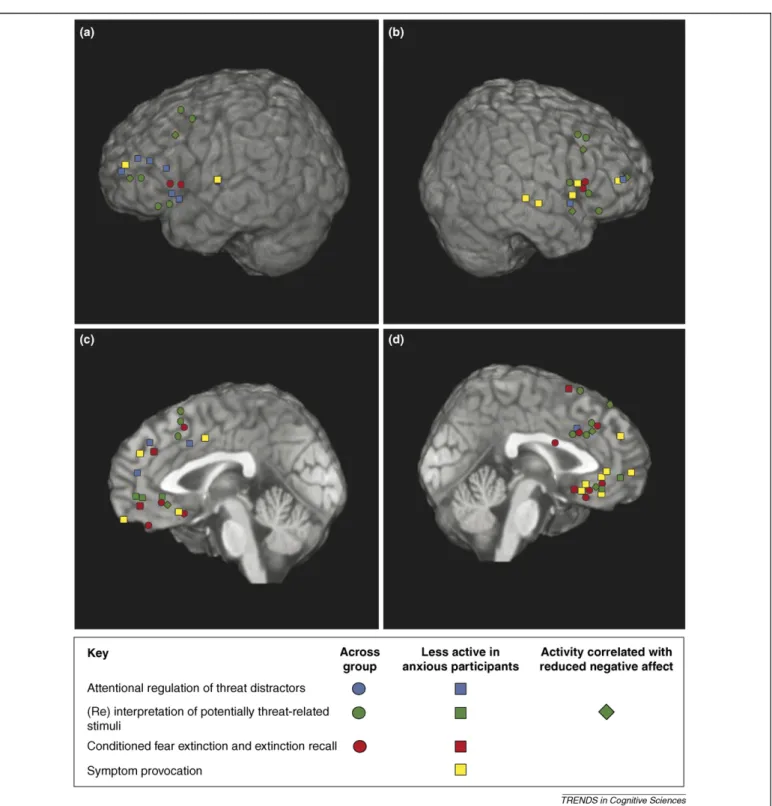
The findings reviewed here provide strong support for a common amygdala–prefrontal circuitry underlying selec-tive attention to threat, interpretation of emotional Figure 3. Prefrontal activation peaks for the neuroimaging studies reviewed in this article. The studies are color coded by paradigm, including regulation of attention to threat distractors, (re) interpretation of potentially threat-related stimuli, conditioned fear extinction and extinction recall, and symptom provocation (in PTSD). Symbol shape indicates whether the activation peak represents activation of the region by the given process across subjects, or as an inverse function of anxiety, or in association with changes in affect. Activations are shown on the AFNI TT_N27 template brain converted to MNI space. The four panels present activation peaks in(a)left lateral PFC,
(b)right lateral PFC,(c)left medial PFC and(d)right medial PFC. All activations with an x of less than 25 are represented on the medial surfaces, activations with an x of 25 or more are represented upon the lateral surfaces.
stimuli, and acquisition and extinction of conditioned fear. Heightened amygdala activity and reduced prefrontal recruitment appear to bias the system towards threat-related responses. At a cognitive level, this is thought to reflect both increased activation of threat-related repre-sentations and a failure to use controlled processing to support the activation of alternate non-threat-related rep-resentations. At a neural level, these effects arguably oper-ate through competing modulatory influences on activity in other brain regions, together with direct opposing influences of prefrontal downregulation of amygdala output[57]and amygdala-driven modulation of prefrontal activity [58]
(Figure I inBox 1). Initial evidence suggests that such a bias towards amygdala hyper-responsivity and prefrontal under-recruitment could be characteristic of anxiety. An interesting question for future research concerns the extent to which genetic and environmental factors contributing to vulnerability to anxiety act through influences upon this circuitry. A closer integration of anxiety research using animal models and studies of neurocognitive function in human volunteers should help to address this and other important remaining questions (Box 3).
Acknowledgements
Thanks to Trevor Robbins and John Duncan for comments. This work was supported by a Career Development Award from the Medical Research Council, and conducted at the University of Cambridge Behavioural and Clinical Neuroscience Institute – cofunded by the Medical Research Council, and the Wellcome Trust – and at the Medical Research Council Cognition and Brain Sciences Unit, Cambridge, UK.
References
1 Bremner, J.D. (2004) Brain imaging in anxiety disorders.Expert Rev. Neurother.4, 275–284
2 Millan, M.J. (2003) The neurobiology and control of anxious states. Prog. Neurobiol.70, 83–244
3 Holmes, A. (2001) Targeted gene mutation approaches to the study of anxiety-like behavior in mice.Neurosci. Biobehav. Rev.25, 261–273 4 Cannistraro, P.A. and Rauch, S.L. (2003) Neural circuitry of anxiety:
evidence from structural and functional neuroimaging studies. Psychopharmacol. Bull.37, 8–25
5 Lang, P.J.et al.(2000) Fear and anxiety: animal models and human cognitive psychophysiology.J. Affect. Disord.61, 137–159
6 MacLeod, C. et al. (2002) Selective attention and emotional vulnerability: assessing the causal basis of their association through the experimental manipulation of attentional bias.J. Abnorm. Psychol. 111, 107–123
7 Mathews, A. and MacLeod, C. (2002) Induced processing biases have causal effects on anxiety.Cogn. Emot.16, 331–354
8 Beaver, J.D.et al.(2005) Emotional conditioning to masked stimuli and modulation of visuospatial attention.Emotion5, 67–79
9 Van Damme, S.et al.(2006) The role of extinction and reinstatement in attentional bias to threat: a conditioning approach.Behav. Res. Ther. 2006, 1555–1563
10 Bouton, M.E. (2002) Context, ambiguity, and unlearning: sources of relapse after behavioral extinction.Biol. Psychiatry52, 976–986 11 Mathews, A. and MacLeod, C. (1994) Cognitive approaches to emotion.
Annu. Rev. Psychol.45, 25–50
12 Mathews, A. and Mackintosh, B. (1998) A cognitive model of selective processing in anxiety.Cogn. Ther. Res.22, 539–560
13 Mogg, K. and Bradley, B.P. (1998) A cognitive-motivational analysis of anxiety.Behav. Res. Ther.36, 809–848
14 Desimone, R. and Duncan, J. (1995) Neural mechanisms of selective attention.Annu. Rev. Neurosci.18, 193–222
15 Richards, A.et al.(2002) Anxiety-related bias in the classification of emotionally ambiguous facial expressions.Emotion2, 273–287 16 Hirsch, C. and Mathews, A. (1997) Interpretative inferences when
reading about emotional events.Behav. Res. Ther.35, 1123–1132
17 Calvo, M.G.et al.(1997) Interpretation bias in test anxiety: the time course of predictive inferences.Cogn. Emot.11, 43–64
18 Calvo, M.G. and Castillo, M.D. (1997) Mood congruent bias in interpretation of ambiguity: strategic processes and temporary activation.Q. J. Exp. Psychol.50, 163–182
19 Lissek, S. et al. (2005) Classical fear conditioning in the anxiety disorders: a meta-analysis.Behav. Res. Ther.43, 1391–1424 20 Maren, S. and Quirk, G.J. (2004) Neuronal signaling of fear memory.
Nat. Rev. Neurosci.5, 844–852
21 Milad, M.R.et al.(2006) Fear extinction in rats: implications for human brain imaging and anxiety disorders.Biol. Psychol.73, 61–67 22 Sotres-Bayon, F.et al.(2004) Emotional perseveration: an update on
prefrontal-amygdala interactions in fear extinction.Learn. Mem.11, 525–535
23 Milad, M.R. and Quirk, G.J. (2002) Neurons in medial prefrontal cortex signal memory for fear extinction.Nature420, 70–74
24 Grace, A.A. and Rosenkranz, J.A. (2002) Regulation of conditioned responses of basolateral amygdala neurons. Physiol. Behav. 77, 489–493
25 Quirk, G.J. et al. (2003) Stimulation of medial prefrontal cortex decreases the responsiveness of central amygdala output neurons. J. Neurosci.23, 8800–8807
26 Anagnostaras, S.G.et al. (2001) Hippocampus and contextual fear conditioning: recent controversies and advances. Hippocampus11, 8–17
27 Delamater, A.R. (2004) Experimental extinction in Pavlovian conditioning: behavioural and neuroscience perspectives. Q. J. Exp. Psychol. B.57, 97–132
28 Gray, J.A. (1982)The Neuropsychology of Anxiety: an Enquiry in to the Functions of the Septo-hippocampal System,Oxford University Press 29 Hermans, D. et al. (2006) Extinction in human fear conditioning.
Biol. Psychiatry60, 361–368
30 LaBar, K.S. and Phelps, E.A. (2005) Reinstatement of conditioned fear in humans is context dependent and impaired in amnesia. Behav. Neurosci.119, 677–686
31 LaBar, K.S. et al. (1998) Human amygdala activation during conditioned fear acquisition and extinction: a mixed-trial fMRI study.Neuron20, 937–945
32 Buchel, C.et al.(1998) Brain systems mediating aversive conditioning: an event-related fMRI study.Neuron20, 947–957
33 Buchel, C.et al.(1999) Amygdala-hippocampal involvement in human aversive trace conditioning revealed through event-related functional magnetic resonance imaging.J. Neurosci.19, 10869–10876
34 Gottfried, J.A. and Dolan, R.J. (2004) Human orbitofrontal cortex mediates extinction learning while accessing conditioned representations of value.Nat. Neurosci.7, 1144–1152
35 Milad, M.R. et al. (2007) Recall of fear extinction in humans activates the ventromedial prefrontal cortex and hippocampus in concert. Biol. PsychiatryDOI: 10.1016/j.biopsych.2006.10.011 (www. sciencedirect.com)
36 Phelps, E.A.et al.(2004) Extinction learning in humans: role of the amygdala and vmPFC.Neuron43, 897–905
37 Kalisch, R.et al.(2006) Context-dependent human extinction memory is mediated by a ventromedial prefrontal and hippocampal network. J. Neurosci.26, 9503–9511
38 Bremner, J.D.et al.(2005) Positron emission tomographic imaging of neural correlates of a fear acquisition and extinction paradigm in women with childhood sexual-abuse-related post-traumatic stress disorder.Psychol. Med.35, 791–806
39 O¨ hman, A. and Wiens, S. (2004) The concept of an evolved fear module and cognitive theories of anxiety. In Feelings and Emotions: The Amsterdam Symposium (Manstead, A.S.R. et al., eds), pp. 58–80, Cambridge University Press
40 Vuilleumier, P.et al.(2001) Effects of attention and emotion on face processing in the human brain: an event-related fMRI study.Neuron 30, 829–841
41 Anderson, A.K. et al. (2003) Neural correlates of the automatic processing of threat facial signals.J. Neurosci.23, 5627–5633 42 Pessoa, L.et al.(2002) Neural processing of emotional faces requires
attention.Proc. Natl Acad. Sci. U. S. A.99, 11458–11463
43 Pessoa, L.et al.(2005) Fate of unattended fearful faces in the amygdala is determined by both attentional resources and cognitive modulation. Neuroimage28, 249–255
44 Bishop, S.J.et al. (2004) State anxiety modulation of the amygdala response to unattended threat-related stimuli.J. Neurosci.24, 10364– 10368
45 Bishop, S.J.et al.(2006) Neural processing of fearful faces: effects of anxiety are gated by perceptual capacity limitations.Cereb. Cortex. DOI: 10.1016/j.biopsych.2006.10.011(cercor.oxfordjournals.org) 46 Eysenck, M.W. and Calvo, M.G. (1992) Anxiety and performance: the
processing efficiency theory.Cogn. Emot.6, 409–434
47 Fox, E. (1994) Attentional bias in anxiety: a defective inhibition hypothesis.Cogn. Emot.8, 165–195
48 Bishop, S. et al. (2004) Prefrontal cortical function and anxiety: controlling attention to threat-related stimuli. Nat. Neurosci. 7, 184–188
49 Shin, L.M.et al.(2001) An fMRI study of anterior cingulate function in posttraumatic stress disorder.Biol. Psychiatry50, 932–942
50 Somerville, L.H. et al. (2004) Human amygdala responses during presentation of happy and neutral faces: correlations with state anxiety.Biol. Psychiatry55, 897–903
51 Kim, H.et al.(2003) Inverse amygdala and medial prefrontal cortex responses to surprised faces.Neuroreport14, 2317–2322
52 Ochsner, K.N.et al.(2002) Rethinking feelings: an FMRI study of the cognitive regulation of emotion.J. Cogn. Neurosci.14, 1215–1229 53 Ochsner, K.N. et al.(2004) For better or for worse: neural systems
supporting the cognitive down- and up-regulation of negative emotion. Neuroimage23, 483–499
54 Phan, K.L.et al.(2005) Neural substrates for voluntary suppression of negative affect: a functional magnetic resonance imaging study.Biol. Psychiatry57, 210–219
55 Kim, H.et al.(2004) Contextual modulation of amygdala responsivity to surprised faces.J. Cogn. Neurosci.16, 1730–1745
56 Ray, R.D.et al.(2005) Individual differences in trait rumination and the neural systems supporting cognitive reappraisal. Cogn. Affect. Behav. Neurosci.5, 156–168
57 Correll, C.M.et al.(2005) Chronic cold stress alters prefrontal cortical modulation of amygdala neuronal activity in rats.Biol. Psychiatry58, 382–391
58 Goldstein, L.E.et al.(1996) Role of the amygdala in the coordination of behavioral, neuroendocrine, and prefrontal cortical monoamine responses to psychological stress in the rat.J. Neurosci.16, 4787–4798 59 Lavie, N. (2005) Distracted and confused? Selective attention under
load.Trends Cogn. Sci.9, 75–82
60 Vuilleumier, P. (2005) How brains beware: neural mechanisms of emotional attention.Trends Cogn. Sci.9, 585–594
61 Derryberry, D. and Reed, M.A. (2002) Anxiety-related attentional biases and their regulation by attentional control. J. Abnorm. Psychol.111, 225–236
62 Kalisch, R.et al.(2006) Levels of appraisal: a medial prefrontal role in high-level appraisal of emotional material.Neuroimage30, 1458–1466 63 Kalin, N.H.et al.(2001) The primate amygdala mediates acute fear but not the behavioral and physiological components of anxious temperament.J. Neurosci.21, 2067–2074
64 Fredrikson, M. and Furmark, T. (2003) Amygdaloid regional cerebral blood flow and subjective fear during symptom provocation in anxiety disorders.Ann. N. Y. Acad. Sci.985, 341–347
65 Shin, L.M. et al. (2006) Amygdala, medial prefrontal cortex, and hippocampal function in PTSD.Ann. N. Y. Acad. Sci.1071, 67–79 66 Rauch, S.L.et al.(2006) Neurocircuitry models of posttraumatic stress
disorder and extinction: human neuroimaging research-past, present, and future.Biol. Psychiatry60, 376–382
67 Hariri, A.R. and Holmes, A. (2006) Genetics of emotional regulation: the role of the serotonin transporter in neural function.Trends Cogn. Sci.10, 182–191
68 Heinz, A. and Smolka, M.N. (2006) The effects of catechol O-methyltransferase genotype on brain activation elicited by affective stimuli and cognitive tasks.Rev. Neurosci.17, 359–367
69 Smolka, M.N.et al.(2007) Gene-gene effects on central processing of aversive stimuli.Mol. Psychiatry12, 307–317
70 Bishop, S.J.et al.(2006) COMT genotype influences prefrontal response to emotional distraction.Cogn. Affect. Behav. Neurosci.6, 62–70 71 Liston, C.et al.(2006) Stress-induced alterations in prefrontal cortical
dendritic morphology predict selective impairments in perceptual attentional set-shifting.J. Neurosci.26, 7870–7874
72 Miracle, A.D.et al.(2005) Chronic stress impairs recall of extinction of conditioned fear.Neurobiol. Learn. Mem.85, 213–218
73 Francis, D.D.et al.(1999) The role of corticotropin-releasing factor– norepinephrine systems in mediating the effects of early experience on the development of behavioral and endocrine responses to stress.Biol. Psychiatry46, 1153–1166
74 Davis, M. (1998) Are different parts of the extended amygdala involved in fear versus anxiety?Biol. Psychiatry44, 1239–1247
75 Spielberger, C.D. (1983)Manual for the State-Trait Anxiety Inventory, Consulting Psychologists Press