Platelet-mimetic strategies for modulating the wound
environment and inflammatory responses
Seema Nandi and Ashley C Brown
Joint Department of Biomedical Engineering, North Carolina State University and University of North Carolina at Chapel-Hill, Raleigh, NC 27606, USA
Corresponding author: Ashley C Brown. Email: [email protected]
Abstract
Platelets closely interface with the immune system to fight pathogens, target wound sites, and regulate tissue repair. Natural platelet levels within the body can be depleted for a variety of reasons, including excessive bleeding following traumatic injury, or diseases such as cancer and bacterial or viral infections. Platelet transfusions are commonly used to improve platelet count and hemostatic function in these cases, but transfusions can be complicated by the contamination risks and short storage life of donated platelets. Lyophilized platelets that can be freeze-dried and stored for longer periods of time and synthetic platelet-mimetic technologies that can enhance or replace the functions of natural platelets, while minimizing adverse immune responses have been explored as alternatives to transfusion. Synthetic platelets typically comprise nanoparticles surface-decorated with peptides or ligands to recreate specific biological characteristics of platelets, including targeting of wound and disease sites and facilitating platelet aggregation. Recent efforts in synthetic platelet design have additionally focused on matching platelet shape and mechanics to recreate the marginalization and clot contraction capabilities of natural platelets. The ability to specifically tune the properties of synthetic platelet-mimetic materials has shown utility in a variety of applications including hemostasis, drug delivery, and targeted delivery of cancer therapeutics.
Keywords: Platelets, hemostasis, artificial platelets, platelet-mimetic, nanoparticles, bionanoscience
Experimental Biology and Medicine2016; 241: 1138–1148. DOI: 10.1177/1535370216647126
Introduction
Platelets provide several key functions during the process of hemostasis and tissue repair including targeting wound sites, facilitating hemostasis, and modulating the repair and regeneration process following injury. In addition to their role in hemostasis, platelets play a large role in the body’s innate immune response to pathogens and other contaminants. Furthermore, platelets also interface with cancer cells and have been shown to contribute to cancer metastasis. The development of non-immunogenic platelet-mimetic technologies has the potential to greatly improve treatment of bleeding and may also have significant poten-tial for targeted drug delivery for treatment of cardiovascu-lar disease and cancer. Recently, researchers have used various platelet-mimetic strategies for such applications. In this review, we present an overview of platelet biology and immune-related functions, and then discuss recent pla-telet-mimetic strategies. Finally, we discuss how these stra-tegies allow for the design of materials that interface with the immune system.
Platelets in coagulation and hemostasis
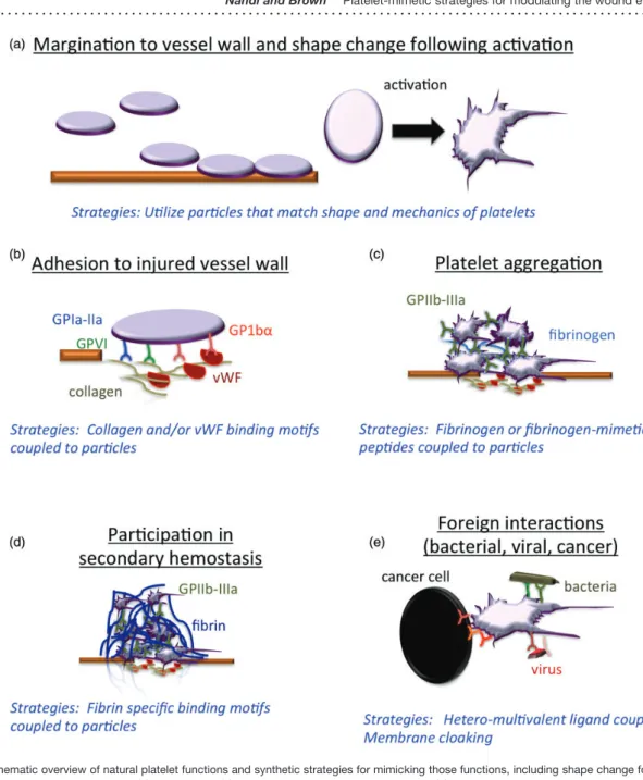
Platelets are small, anuclear blood cells that play a primary role in coagulation and hemostasis following tissue injury. Platelets are derived from megakaryocytes, which become polyploid and form a demarcation membrane system (DMS) as they mature. This DMS allows for storage of membranes that can be released into proplatelet protrusions that then develop into mature platelets upon release and circulation through the bloodstream.1,2Their unique struc-tural and biological components enable platelets to promote hemostasis and wound repair following injury (Figure 1). Platelets circulate in an inactive state; following initiation of the coagulation cascade, platelets become active, which stimulates adhesion of platelets to the injured vessel wall, platelet aggregation, and release of growth factors from platelet granules. In their inactive, or quiescent, state, plate-lets are stiffer than red blood cells. This stiffness causes them to be pushed towards the vessel wall during circula-tion in a process known as marginacircula-tion, which in turn gives them the proximity necessary to interact with and aggregate
ISSN: 1535-3702 Experimental Biology and Medicine2016;241: 1138–1148
at injury sites along vascular vessel walls.3–5Upon activa-tion, platelets undergo a shape change, deform extensively and ‘‘spread’’ along the vascular wall at injury sites.3 The thrombogenic properties of endothelial collagen and the hemostatic behavior of natural platelets interact to form the basis for the hemostatic activity and clot contraction that occurs at the vascular wall.3,6
Hemostasis occurs in two phases, known as primary and secondary hemostasis. During primary hemostasis, plate-lets target and aggregate at the injury site to form a ‘‘plug’’ of sorts.7Platelets bind to exposed sub-endothelial
collagen at the injury site via the von Willebrand factor
(VWF), which is released from the Weibel–Palade bodies of injured vessel walls.8 The A1 domain of the secreted VWF interacts with GPIba, a ligand-binding protein within the glycoprotein GP-Ib-V-IX complex on the plate-let’s surface, allowing the platelets to adhere to the damaged vascular wall.9–11GPIba also allows platelets to
activate under high-shear conditions, which facilitates platelet aggregation at wound sites.10The activated plate-lets then spread along the wall and adhere to each other, forming a platelet plug to stem initial blood loss. Secondary hemostasis is characterized by the activation of the blood coagulation cascade and results in the formation
of a fibrin clot.7 These events occur alongside primary hemostasis, but rather than facilitating platelet aggregation, they induce the formation of an insoluble fibrin clot through the binding of the pro-coagulant tissue factor (TF) molecule to coagulation factor fVII, forming a TF/fVII complex that can cleave factorX into factorXa, a molecule that generates thrombin. Thrombin activates the adherent platelet aggre-gate, allowing several other pro-coagulant factors to bind to the surface of the aggregate. Thrombin also activates fibrinogen, which in turn produces a cross-linked fibrin mesh. Platelet activation also results in an increase of GPIIb-IIIa, a platelet surface integrin that serves as a recep-tor for fibrinogen and carries out mediated fibrinogen bind-ing durbind-ing the fibrin-platelet crosslinkbind-ing process.10,12The fibrin mesh and platelet aggregate form a complete clot that can prevent further blood loss.7,13
Following the cessation of bleeding, cytoplasmic motility proteins, including actin and myosin, facilitate platelet clot contraction, resulting in expulsion of serum from the clot.14 The contractile forces generated by the platelet-rich plasma within the clot increases with time, forming a seal at the clot site to fortify the hemostatic capabilities of the clot. Local fibrin concentration and clot stiffness increase as clot con-traction occurs over time; therefore, it is likely that platelets actively increase their contractile forces in response to the changing fibrin dynamics over time. Indeed, atomic force microscopy experiments with single platelets attached to fibrinogen-coated surfaces demonstrate that force gener-ation by individual platelets increases in response to increased cantilever stiffness.15 Additionally, platelets become increasingly activated on increasingly stiff fibrino-gen-coated surfaces.16 Clot contraction promotes wound healing by decreasing the clot surface area, which allows greater blood flow to the healing tissue.14Clot contraction is supplemented by fibrinolysis, the process by which clots are degraded, following contraction in order to prevent thrombosis. Interestingly, clot retraction plays a role in the dynamics of fibrinolysis, by increasing clot stability and decreasing clot susceptibility to fibrinolysis.17,18
Platelets and the immune system
The unique structure and composition of platelets allow them to interface with the immune system after injury by targeting wound sites, facilitating healing, and preventing infection or contamination from occurring at the site (Figure 1). Platelets are involved in several of the mechanisms com-prising the first lines of defense against pathogenic sub-stances in the body; circulating platelets have scavenger receptors, including CD36, on their surfaces that constantly scan the surrounding area for the presence of potentially dangerous molecules or molecular patterns that could be indicative of pathogenic activity.19 As part of the innate immune system, platelets have a significant role in the inflammatory response at sites of injury and/or disease through activation of and close interaction with leukocytes (regulated by cathepsin G), secretion of chemokines and cytokines that attract other immune cells, and through add-itional mechanisms that influence the body’s adaptive immunity.10,20–23 Tissue inflammation and healing are
further modulated by protease-activated receptors (PARs) found on platelet surfaces; these PARs stimulate the release of alpha granules, which contain various growth factors and angiogenic factors that aid in tissue repair.24,25Many glycoproteins found on the platelet surface membrane, such as CD55 and CD59, are a part of the complement system, which eliminates particulate invaders by providing recog-nition mechanisms for phagocytes that can then clear inva-ders from the body via phagocytosis.4 Other glycoprotein receptors on the platelet surface membrane, such as (GP)IIb-IIIa, GPIba, FcgRIIa, and toll-like receptors (TLRs), are triggered when bacteria bind to the platelet surface; these receptors, in turn, induce the secretion of antimicro-bial peptides known as platelet microbicidal proteins, which mediate chemotaxis of phagocytes, and cause the platelet to shift from a quiescent to an activated state.10,26,27 Additionally, TLRs are critical to thrombosis, activation of the immune system cells, and determination of both non-specific and specific immune responses to vari-ous pathogens.28,29 Platelets also express Fc-receptors for
antibodies, which, together with the complement receptors, allow them to bind virus-antibody complexes.30
Interactions between pathogens and platelet surface receptors can trigger platelet activation and thrombus for-mation. Bacterial binding can occur via direct or indirect methods, including binding directly to a receptor on the platelet’s surface, binding of secreted bacterial toxins to platelet surface receptors, or binding to a plasma protein that functions as a ligand for a platelet membrane recep-tor.10Platelets can also interface with viral particles as part of the body’s immune response; however, in the case of certain viral infections such as HIV, dengue virus, and hepa-titis C, this interaction can result in the destruction of plate-lets or platelet function and can lead to potentially fatal levels of thrombocytopenia and bleeding.30,31
Despite the variety of capabilities that platelets possess to fight pathogenic agents within the body, interactions between platelets and pathogens can also create severe problems for the immune system. For example, the throm-botic response of platelets in the presence of bacteria can lead to events such as strokes or pulmonary embolisms if the thrombus forms on a heart valve. Even microthrombi can present large problems if they result in blockade of the body’s microvasculature.10Additionally, in cancer patients, the innate behavior of platelets that causes them to adhere to pathogens and injured vasculature results in the aggre-gation of platelets around tumor cells. This can have the unintended effect of promoting tumor survival in the bloodstream without creating a noticeable immune response, allowing the tumor cells to metastasize and extravasate to tissues or organs other than their tissue of origin.32
The need for platelet-mimetic materials
during viral infections can lead to severe and potentially fatal bleeding, demonstrating the need for platelet-mimetic materials for the treatment of diseases such as HIV and Hepatitis C.30 In cases of traumatic injury, patients often bleed out in the pre-hospital phase; therefore, platelet-mimetic materials could provide sufficient stabilization in traumatic injury victims to facilitate survival until hospital care is received.33In cancer patients, chemotherapy cantly decreases platelet counts and often results in signifi-cant bleeding problems, requiring multiple platelet transfusions; these immunocompromised patients would benefit greatly from synthetic platelet strategies.34,35
Platelet-mimetic materials could also improve targeted drug delivery, since current targeting technologies are ham-pered by nonspecific binding and insufficient margination and circulation and thus cannot target specific molecules or structures effectively.36 Since platelets have the ability to marginate to the vascular wall, target injury sites, and can effectively circulate throughout the body, the use of platelet-mimetic technologies that mimic these features could allow researchers and clinicians to more effectively deliver drugs to specified locations within the body for improved thera-peutic action. These agents could be used to treat bleeding and hemorrhages, as well as thrombosis, atherosclerosis, and cancer.
There are a wide variety of applications for platelet-mimetic materials, and current strategies typically focus on replicating various biochemical or mechanical properties of platelets to replicate specific desired functions of natural platelets, such as stabilization and augmentation of clot for-mation (Figure 1).36–38Current research in the field of plate-let-mimetic technologies utilizes a wide variety of strategies including both naturally derived platelet derivatives and synthetic platelet strategies. In subsequent sections, we review various strategies that encompass a large range of approaches to achieve various goals such as wound target-ing, drug delivery, and immune system evasion through platelet membrane cloaking.
Platelet-mimetic strategies and technologies
Natural platelet derived approaches
Allogeneic platelet transfusion is the most commonly used platelet therapy in the clinic. In clinical cases involving low platelet counts, platelet transfusions are often used in order to supplement or replace the activity of the body’s own natural platelets.39Platelet transfusion has the advantage of utilizing natural platelets that innately possess the ability to biologically, biochemically, and mechanically interface with the immune system, endothelial cells, and other plate-lets to the required degree to supplemental lost platelet function, especially platelet functions required for hemosta-sis. However, platelet transfusion is limited by the relative lack of donors, storage cost, and short shelf life of donated platelets, and the fact that most platelet therapy is done prophylactically rather than therapeutically.40,41The short shelf life of stored platelets makes the development of new therapeutic natural platelet-based technologies difficult.42 Even though recent technologies have improved the shelf life of natural platelets to up to seven days, this shelf life
does not allow for a ‘‘bank’’ of platelets to be generated, which may be necessary for therapeutic applications requir-ing large volumes of platelets; furthermore, stored platelets are placed at an increasing risk of bacterial contamination that could result in a severe immunologic response if used in a transfusion.43,44 Additionally, studies by Ponschab et al.44demonstrated that platelets increasingly lose their ability to aggregate over the short period of time in which they are kept in storage, further complicating the use of natural platelets as a viable means of treatment for vascular injury or disease. Platelet transfusion also has the inherent risk of transmission of blood-borne diseases; polymorph-ism of human platelet antigen (HPA) and human leukocyte antigen (HLA) after transfusion can also lead to complica-tions such as transfusion refractoriness, which is the failure to achieve the desired level of platelets following transfu-sion, and von Willebrand’s disease.22,43,45Platelet transfu-sion can also be potentially pro-inflammatory or highly immunogenic; recipients of donated platelets run the risk of alloimmunization, exposure to the transmission of infec-tions, graft-vs-host disease, or transfusion-related immuno-suppression, which could increase their vulnerability to a host of other harmful pathogens.20,46
Therefore, the short shelf life of natural platelets poses a considerable restriction on the practicality of platelet trans-fusion therapy. This shortcoming is being combated through the use of lyophilized platelets, which can be dehy-drated and freeze-dried in order to improve the length of their shelf life.46Freeze-dried platelets can be prepared by paraformaldehyde-treating platelets, washing in citrated saline prior to freezing, and rehydrating in citrated saline upon thawing. This freezing and thawing process allows the lyophilized platelets to retain their hemostatic function-ality, unlike previously investigated cryopreserved lyophi-lized platelets washed in standard saline that were unable to form the characteristic primary hemostatic platelet plug after reconstitution.47However, in vivo studies in animal models indicate that these freeze-dried lyophilized plate-lets, although capable of forming a hemostatic plug, have a short duration of circulation, lasting only 9.5 min prior to being cleared from the bloodstream.48 These lyophilized platelets have also been shown to have the potential to increase thrombogenicity and antigenicity, and aggregate in the spleen.46Therefore, the use of lyophilized platelets lacks the reliability that is required for large scale clinical application of naturally derived platelet replacements.
and unreliable for use in emergency trauma-related treat-ments.45As an alternative approach, studies performed by Nguyen et al.24demonstrated that the body’s existing plate-lets can be differentially stimulated using the protease acti-vated receptor agonists PAR-1-agonist and PAR-4-agonist
to selectively secrete VEGF, to promote angiogenesis, or endostatin, an anti-angiogenic factor. While the conclusions they drew from this study need to be tested under more applicable physiological conditions, such as in the presence of microbial agents, this method may present a useful
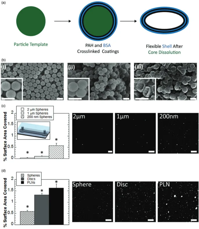
Figure 2 Synthesis and analysis of platelet-like nanoparticles developed by Anselmo et al.36
future method of utilizing natural platelets and their exist-ing immunological and healexist-ing mechanisms on an ampli-fied scale without the need for platelet transfusions.
Artificial platelets
Artificial platelet-mimetic technologies address the immunogenicity issues raised by natural platelet-based treatments, because artificial materials do not need to be blood-matched prior to therapeutic use, but must still be evaluated for biocompatibility and other potential adverse effects, such as thrombotic responses.49The use of artificial platelet-mimetic materials prevents adverse immune responses from developing within the body as well as decreases clearance of targeted therapeutic nanoparticles from the bloodstream. Clearance of nanoparticle-based technologies is low because they are generally not
recognized or marked by the immune system, and clear-ance can be further modified through nanoparticle size or material parameters.36,50 Synthetic materials have further advantages over natural platelets due to their longer shelf life, scale-up potential, and ease of manufacturing and reproducibility.3 The material properties of artificial plate-let-mimetic devices can also be tuned to reflect the mech-anical properties of natural platelets in both their resting and active conformations, giving researchers a greater level of control over obtaining specific desirable character-istics, such as clearance time, for various particular clinical applications.
A multitude of design strategies have been utilized to create artificial platelets and typically entail the coupling of a particle or polymer platform with a binding motif that facilitates interactions at a wound site (Figure 1). Particle platforms previously utilized for artificial platelets have included liposomes, albumin microparticles, latex par-ticles, and even erythrocytes.3 Wound targeting strategies vary widely and having included targeting elements which bind to the subendothelial matrix, platelets, both the subendothelial matrix and platelets, and fibrin. Examples include coupling of full-length fibrinogen, fibrinogen-derived RGD-peptides and other fibrinogen-mimetic pep-tides, platelet surface glycoproteins, and fibrin-specific single domain variable fragment antibodies (sdFvs). Past and state-of-the art strategies to create artificial platelets have been reviewed in detail by others recently.3,51,52 Several recent attempts to develop artificial platelet technol-ogies have focused on designing particles capable of mimicking the shape and mechanical properties of natural platelets; we primarily focus this review on such attempts. Platelets are generally found in the bloodstream in their inactive, or quiescent, state, and only shift to their active, spread conformation in the presence of an injury or patho-gen.36Attempts to mimic natural platelet shape and mech-anics could potentially lead to particles that are capable of replicating the shape change of natural platelets following activation. Efforts in this area include the creation of par-ticles capable of mimicking the mechanical properties of active platelets, then coupling with various binding agents to confer specificity for molecules present in active wound sites.11,36,38Doshi et al.11formed synthetic
platelet-like particles by stretching polymeric particles into a discoid shape reminiscent of the true shape of natural platelets, crosslinking alternating layers of bovine serum albumin and polyelectrolytes over the stretched discoid particles, then degrading the polymeric core to leave a flexible dis-coid capable of imitating the mechanical stretching of natural platelets.11 Anselmo et al.36 also made use of a layer-by-layer synthesis method with a degraded core to imitate innate platelet deformability while synthesizing their own platelet-like nanoparticles (Figure 2).36In an alternative approach, Brown et al.38,53used soft pNIPAM-based
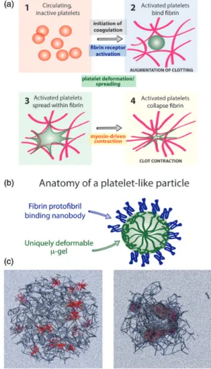
micro-gels synthesized via precipitation polymerization to yield ultra-low cross-linked particles with a high degree of flexibil-ity that allow for extensive spreading within fibrin clots.
Following structural design development, platelet-like particles with appropriate mechanical properties can be functionalized with various molecules to confer targeting
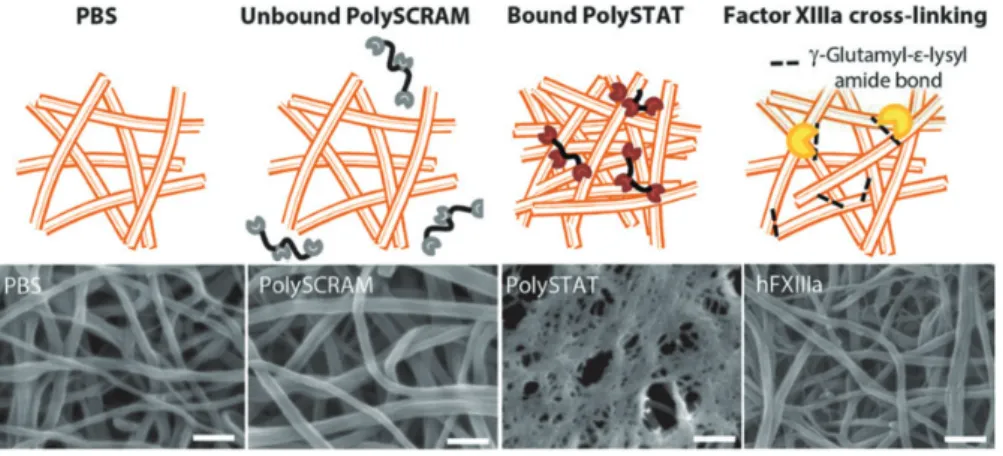
capabilities onto the particle surface. This can be done via a variety of targeting methods, including peptides, antibo-dies, and growth factors. Doshi et al.11conferred targeting abilities to their particles via the VWF-A1 domain or GPIbaN (an amino terminal domain of GPIba), which resulted in particles that adhere to injured endothelial vas-cular walls under high shear stress, and to each other to form an aggregate at the injury site. Particles synthesized by Anselmo et al.36were heteromultivalently surface-deco-rated with peptides to give them binding specificity for exposed collagen and VWF in the vascular injury site and GPIIb-IIIa on activated natural platelets. This method allows for simultaneous targeting of the wound site and aggregation of platelets to each other, increasing the efficacy of the particles in targeting wound sites and decreasing bleeding time. A study performed by Ravikumar et al.54,55 also involved particles surface-decorated with VWF and collagen-binding ligands, with the purpose of promoting binding to VWF under shear flow and binding to collagen independent of shear flow in order to imitate the dual-bind-ing adhesion and aggregation capability of natural platelets. The platelet-like particles developed by Brown et al.38use sdFvs, identified through phage display techniques, to obtain a high specificity for fibrin, allowing the particles to target fibrin clots, and therefore secondary hemostasis (Figure 3). These platelet-like particles were found to spread within the fibrin network, and induce collapse of the fibrin network, without binding fibrinogen present in the blood stream or in areas without injured vasculature. The particle-mediated clot collapse was found to be dependent on the deformability of the particles coupled with high affinity for fibrin, conferred by the sdFv.38 Fibrin monomers cleaved from fibrinogen by thrombin enzymes also possess the ability to self-polymerize and assemble into a three-dimensional scaffold. Transglutaminase factor XIIIa supplements the scaffold by increasing the fiber density and helps to stabilize the clot by creating crosslinks between the fibers.56 Pun and her co-workers developed a synthetic hemostatic polymer
known as PolySTAT that mimics the crosslinking behavior of factor XIIIa to stabilize clots and inhibit fibrinolysis via incorporation of polymers that are resistant to plasmin enzyme degradation (Figure 4). PolySTAT was synthesized from poly(HEMA) copolymerized with NHSMA mono-mers to create a polymer capable of a high degree of peptide grafting. Once inserted into the body, PolySTAT bound noncovalently to fibrin monomers to strengthen clots. Haji-Valizadeh et al.8developed an alternative strategy for creating artificial platelets, involving the use of self-assembled peptide-lipid nanoconstructs that can adhere to injury sites and facilitate aggregation of active platelets. The nanoconstructs were coated in a factor FVIII-VWF binding peptide that was able to promote the injury-site adhesion component of the design, while additional fibrinogen-mimetic peptides on the nanoconstruct surface functioned to promote platelet aggregation. The use of the factor FVIII-VWF binding peptide allowed the nanovehicles to target and bind to injury sites without obstructing the binding mechanisms of natural platelets, which resulted in a greater degree of overall aggregation than was possible with the synthetic or natural platelets alone.8 Overall, these recent approaches have resulted in platelet-mimetics that more closely recapitulate natural platelet mechanics and shape.
Cloaking and active targeting mechanisms for
drug delivery
membrane-cloaked nanoparticles can be used in a similar manner to deliver drugs to specific structures, such as wound sites or thrombi, in the body without causing a severe immune response, which could prevent the drug capsules from being delivered effectively. This
membrane-encapsulated strategy allows for greater circulation time, since high clearance rates are often a hindrance to effect-ively targeted drug delivery, decreased immunogenicity, and improved specific targeting.59Cloaked drug delivery can also suppress the complement system, increasing the
Figure 5 (a) Schematic of platelet membrane-enclosed nanovehicle drug delivery mechanism developed by Hu et al.59
antimicrobial efficacy of the system, and has several poten-tial applications in the field of cancer nanomedicine due to the natural aggregation of platelets around metastatic tumors.32,59,60
The Gu research group has developed a platform for delivering anticancer drugs within a platelet membrane-enclosed nanovehicle (Figure 5(a)). The outer platelet mem-brane shell of the particle is coated with proteins such as P-selectin, which allow the particle to bind CD44 receptors on cancer cells, and the inner nanogel is loaded with doxo-rubicin, a small-molecule anticancer drug that can be released upon internalization and subsequent digestion of the particle by cancer cells.59In vivo testing of the particles in MDA-MB-231 tumor-bearing mice revealed that the par-ticles exhibit significant antitumor effects with minimal immunogenicity.59 Hu et al.32 developed a nanoparticle drug delivery system cloaked in platelet plasma membranes to reduce clearance of the nanovehicles by macrophages in the immune system (Figure 5(b)).32 In their design, polymeric nanoparticles were loaded with drugs and enclosed in platelet plasma membranes that had previously been treated to remove thrombotic mol-ecules, thus decreasing the chance of an immune response when inserted into the body.32
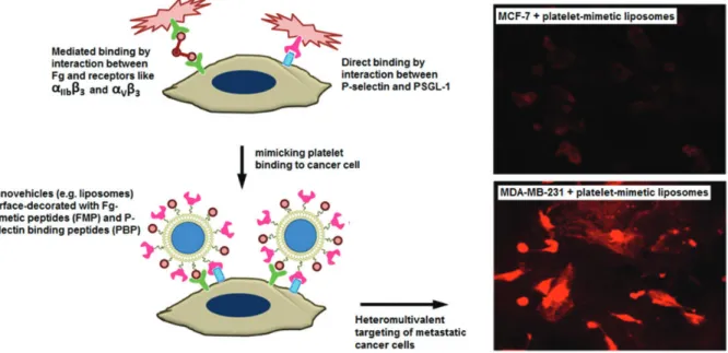
Alternative methods of active targeting involve nano-constructs that have been surface-decorated with various peptides or ligands that can bind to areas on active platelets, injured vessel walls, or cancer cells. Modery et al.61 devel-oped a platform for targeting injury sites using surface-decorated nanoconstructs. The nanoconstructs are coated with peptides that have a high affinity for GPIIb-IIIa and P-selectin, which bind active platelets in sites of vascular disease or injury. These peptides allow the nanoconstructs to target specific wound sites with a high level of specificity and release thrombolytic or anti-proliferative therapeutic
drugs at sites of injury, thrombosis, or vascular disease such as atherosclerosis.61 Similar heteromultivalent ligand-receptor pathway surface-decorated nanoconstructs were later used by this group to target and deliver antic-ancer drugs to metastatic MDA-MB-231 and MCF-7 cantic-ancer cells (Figure 6).37
Conclusion
Platelets have a multitude of functions in addition to their vital role in coagulation and hemostasis, including modu-lating immune functions through interactions with patho-gens and cytokine and growth factor release, immune cloaking, and modulation of cancer metastasis. The design of platelet mimetic technologies has shown promise for augmentation of hemostasis following traumatic injury, tar-geted delivery of cancer therapeutics, and improved immu-nocompatibility of nanoparticle platforms. As researchers continue to develop more sophisticated strategies for mimicking platelet features and functions, and as we learn more about platelet biology, we can likely expect to continue to see additional application of artificial platelet technologies. Thus, while artificial platelet technologies emerged in an effort to merely supplement the hemostatic function of platelets, the potential application of artificial platelet technologies clearly extends into numerous additional areas including targeted drug delivery, cancer therapeutics and immune modulation.
Author Contribution:SN and ACB wrote the manuscript and prepared figures.
ACKNOWLEDGEMENTS
This work was supported by North Carolina State/University of North Carolina at Chapel Hill Start-up Funding to ACB.
DECLARATION OF CONFLICTING INTERESTS
The author(s) declared no potential conflicts of interest with respect to the research, authorship, and/or publication of this article.
REFERENCES
1. Sui Z, Nowak R, Sanada C, Halene S, Krause D, Fowler V. Regulation of actin polymerization by tropomodulin-3 controls megakaryocyte actin organization and platelet biogenesis.pdf.Blood2015;126:520–30 2. Geddis AE. The regulation of proplatelet production.Haematologica
2009;94:756–9
3. Modery-Pawlowski CL, Tian LL, Pan V, McCrae KR, Mitragotri S, Sen Gupta A. Approaches to synthetic platelet analogs.Biomaterials
2013;34:526–41
4. Moghimi SM, Hunter AC, Peer D. Platelet mimicry: the emperor’s new clothes?Nanomed Nanotechnol Biol Med2015, www.sciencedirect.com/ science/article/pii/S1549963415001793 (accessed 6 November 2016 5. Modery-Pawlowski CL, Kuo H-H, Baldwin WM, Gupta AS. A
platelet-inspired paradigm for nanomedicine targeted to multiple diseases.
Nanomed2013;8:1709–27
6. Browning MB, Dempsey D, Guiza V, Becerra S, Rivera J, Russell B, Ho¨o¨k M, Clubb F, Miller M, Fossum T, Dong JF, Bergeron AL, Hahn M, Cosgriff-Hernandez E. Multilayer vascular grafts based on collagen-mimetic proteins.Acta Biomater2012;8:1010–21
7. Yau JW, Teoh H, Verma S. Endothelial cell control of thrombosis.BMC Cardiovasc Disord2015;15:130. www.biomedcentral.com/1471-2261/15/ 130 (accessed 6 November 2015)
8. Haji-Valizadeh H, Modery-Pawlowski CL, Sen Gupta A. A factor VIII-derived peptide enables von Willebrand factor (VWF)-binding of arti-ficial platelet nanoconstructs without interfering with VWF-adhesion of natural platelets.Nanoscale2014;6:4765.
9. Sirotkina OV, Laskovets AB, Goldobin VV, Topanova AA, Karelov DV, Vavilova TV. The molecular mechanisms of platelet activation in patients with cerebrovascular disease.Biochem Mosc2015;9:79–85 10. Cox D, Kerrigan SW, Watson SP. Platelets and the innate immune
system: mechanisms of bacterial-induced platelet activation: Platelets and the innate immune system.J Thromb Haemost2011;9:1097–107 11. Doshi N, Orje JN, Molins B, Smith JW, Mitragotri S, Ruggeri ZM. Platelet
mimetic particles for targeting thrombi in flowing blood.Adv Mater
2012;24:3864–9
12. Chang S-T, Chung C-M, Chu C-M, Yang T-Y, Pan K-L, Hsu J-T, Hsiao J-F. Platelet glycoprotein IIb/IIIa inhibitor tirofiban ameliorates cardiac reperfusion injury.Int Heart J2015;56:335–340. http://jlc.jst.go.jp/DN/ JLC/92000021505?from¼Google (accessed 2 November 2015) 13. Schlimp CJ, Solomon C, Ranucci M, Hochleitner G, Redl H, Scho¨chl H.
The effectiveness of different functional fibrinogen polymerization assays in eliminating platelet contribution to clot strength in throm-boelastometry.Anesth Analg2014;118:269–76
14. Cines DB, Lebedeva T, Nagaswami C, Hayes V, Massefski W, Litvinov RI, Rauova L, Lowery TJ, Weisel JW. Clot contraction: com-pression of erythrocytes into tightly packed polyhedra and redistribu-tion of platelets and fibrin.Blood2014;123:1596–603
15. Lam WA, Chaudhuri O, Crow A, Webster KD, Li T-D, Kita A, Huang J, Fletcher DA. Mechanics and contraction dynamics of single platelets and implications for clot stiffening.Nat Mater2011;10:61–6
16. Qiu Y, Brown AC, Myers DR, Sakurai Y, Mannino RG, Tran R, Ahn B, Hardy ET, Kee MF, Kumar S, Bao G, Barker TH, Lam WA. Platelet mechanosensing of substrate stiffness during clot formation mediates adhesion, spreading, and activation.Proc Natl Acad Sci2014;111:14430–5 17. Katori N, Tanaka KA, Szlam F, Levy JH. The effects of platelet count on clot retraction and tissue plasminogen activator-induced fibrinolysis on thrombelastography.Anesth Analg2005;100:1781–5
18. Kunitada S, FitzGerald GA, FitzGerald DJ. Inhibition of clot lysis and decreased binding of tissue-type plasminogen activator as a conse-quence of clot retraction.Blood2015;79:1420–7
19. Silverstein RL. Disabling the platelet’s brakes to promote thrombosis.
Blood2015;125:2591–3
20. Garraud O. Editorial: platelets as immune cells in physiology and immunopathology.Front Immunol2015;6 :274.http://jour-nal.frontiersin.org/Article/10.3389/fimmu.2015.00274/abstract (accessed 2 November 2015)
21. Hamzeh-Cognasse H, Damien P, Chabert A, Pozzetto B, Cognasse F, Garraud O. Platelets and infections – complex interactions with bac-teria.Front Immunol2015;6:82. http://journal.frontiersin.org/Article/ 10.3389/fimmu.2015.00082/abstract (accessed 2 November 2015) 22. Cognasse F. The inflammatory role of platelets via their TLRs and Siglec
receptors.Front Immunol2015;6: 83.http://journal.frontiersin.org/ Article/10.3389/fimmu.2015.00083/abstract (accessed 2 November 2015)
23. Kinlough-Rathbone RL. Effects of cathepsin G pretreatment of platelets on their subsequent responses to aggregating agents.Thromb Res
1999;95:315–23
24. Nguyen KA, Hamzeh-Cognasse H, Laradi S, Pozzetto B, Garraud O, Cognasse F. Specific activation, signalling and secretion profiles of human platelets following PAR-1 and PAR-4 stimulation.Platelets
2015;26:795–8
25. Martı´nez CE, Smith PC, Palma Alvarado VA. The influence of platelet-derived products on angiogenesis and tissue repair: a concise update.
Front Physiol2015;6: 290. http://journal.frontiersin.org/Article/ 10.3389/fphys.2015.00290/abstract (accessed 6 November 2015) 26. Yeaman MR, Bayer AS, Koo S-P, Foss W, Sullam PM. Platelet
microbi-cidal proteins and neutrophil defensin disrupt the Staphylococcus aureus cytoplasmic membrane by distinct mechanisms of action.J Clin Invest1998;101:178
27. Yount NY, Gank KD, Xiong YQ, Bayer AS, Pender T, Welch WH, Yeaman MR. Platelet microbicidal protein 1: structural themes of a multifunctional antimicrobial peptide.Antimicrob Agents Chemother
2004;48:4395–404
28. Trzeciak-Ryczek A, Tokarz-Deptula B, Deptula W. Platelets – an important element of the immune system.Pol J Vet Sci2013;16: 407–413. (www.degruyter.com/view/j/pjvs.2013.16.issue-2/pjvs-2013-0058/ pjvs-2013-0058.xml (accessed 6 November 2015)
29. Li C, Li J, Li Y, Lang S, Yougbare I, Zhu G, Chen P, Ni H. Crosstalk between platelets and the immune system: old systems with new dis-coveries.Adv Hematol2012;2012:1–14
30. Chabert A, Hamzeh-Cognasse H, Pozzetto B, Cognasse F, Schattner M, Gomez RM, Garraud O. Human platelets and their capacity of binding viruses: meaning and challenges?BMC Immunol2015;16: 26. www.bio medcentral.com/1471-2172/16/26 (accessed 2 November 2015) 31. Assinger A. Platelets and infection – an emerging role of platelets in
viral infection.Front Immunol2014;5:649. http://journal.frontiersin. org/article/10.3389/fimmu.2014.00649/abstract (accessed 9 December 2015)
32. Hu C-MJ, Fang RH, Wang K-C, Luk BT, Thamphiwatana S, Dehaini D, Nguyen P, Angsantikul P, Wen C, Kroll A, Carpenter C, Ramesh M, Qu V, Patel S, Zhu J, Shi W, Hofman F, Chen C, Gao W, Zhang K, Chien S, Zhang L. Nanoparticle biointerfacing by platelet membrane cloaking.Nature2015;526:118–21
33. Evans JA, van Wessem KJP, McDougall D, Lee KA, Lyons T, Balogh ZJ. Epidemiology of traumatic deaths: comprehensive population-based assessment.World J Surg2010;34:158–63
34. Wandt H, Schaefer-Eckart K, Wendelin K, Pilz B, Wilhelm M, Thalheimer M, Mahlknecht U, Ho A, Scaich M, Karmer M, Kaufmann M, Leimer L, Schwerdtfeger R, Conradi R, Do¨lken G, Klenner A, Ha¨nel M, Herbst R, Junghanss C, Ehninger G. Therapeutic platelet transfusion versus routine prophylactic transfusion in patients with haematological malignancies: an open-label, multicentre, rando-mised study.Lancet2012;380:1309–16
35. Stanworth SJ, Estcourt LJ, Powter G, Kahan BC, Dyer C, Choo L, Bakrania L, Llewelyn C, Littlewood T, Soutar R, Norfolk D,
36. Anselmo AC, Modery-Pawlowski CL, Menegatti S, Kumar S, Vogus DR, Tian LL, Chen M, Squires TM, Sen Gupta A, Mitragotri S. Platelet-like nanoparticles: mimicking shape, flexibility, and surface biology of platelets to target vascular injuries.ACS Nano2014;8:11243–53 37. Modery-Pawlowski CL, Master AM, Pan V, Howard GP, Sen Gupta A.
A platelet-mimetic paradigm for metastasis-targeted nanomedicine platforms.Biomacromolecules2013;14:910–9
38. Brown AC, Stabenfeldt SE, Ahn B, Hannan RT, Dhada KS, Herman ES, Stefanelli V, Guzzetta N, Alexeev A, Lam WA, Lyon LA, Barker TH. Ultrasoft microgels displaying emergent platelet-like behaviours.Nat Mater2014;13:1108–14
39. Estcourt LJ, Stanworth S, Doree C, Trivella M, Hopewell S, Blanco P, Murphy MF. Different doses of prophylactic platelet transfusion for preventing bleeding in people with haematological disorders after myelosuppressive chemotherapy or stem cell trans-plantation.Cochrane Database Syst Rev2015;10:CD010984. http://onlinelibrary.wiley.com/ doi/10.1002/14651858.CD010984.pub2/epdf/standard (accessed 9 November 2015)
40. Hux BD, Martin LG. Platelet transfusions: treatment options for hem-orrhage secondary to thrombocytopenia: Platelet transfusions in thrombocytopenia.J Vet Emerg Crit Care2012;22:73–80
41. Davidow EB, Brainard B, Martin LG, Beal MW, Bode A, Ford MJ, Ramsey N, Fagella A, Jutkowitz A. Use of fresh platelet concentrate or lyophilized platelets in thrombocytopenic dogs with clinical signs of hemorrhage: a preliminary trial in 37 dogs: lyophilized platelets in bleeding thrombocytopenic dogs.J Vet Emerg Crit Care2012;22:116–25 42. Montecinos VP, Morales CH, Fischer TH, Burns S, San Francisco IF,
Godoy AS, Smith GJ. Selective targeting of bioengineered platelets to prostate cancer vasculature: new paradigm for therapeutic modalities.
J Cell Mol Med2015;19:1530–7
43. Modery-Pawlowski CL, Tian LL, Ravikumar M, Wong TL, Gupta AS. In vitro and in vivo hemostatic capabilities of a functionally integrated platelet-mimetic liposomal nanoconstruct.Biomaterials2013;34:3031–41 44. Ponschab M, Schlimp CJ, Zipperle J, Gabriel C, Su¨ssner S, Cadamuro J,
Gratz J, Redl H, Scho¨chl H. Platelet function in reconstituted whole blood variants: an observational study over 5 days of storage time.
J Trauma Acute Care Surg2015;79:797–804
45. Dunn PPJ. Recent developments in transplantation and transfusion medicine.Ann Transplant2015;20:424–9
46. Cap AP, Perkins JG. Lyophilized platelets: challenges and opportu-nities.J Trauma Inj Infect Crit Care2011;70:S59–60
47. Read MS, Reddick RL, Bode AP, Bellinger DA, Nichols TC, Taylor K, Smith SV, McMahon DK, Griggs TR, Brinkhous KM. Preservation of hemostatic and structural properties of rehydrated lyophilized plate-lets: potential for long-term storage of dried platelets for transfusion.
Proc Natl Acad Sci1995;92:397–401
48. Fischer TH, Merricks E, Bellinger DA, Hayes PM, Smith RS, Raymer RA, Read MS, Nichols TC, Bode AP. Splenic clearance
mechanisms of rehydrated, lyophilized platelets.Artif Cells Blood Substit Biotechnol2001;29:439–51
49. Huangfu P, Gong M, Zhang C, Yang S, Zhao J, Gong Y. Cell outer membrane mimetic modification of a cross-linked chitosan surface to improve its hemocompatibility.Colloids Surf B Biointerfaces
2009;71:268–74
50. Merkel TJ, Jones SW, Herlihy KP, Kersey FR, Shields AR, Napier M, Luft JC, Wu H, Zamboni WC, Wang AZ, Bear JE, DeSimone JM. Using mechanobiological mimicry of red blood cells to extend circulation times of hydrogel microparticles.Proc Natl Acad Sci2011;108:586–91 51. Myerson JW, Anselmo AC, Liu Y, Mitragotri S, Eckmann DM,
Muzykantov VR. Non-affinity factors modulating vascular targeting of nano- and microcarriers.Adv Drug Deliv Rev2015;99(Pt A): 97–112. http://linkinghub.elsevier.com/retrieve/pii/S0169409X15002367 (accessed 9 December 2015)
52. Chan LW, White NJ, Pun SH. Synthetic strategies for engineering intravenous hemostats.Bioconjug Chem2015;26:1224–36
53. Bachman H, Brown AC, Clarke KC, Dhada KS, Douglas A, Hansen CE, Herman E, Hyatt JS, Kodlekere P, Meng Z, Saxena S, Spears Jr MW, Welsch N, Lyon LA. Ultrasoft, highly deformable microgels.Soft Matter
2015;11:2018–28
54. Ravikumar M, Modery CL, Wong TL, Dzuricky M, Sen Gupta A. Mimicking adhesive functionalities of blood platelets using ligand-decorated liposomes.Bioconjug Chem2012;23:1266–75
55. Ravikumar M, Modery CL, Wong TL, Sen Gupta A. Peptide-decorated liposomes promote arrest and aggregation of activated platelets under flow on vascular injury relevant protein surfaces in vitro.
Biomacromolecules2012;13:1495–502
56. Chan LW, Wang X, Wei H, Pozzo LD, White NJ, Pun SH. A synthetic fibrin cross-linking polymer for modulating clot properties and indu-cing hemostasis.Sci Transl Med2015;7:277ra29
57. Greineder CF, Brenza JB, Carnemolla R, Zaitsev S, Hood ED, Pan DC, Ding B-S, Esmon CT, Chacko AM, Muzykantov VR. Dual targeting of therapeutics to endothelial cells: collaborative enhancement of delivery and effect.FASEB J2015;29:3483–92
58. Lee S-Y, Ferrari M, Decuzzi P. Design of bio-mimetic particles with enhanced vascular interaction.J Biomech2009;42:1885–90
59. Hu Q, Sun W, Qian C, Wang C, Bomba HN, Gu Z. Anticancer platelet-mimicking nanovehicles.Adv Mater2015;27:7043–50
60. Modery-Pawlowski CL, Sen Gupta A. Heteromultivalent ligand-decoration for actively targeted nanomedicine.Biomaterials
2014;35:2568–79