STRUCTURAL ANALYSES OF AvrRpm1 AND HopBA1: TWO TTSS EFFECTORS FROM THE PLANT PHYTOPATHOGEN Pseudomonas syringae.
Karen Ann Cherkis
A dissertation submitted to the faculty of the University of North Carolina at Chapel Hill in partial fulfillment of the requirements for the degree of Doctor of Philosophy in the
Curriculum of Genetics and Molecular Biology.
Chapel Hill 2012
Approved by:
Jeffery L. Dangl, PhD
John Sondek, PhD
Sarah Grant, PhD
Kevin Slep, PhD
ABSTRACT
KAREN ANN CHERKIS: Structural Analyses of AvrRpm1 and HopBA1: Two TTSS Effectors from the Plant Phytopathogen Pseudomonas syringae.
(Under the direction of Jeffery L. Dangl, PhD and John Sondek, PhD)
Plants recognize microbes via specific pattern recognition receptors that are
activated by microbe-associated molecular patterns (MAMPs), resulting in
MAMP-triggered immunity (MTI). Successful pathogens bypass MTI in genetically diverse
hosts via deployment of effectors (virulence factors) that inhibit MTI responses,
leading to pathogen proliferation. Plant pathogenic bacteria like Pseudomonas
syringae utilize a type III secretion system to deliver effectors into cells. These
effectors can contribute to pathogen virulence or elicit disease resistance, depending
upon the host plant genotype. In disease resistant genotypes, intracellular immune
receptors, typically belonging to the nucleotide binding leucine-rich repeat family of
proteins, perceive bacterial effector(s) and initiate downstream defense responses
(effector triggered immunity) that include the hypersensitive response, and
transcriptional re-programming leading to various cellular outputs that collectively
halt pathogen growth. Nucleotide binding leucine-rich repeat sensors can be
indirectly activated via perturbation of a host protein acting as an effector target.
host cell, AvrRpm1 is directed to the plasma membrane, where it contributes to
virulence. This is correlated with phosphorylation of Arabidopsis RIN4 in vivo. The
RPM1 nucleotide binding leucine-rich repeat sensor perceives RIN4 perturbation in
disease resistant plants, leading to a successful immune response. Here, we
demonstrate that AvrRpm1 has a fold homologous to the catalytic domain of
poly(ADP-ribosyl)polymerase. Site-directed mutagenesis of each residue in the
putative catalytic triad, His63-Tyr122-Asp185 of AvrRpm1 results in loss of both
AvrRpm1- dependent virulence and AvrRpm1-mediated activation of RPM1, but,
surprisingly, causes a gain of function: the ability to activate the RPS2 nucleotide
binding leucine-rich repeat sensor. Additionally, we determined the crystal structure
of HopBA1. We were able to show that despite low sequence similarity, HopBA1
shares structural homology to the ChaN/ EreA-like superfamily of proteins. Through
structural analysis of HopBA1 we generated several missense mutations that are
critical for recognition inside the host. We were also able to putatively classify two
additional type III effectors, HopB1 and HopAC1, from P. syringae as additional
For my parents Rita Lee and John,
I would like to thank all of the members of the Dangl-Grant and Sondek labs,
past and present, for their help and guidance throughout my graduate career. I
“You are a wonderful creation. You know more than you think you know, just as you know less than you want to know.”[7]
Table of Contents
List of tables ... viii
List of figures ... ix
List of abbreviations ... xii
CHAPTER ONE: Introduction ... 1
Introduction ... 1
The Strucutral Biology of TTSS Effectors and the Functional Insights Provided ... 10
Summary ... 21
Chapter one figures ... 23
CHAPTER TWO:AvrRpm1 missense mutations weakly activate RPS2-mediated immune response in Arabidopsis thaliana ... 28
Introduction ... 28
Materials and methods ... 32
Results ... 37
Discussion ... 46
Chapter two figures ... 51
CHAPTER THREE: Structural and functional analysis of the type III effector HopBA1 ... 61
Introduction ... 61
Methods and results ... 62
Discussion ... 72
Chapter three figures ... 74
CHAPTER FOUR: Contributions to other works ... 85
CHAPTER FIVE: Conclusions and future directions ... 92
Introduction ... 92
LIST OF TABLES
Table 3.1. Refinement Statistics for HopBA1. ... 75
Table 3.2. Coordinated metals present in structures deposited in the PDB and their distances (Å) from the
coordinating residues. ... 77
Table 3.3. The catalytic tetrad from Bcr136 is conserved among other EreA-like/ChaN superfamily members and
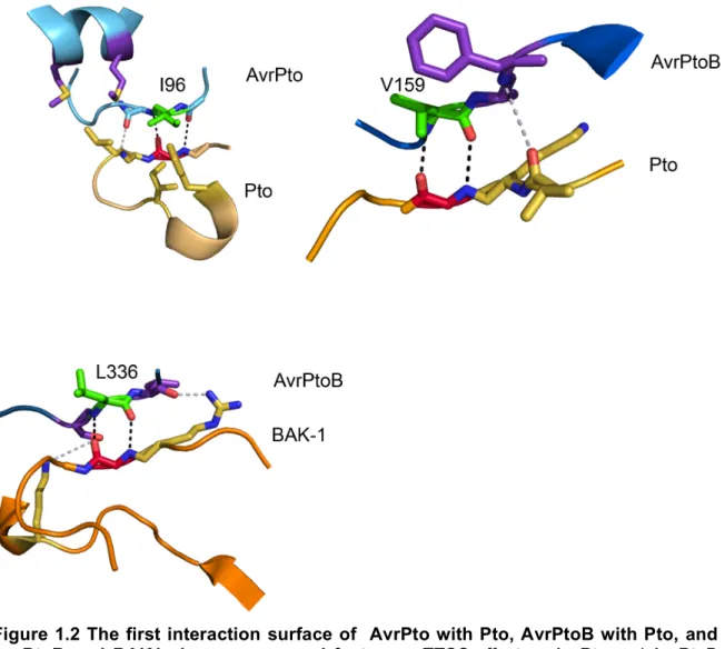
LIST OF FIGURES Figure 1.1. The complex host-pathogen interactions
involved in plant immune response. ... 23 Figure 1.2 The first interaction surface of AvrPto with
Pto, AvrPtoB with Pto, and AvrPtoB and BAK1 shows
conserved features. ... 24 Figure 1.3. Interaction surfaces vary both in location
and in number when comparing the three TTSS
effector: target interactions; AvrPto with Pto, AvrPtoB
with Pto and AvrPtoB with BAK1. ... 25 Figure 1.4. Structural conservation exists among
SopE and the WXXXE family of TTSS effectors with target specificity for RhoGTPases arising on the α4/α6
helical interface. ... 26 Figure 1.5. Ribosylating toxins show varying degrees of
structural similarity and have evolved different structural
elements for a conserved function. ... 27 Figure 2.1 (previous page). AvrRpm1 exhibits structural
homology to the catalytic domain of Poly-ADP-ribosyl
polymerase (PARP). ... 51 Figure 2.2. Alignment of AvrRpm1 alleles. ... 52 Figure 2.3. Missense mutants of AvrRpm1 do not elicit
an RPM1-mediated hypersensitive response, but can
be translocated. ... 53 Figure 2.4. Pto DC3000 expressing AvrRpm1 missense
mutations cannot grow on wild type plants. ... 54 Figure 2.5. Putative catalytic triad residues are required
for AvrRpm1 virulence that is inhibited via weak
Figure 2.6. A mislocalized AvrRpm1 double mutant,
G2A D185A does not limit virulence. ... 56 Figure 2.7. Generation of new antibody using RIN4
specific peptide. ... 56 Figure 2.8. AvrRpm1 mutants do not exhibit increased
interference with AvrRpt2-mediated cleavage of RIN4. ... 57 Figure 2.9. AvrRpm1 does not preferentially ribosylate
Arabidopsis proteins, or RIN4. ... 58 Figure 2.10. Soluble, purified AvrRpm1 is highly
susceptible to degradation and lacks defined
secondary structure. ... 59 Figure 2.11. AvrRpm1 lead to a loss of HR mediated
by the resistance protein RPM1 while simultaneously
triggering RPS2 mediated growth suppression. ... 60
Figure 3.1. HopBA1 forms crystals large enough for
diffraction analysis in slightly different conditions. ... 74
Figure 3.2. HopBA1 is an α/β protein with a β core. ... 76 Figure 3.3. HopBA1 crystal structure contains an
inferred potassium ion held by carbonyl groups. ... 77 Figure 3.4. HopBA1 binds an unknown compound in
its central cleft. ... 78 Figure 3.5. EreA/ChaN superfamily members contain
regions that are variable compared to the core fold
represented by HopBA1. ... 79 Figure 3.6. HopBA1 shows structural homology to
the heme scavenger protein ChaN from the pathogen
Campylobater jejuni. ... 80
Figure 3.7. HopBA1 induces only a modest shift in
heme absorption and thus does not appear to bind heme. ... 81 Figure 3.8. HopBA1 mutated within its hydrophobic
Figure 3.9. HopBA1 mutants that are translocation
competent are also thermal stable. ... 83 Figure 3.10. Functional patches of HopBA1 and
Bcr136 share considerable spatial overlap. ... 84 Figure 3.11. Two additional TTSS effectors HopB1
and HopAC1 are predicted to share structural homology
with the PMT C2 domain. ... 85 Figure 3.12. The N-terminal fold of HopAC1 is
predicted to be structurally homologous to the entire C2
LIST OF ABBREVIATIONS ADPRT- ADP-ribosyl transferase
CD- circular dichroism
ETI- effector triggered immunity GEF- G-protein exchange factor HR- hypersensitive response
hrp- hypersensitive reaction and pathogenicity genes
MAMP- microbe associated molecular patterns mART- mono(adp-ribosyl) transferase
MTI- MAMP triggered immunity
NB-LRR- nucleotide-binding leucine-rich repeat resistance protein NMR- nuclear magnetic resonance
PAR- poly(ADP-ribose)
PARG- poly(ADP-ribose) glycohydrolase PARP- poly(ADP-ribosyl) polymerase PDB- protein data bank
PR1- Pathogenesis Related 1; early defense response gene PRR-pattern recognition receptors
pv.- pathovar
CHAPTER ONE INTRODUCTION Preface
Understanding the molecular mechanisms by which pathogens are able to
evade the immune system of their host is an area of research undergoing significant
growth. Type three secretion system (TTSS) effectors, which are the focus of this
dissertation, are used by pathogens to bypass the host immune system by various
and often unknown mechanisms. The advent of high-throughput sequencing
initiatives coupled with promoter motif searches have enabled the identification in
the Pseudomonas syringae genome of multiple families of TTSS effectors. Despite
these technological advances, we lack the ability to accurately assign enzymatic
functions to these proteins. Complicating these studies, TTSS effectors show very
little overall sequence identity with proteins of known function, making attempts at
classification through sequence similarity contra spem spero, hope against hope.
Instead, we rely on characterization of disease outputs once these TTSS effectors
are inside the host. However, disease outputs are not direct read-outs for enzymatic
mechanisms of TTSS effectors. Accepting the unique challenges of studying TTSS
effectors, there is a significant drive toward structural-based initiatives to
characterize the effectors. In this research, we applied a structural-based approach
characterized by identification of effector folds allowing for effector protein family
complete our analysis by tying these structural initiatives to two different, biologically
relevant contexts, resulting in an increased understanding of the mechanisms
employed by these two TTSS effectors. This will be discussed in the following
chapters.
Pseudomonas syringae
Pseudomonas syringae is a model pathogenic tool for studying host-pathogen
interaction because of the current efforts in sequencing the genomes of the different
pathovars and for its ability to infect the model plant Arabidopsis thaliana. In
addition, the genomes of different accessions (inbreds) of Arabidopsis thaliana that
show varying disease resistance phenotypes to Pseudomonas syringae infection are
currently being sequenced [9]. Pseudomonas syringae is a Gram-negative
phytopathogen. Typically it lives first as an epiphyte on the surface of the host plant
and then switches to invade and colonize the apoplastic space [10]. Pseudomonas
syringae is responsible for significant amounts of crop damage every year making it
a critical phytopathogen to target for research. Each pathovar or race of
Pseudomonas syringae exhibits a high degree of host specificity infecting only a few
host species or only a few cultivars within a species [9,10]. This altered specificity is
due in large part to the TTSS and the varied repertoire of effectors the bacterium
Plant Innate Immunity
When observing host-pathogen dynamics, the first process to ward off
infection begins with the innate immune response. Plant innate immunity is
triggered upon recognition of molecules associated with pathogens [11]. These
molecules, known as microbe associated molecular patterns (MAMPs), are
evolutionarily conserved and slow to evolve [12,13]. MAMPs include the 22 residue
flagellar peptide flg22 and the 18 residue acetylated peptide from EF-TU, elongation
factor elf18, both conserved in bacterial pathogens including P. syringae [14,15].
Recognition on Arabidopsis thaliana occurs through surface exposed pattern
recognition receptors (PRR)/ receptor like kinases (RLK), such as the recognition of
flg22 or elf18 by the transmembrane host receptors FLS-2 and EFR respectively
[16,17]. This recognition leads to early defense responses, such as transcriptional
reprogramming of defense genes like Pathogenesis Related 1 (PR1) and activation
of the kinase signaling cascade (MKK4/MKK5 and MPK3/MPK6 for flg22/FLS2)
[14,16,18,19]. Later responses include callose deposition, a thickening of the cell
wall that serves as a physical barrier between uninfected cells and the site of
infection [14]. The sum of these responses is known as MAMP triggered immunity
(MTI), and it is initiated to inhibit pathogen growth, colonization and disease
occurrence once initial contact has been made (Figure 1.1A) [12].
Type Three Secretion System and Effectors
P. syringae has acquired a TTSS system for subverting the host innate
mechanism for delivery of virulence proteins among many Gram-negative
pathogenic bacteria, including Salmonella typhimurium, Shigella flexneri,
Pseudomonas aeruginosa, and Pseudomonas syringae [20,21]. The TTSS
apparatus is responsible for piercing the host cell membrane and injecting the
effectors into the host (Figure 1.1B) [20,22]. The TTSS in P. syringae and other
Gram-negative plant pathogenic bacteria is encoded by the hypersensitive reaction
and pathogenicity (hrp) genes, which are induced when the Pseudomonad enters
the apoplastic space [23,24]. These genes encode the TTSS apparatus; effectors
can be linked to the hrp cluster, encoded elsewhere on the chromosome, or found
on plasmids [20,24]. The TTSS apparatus genes are quite conserved while the
effector genes are generally more divergent [20,24].
A single pathovar of Pseudomonas syringae is capable of simultaneously
delivering 15-30 different effectors into the host cell although a total of ~50 different
effector families have been identified across all pathovars [10,25]. Delivery of these
effectors through the TTSS apparatus is dependent upon the N-terminal 15 amino
acids. Some effectors carry targeting sequences and rely on host post-translational
modifications for proper localization and function once inside the host [6,26]. Other
TTSS effectors require additional modification, for example, the TTSS effector
protein AvrRps4 is cleaved from a 32.5 kDa protein into a shorter 11kDa protein
once inside the host, and this shorter protein carries the virulence function [26].
Once inside the host, bacterial growth is due to the virulence factors encoded
by a given P. syringae strain; this is thought to be accomplished through two
nutrients from the cellular compartments into the apoplastic space and b)
manipulation of host targets that allows the bacterium to elude MTI (Figure 1.1C left
side) [10]. A specific mutant, P. syringae pv. tomato DC3000hrcC is deficient in the
ability to secrete TTSS effectors (via mutation to the TTSS apparatus) and cannot
overcome MTI to promote bacterial growth [27]. The repertoire of the effectors
delivered into the host cells is thought to include redundant functions and, to the
extent that some effectors can block the perception of other effectors, antagonistic
functions [10]. Thus, loss of any single effector often does not result in loss of
virulence [10]. Rather, it can manifest as only an attenuation of symptoms within the
host [10,28]. Additionally, it is also important to consider that acquiring multiple
effectors could be a means of increasing host range specificity rather than a need for
overlapping function [28].
Disease Resistance Proteins
In an effort to combat the manipulation of host targets by TTSS effectors,
plants have evolved disease resistance proteins (R proteins) [27]. R proteins are
responsible for both recognition of the manipulation on host cellular targets by TTSS
effectors, and initiation of downstream defense responses (Figure 1C right side)
[27,29]. R proteins can monitor the targets of effectors rather than the effectors
themselves and as such are capable of monitoring more than one effector [12]. A
caveat to this is the direct recognition of the effectors AvrPto and AvrPtoB by the R
protein Pto that is further facilitated by an additional R protein Prf, whose structural
recognizes effector function, a defense response called effector triggered immunity
(ETI) is mounted [12]. R proteins determine the resistance portfolio of the plant in
that not all accessions have the same complement of available R proteins that will
enable them to equally recognize the population of effectors [27]. When an effector
goes unrecognized by the host, the host is said to be disease susceptible; when an
effector is recognized the host is disease resistant. Activation of R proteins leads to
an influx of calcium ions, an extracellular oxidative burst, altered transcriptional
profiles, and localized cell death around the site of infection known as the
hypersensitive response (HR) [27,31]. ETI responses are quicker and quantitatively
stronger than MTI responses. These cumulative observations further the need to
study individual TTSS effectors, their targets and the R proteins involved in
recognition, in a structural context away from the ambiguous and often redundant
phenotypes observed in plants.
Advances in Identification of Protein Folds
Proteins take on dynamic, three dimensional conformations allowing for
unique and tunable interactions with other cellular components, including small
molecules, nucleic acids, and other proteins. These interactions are the platform for
essential cellular processes. Disruption and reorganization of these interactions by
TTSS effectors is a critical means to manipulate host targets, promote virulence of
the pathogen and evade detection by the host [32]. In an effort to characterize how
determining it with either X-ray crystallography or nuclear magnetic resonance
(NMR) or by estimating it with computational homology modeling.
X-ray crystallography and NMR are the only two spectroscopic techniques
capable of producing high resolution, multidimensional coordinates of proteins.
Other techniques are limited in that they can only provide auxiliary information and
are frequently dependent on these coordinates for interpretation [33].
Aiding overall structural characterization of proteins is the growth of publicly
available structural data found in resources like the protein data bank (PDB).
Additionally, there have been vast improvements made to computational analyses
used to derive structural information from experimental data [34], the emergence of
synchrotron sources for radiation [35], incorporation of anomalous signals into the
peptide chain [36], and the efforts of structural genomics consortia (SGC). These
advances are not without caveats. For example, not every structure deposited has
an annotated function. It is estimated that nearly 30-50% of all deposited structures
from SGC lack a functional annotation [35]. While contributions of the SGC cannot
be diminished, it is interesting to note that papers linking structural biology to a
functional output are cited with much greater frequency [35].
To overcome this barrier of ‘folds without function and function without folds’,
computational approaches are being used. These computational approaches
include structure alignment, identification of homologous folds and homology
modeling, and functional analysis based on tertiary structure. The computational
approach can give “strong” functional clues when sequence similarity is greater than
homologous folds allows for the investigation of divergent evolutionary relationships
and the possibility of a shared ancestral function. Additionally, the presence of
common motifs, ligand binding sites, and interaction platforms across dissimilar folds
can also aid in functional characterization [35]. Concomitant with computational
approaches to predict protein structure are computational approaches that use
structural data such as quantum mechanical dynamics for enzyme reactions,
molecular dynamics for membrane protein structures, and simulated docking, which
can be used to interrogate protein-protein and protein-ligand interactions. In the
end, all of these computational techniques must be experimentally validated by
traditional biochemical, genetic, and proteomic methods in order to properly assess
their accuracy.
Simply stated, homology modeling involves taking the sequence of a protein
for which the structure is unknown and calculating an estimated structure using a
known and homologous structure as a template. The first homology models were
created in the late 1970s [33]. The steps for generating a comparative model are as
follows: finding a suitable template protein or proteins for the target, aligning the
target to the template protein(s), identifying structurally conserved and predicting
structurally variable regions, modeling the side chains, and evaluating the model
[37]. The models built today with a fully automated web server are continually
increasing their accuracy [33]. There are three commonly used methods for
generating homology models: rigid-body assembly, modeling by segment matching,
and modeling by satisfaction of spatial restraints. Rigid-body assembly was the first
structure. In rigid body assembly, conserved structural elements (rigid bodies) from
homologous proteins are used to generate a map of secondary structure [37,38].
This two dimensional map of rigid bodies is then fit onto a template structure. The
second approach, modeling by segment matching uses a database of short
structural elements, energy, and/or geometry rules and then approximates the
position of conserved residues from templates. Lastly, modeling by satisfaction of
spatial restraints, uses distance geometries or optimization techniques to satisfy
spatial restraints obtained from the alignment of the target sequence to the known
structure [38]. Errors in comparative modeling come in different types: distortions in
correctly aligned regions, distortions in insertions, and distortions in incorrectly
aligned regions [38]. Evaluation can typically identify the inaccurately modeled
regions and because of this using a model is still generally better than using only the
homologous template structure when trying to characterize a protein [38]. Explicit
modeling of variable regions or regions that may contain differences between
homologous folds allows for identification of novel functionality or regions of
specificity [37]. Homology modeling gives a researcher great power when
investigating protein structure. In 1997, approximately one third of the known protein
sequences were related to at least one of the known protein structures; suggesting
that at that time the number of sequences that could be accurately modeled was an
order of magnitude larger than the number of experimentally determined protein
structures [38]. Additionally, according to statistics available on the PDB website,
the average number of new folds reported annually hovered around 100 in 1997.
folds). This drop in new folds has occurred even though there was continuous
growth in the number of new structures reported with 8,093 in 2011.
The Strucutral Biology of TTSS Effectors and the Functional Insights Provided
We analyzed the interactions of three sets of TTSS effectors whose
structures had been previously determined as examples of how structural biology
and homology could inform functional studies. These examples serve as a prelude
to the experimental work described in subsequent chapters. Additionally these
examples highlight both the achievements and inherent complications of TTSS
effector driven structural biology.
AvrPto / AvrPtoB and their Kinase Host Targets: Decoys and Virulence Targets P. syringae pv. tomato includes two effector proteins in its repertoire AvrPto
and AvrPtoB that directly interact with the Pto kinase R protein, and is perceived by
an additional nucleotide-binding leucine-rich repeat R protein Prf, resulting in ETI
[39]. The necessity for Prf activity in ETI, coupled with the lack of a role for Pto in
MTI, gave rise to the hypothesis that Pto may function as a molecular decoy for the
virulence functions for both AvrPto and AvrPtoB [39,40]. The determination of the
complex structures of AvrPto/Pto, AvrPtoB/Pto, and AvrPtoB/BAK1 provide insights
into a common mechanism for interaction with host immune kinase domains [41-44].
AvrPto is a single domain protein whereas AvrPtoB contains an N- and a C-terminal
domain, with the N-terminal domain binding to both Pto and BAK1 [41,43].
Pto reveal both common and divergent structural contacts. The first interaction
interface of AvrPto with Pto occurs when the partially hydrophobic GINP loop of
AvrPto interacts with the P+1 loop of Pto (Figure 1.2), and the second being when
the end of the helical bundle of AvrPto interacts with the loop immediately preceding
the first β sheet of Pto (Figure 1.3). Owing to the fact that these interactions occur
on fringe regions of AvrPto, this protein remains largely unchanged when complexed
with Pto, except the GINP motif that undergoes a local change to a more ordered
state [44]. Hydrogen bonding between the main chains of the two proteins, and
hydrophobic packing of I96 from AvrPto into a hydrophobic pocket of Pto dictates the
interactions in the first interface (Figure 1.2). Interestingly, the N-terminal domain of
AvrPtoB is capable of interacting with both Pto and BAK1, an RLK showing
homology with Pto in its kinase domain, while the C-terminal domain of AvrPtoB is
an E-3 ubiquitin ligase [42,45]. The N-terminal domain can be subdivided into two
helical bundles, the first (117-206) is an interaction platform for Pto and the second
(250-359) is an interaction platform for BAK1 [41,42]. These two kinase interacting
domains share only 20% sequence identity but are believed to have arisen from a
duplication event [41]. The interaction between AvrPtoB and Pto occurs at two
interfaces, the first being the analogous region to the GINP motif (VAFS in AvrPtoB),
where again the primary interaction is hydrogen bonding that occurs between the
hydrophobic V159 of AvrPtoB and G203 of Pto (P+1 Loop) (Figure 1.2 and 1.3) [42].
The second site of interaction is composed of extensive hydrophobic interactions
(Figure 1.3) [42]. The interaction between BAK1 and AvrPtoB is unique in that there
AvrPtoB and Pto (Figure 1.3) [41,42]. Also, the orientation of the individual helical
bundles of AvrPtoB must switch (180° rotation) in order for the appropriate
interaction to occur. Again, the first interaction surface is between the hydrophobic
L336 in AvrPtoB and G454 in BAK1 (P+1 loop) (Figure 1.2) [41]. The second
interface is dominated by interactions formed between R271 of AvrPtoB and
residues in BAK1 while the third interface consists of primarily hydrophobic
interactions of AvrPtoB with α2 and α3 of BAK1 [41].
An additional requirement for interaction between Pto and the effectors
AvrPto and AvrPtoB is the phosphorylation of Pto at T199, confirmed in both the
crystal structures and through mass spectroscopy [42,44]. This phosphorylation
event maintains the Pto activation loop in a conformation needed for interaction.
The authors also show that AvrPto is capable of inhibiting Pto kinase function but
that this function is not critical for the hypersensitive response [44]. They reason this
inhibition of activity could be the virulence mechanism for AvrPto; thus, it may
universally down-regulate kinase function through a common mechanism and
achieve overall increased virulence. Subsequently, it was shown that AvrPto and
AvrPtoB can interact with other PRR kinase domains including FLS2 (AvrPto and
AvrPtoB), EFR (AvrPto), BAK1 (AvrPto and AvrPtoB), and CERK1 (AvrPtoB)
[46-49]. The kinase domains of these PRRs also display a degree of sequence similarity
and putative structural similarity (data not shown) illustrating a close relationship and
possibly a global fold represented by the decoy Pto. The various PRRs and
TTSS effectors on their host virulence targets while maintaining the eventual capture
by Pto [50,51].
WXXXE families of TTSS effectors: Diverse Function for a Common Motif?
Small molecular weight GTPase proteins are common targets of TTSS
effectors due to their integral roles in host signal transduction pathways, membrane
trafficking, cytoskeletal dynamics, nuclear import, and other physiological processes
[52]. GTPase proteins act as molecular switches existing in two confirmations, off
(GDP bound) and on (GTP bound). The conversion between these states is
controlled by GTPase activating proteins (GAPs) (turns off) and guanine nucleotide
exchange factors (GEFs) (turns on). Over the past fifteen years a family of TTSS
effectors has risen to the forefront of GTPase biology, providing insight into effector
mimicry and target specificity.
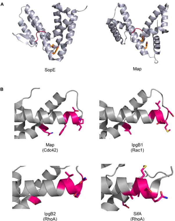
The progenitor of this family is the Salmonella typhimurium TTSS effector
SopE, which displayed GEF activity in the presence of the Rho-family GTPase
Cdc42 [53]. SopE is a small protein with six α-helices arranged in two, V-shaped,
helical bundles (Figure 1.4A) [54]. SopE is critical for virulence and invasion of the
pathogen, which is dependent on the activity on Cdc42 [53]. However, SopE did not
structurally resemble any known eukaryotic RhoGEFs (Figure 1.4A) [54]. When
bound to Cdc42 the vast majority of SopE remains unchanged, but a small “catalytic
loop” inserts in the switch 1 region of Cdc42 allowing for large rotations of the side
active site loop shown in red). Switch 2 of Cdc42 moves closer toward the “catalytic
loop” of SopE allowing for greater stability of the exchange reaction [54].
More recently, a seemingly independent family of TTSS effector proteins from
several species of mammalian pathogens (Salmonella, enteropathogenic and
enterohemorrhagic E. coli, and Shigella) was identified as possessing Rho-family
GTPase signaling capabilities [55]. This family was defined by an internal WXXXE
motif [55]. This motif is required for proper function as missense mutations result in
a loss of function for the effectors, but structurally conserved mutations allow for
conservation of activity (W to Y and E to D) [55-57]. Initially, this family was believed
to be molecular mimics of Rho-GTPase activity [55]. However, later studies revealed
the structure of one of the family members, SifA, was solved and shown to posses a
domain with a homologous fold to SopE, a known GEF [58]. Further research and
structural determination has shown that other effectors in the WXXXE family also
adopt the SopE fold and function as RhoGEFs (Figure 1.4A) [59,60]. While SopE
lacks this WXXXE motif, it does possess a YXXXT motif with relative spatial
conservation making it an intriguing possibility for either a degenerate motif or
additional expansion of the signature of this motif (Figure 1.4A).
WXXXE effectors utilize their GEF activity to subvert actin dynamics and gain
entry into the host cell. Injection of one of these members Map from EHEC/EPEC E.
coli induces filopodia formation under the invading pathogen [61]. SifA is involved in
proliferation and maintenance of the Salmonella-containing vacuole [62].
through Rac1 to evoke a fever and eliminate other competing bacteria and allow for
easier invasion of the host [57].
Analysis of the WXXXE family of proteins in comparison to eukaryotic
RhoGEFs has also provided insight into defining specificity and selectivity for
RhoGEFs among the TTSS effectors [59]. Specificity patches are defined in the β
2-β3 hairpin of the Rho-family GTPases and the α4/α6 helices of the WXXXE effectors
[59]. Swapping of these regions allows for interchange of the individual specificity of
each effector for each GTPase (Figure 1.4B) [59].
Hyun Ham et al. investigated the AvrE family of TTSS effectors from P.
syringae for their relationship to the WXXXE effectors [63]. They found that each
member of the AvrE family of effectors possesses at least one WXXXE motif, and
that most posses two [63]. When missense mutations were made for both the
tryptophan (W) and the glutamate (E), they saw a loss of function for the double
mutant and motif deletion mutant, but not for either of the single mutants. This result
differs from the findings analyzing several of the mammalian WXXXE TTSS effectors
[55-57,63].
Noting this discrepancy, we compared the relative spacing of the WXXXE
motif and looked for a “catalytic loop” and specificity patches within AvrE. We were
unable to find either using clustal X to align the sequences, the method used by
Huang et al. 2009, or by manual analysis. We then attempted to find structural
homology by defining loose borders around the WXXXE motifs approximately
correlating to the size (200aa) of the known WXXXE effectors (SopE, SopE2, BopE,
these sequences using HHpred and found that instead of showing structural
homology toward the WXXXE family of RhoGEFs, AvrE shows homology to β
-propeller containing proteins and that the WXXXE motif aligns to a small helix in this
domain. While a single mutation likely would have no effect, a double mutation or
deletion of this region could cause local instability resulting in disorganization of the
propeller. This finding does not negate the hypothesis that AvrE participates in
G-protein mediated signaling; in fact β-propeller proteins often serve as molecular
scaffolds participating in G-protein signaling [64,65]. Additionally, the WXXXE motif
is not a functional motif, but a structural motif forming a molecular clasp for the hinge
between one to the helical bundles in the WXXXE family members [59]. This could
be an example of a structural mechanism/motif that is more common than we had
previously believed and should not be used as the exclusive definition of TTSS
RhoGEFs.
ADP-ribosylating Proteins: A Different Folds With a Common Function
ADP-ribosylating toxins can be divided into 4 classes due to shared domain
organization, mutual targets, and structural variance (Figure 1.5) [66]. While class
members share structural features and modalities, sequence similarity can be low
and restricted to motifs implicated in catalytic activity [1]. Eukaryotic ribosylating
proteins also share structural identity with their prokaryotic counterparts. The
catalytic domain of poly(ADP-ribosyl) polymerase family shows homology to the
catalytic domain of Diphtheria toxin and this homology is extended to the TTSS
ecto-enzyme ART.2 shares structural homology with VIP2 from Bacillus cereus [68].
All ADP-ribosyl transferases are denoted by a conserved core of 5 β-strands
arranged as two abutting β-sheets [68]. The DT family introduces an additional β
-strand, an α-helix and a conserved H-Y-E catalytic triad whereas the VIP2 family
introduces a seventh β-strand and an R-S-E catalytic triad [66,68].
ADP-ribosyl transferases are capable of catalyzing the removal of the
nicotinamide moiety from NAD and facilitating the addition of the ADP-ribose moiety
onto target substrates including H2O, antibiotics, RNA, DNA, and proteins [1,68,69].
This reaction is known as ribosylation. Ribosylation effects protein function and can
be promiscuous in nature, as in Pseudomonas aeruginosa Exotoxin S, or quite
specific like Corynebacterium diphtheriae DT for the singular target elongation factor
2 (eEF2) [68]. DT activity on eEF2 causes cessation of protein function whereas
ADP-ribosylation of adenylate cyclase leads to constitutive activation [69].
Ribosylation is targeted toward specific amino acid residues when proteins are the
targets: R,C,N,E, and the histidine analog dipthamide [68,70]. Ribosylation is also
reversible by a class of enzymes known as ADP-ribosyl glycohydrolases [69,71].
ADP-ribosyl transferases are found in two varieties; those capable of catalyzing the
addition of one ADP-ribose moiety (mono-ADPRTs, MARTS) or those capable of
catalyzing the addition of multiple ADP-ribose moieties (poly-ADPRTs, PARTs,
PARPs) [1,68-70]. ADP-ribosyl transferases are localized to many cellular regions:
cell membrane, nucleus, secretory pathways, and cytoplasm [69]. ADP-ribosyl
transferases are associated with diverse regulatory domains, DT must be cleaved
DNA binding domain and an auto-modification domain and is not a competent
enzyme until it is bound to DNA and has undergone auto-ribosylation [69].
The ADP-ribosylation event has previously been implicated in plant immune
responses. When looking at the expression profiles elicited by avr-R gene
interactions, genes encoding proteins responsible for the removal of ADP-ribose as
well as the degradation of free ADP-ribose were up-regulated [73]. HopF1 (formerly
AvrPphF) was crystallized and shown to have homology to ADP-ribosylating toxins
but functional assays were indeterminate [74]. However, a homolog HopF2 has
recently been shown to ribosylate Map Kinase Kinase 5 (MKK5) in the host to
interfere with MTI initiated signaling events [75]. Additionally the TTSS effector
HopU1 is able to ribosylate the RNA binding protein GRP7 in order to suppress MTI
[76]. Again, we find that structural biology analyses give us insight into the
molecular mechanisms by which TTSS effectors act on host targets.
The dynamic interaction between the R proteins RPM1 and RPS2, their guardee RIN4, and the TTSS effector proteins AvrRpm1
One main focus of the research presented in this dissertation is on the TTSS
effector protein AvrRpm1. AvrRpm1 was originally isolated from Pseudomonas
syringae pv. maculicola [5,77]. Upon entering the host cell, AvrRpm1 is localized to
the plasma membrane via myristoylation of the second (glycine) residue at its
N-terminus [6]. Transgenic over expression of AvrRpm1 allows for the TTSS deficient
strain Pto DC3000hrcC to grow to increased levels on susceptible plants and
MTI [78]. AvrRpm1 inhibits basal defense responses, such as callose deposition
and early transcriptional activation of defense related genes GST6 and PR1 [79].
Once localized, AvrRpm1 both associates with RIN4, as determined by
co-immunoprecipitation, and leads to its phosphorylation [80]. Phosphorylation of RIN4
at T166 leads to the activation of RPM1 and further downstream defense responses
that trigger the hypersensitive response and lead to partial suppression of
PtoDC3000 (avrRpm1) growth [81]. In a disease susceptible plant (rpm1), AvrRpm1
can contribute to the overall virulence of the pathogen [5]. AvrRpm1 can still
contribute to virulence in the absence of RIN4, indicating that RIN4 is not the only
virulence target in the host [82]. Further, when RPM1, is absent, conditionally
over-expressed AvrRpm1 elicits weak ETI by ‘off target’ RPS2 activation [83]. RPS2 is an
R protein that monitors the cysteine protease cleavage activity of the TTSS effector
AvrRpt2 on RIN4 [84]. The fact that cross-talk exists between two ETI pathways
(RPM1 and RPS2) provides an intriguing framework for analysis into the nature of
how Arabidopsis thaliana immune signaling complexes are structured; how R
proteins relay signals they receive from their prospective guardees; how many
guardees a single R protein is capable of monitoring; and how effectors like
AvrRpm1 functionally subvert one ETI mechanism to be ensnared by another.
These questions will be addressed in the second chapter of this dissertation.
Development of a “medium-high-throughput” proteomics pipeline leads to structural determination of HopBA1
The second main topic of the research presented here is the structural analysis
high-throughput screens for effector identification to a simplified structural genomics
project akin to SGCs [25,85,86]. TTSS effectors show low sequence identity with
proteins of known function, making functional predictions difficult. In order to
identity the functions of P. syringae effector proteins, we applied structural
characterization techniques to the TTSS effectors identified by sequencing
initiatives with the goal that it would facilitate characterization of effector folds and
identification of putative active site residues. Beyond homology modeling we also
tried to purify and crystallize some of these effectors, an effort that led to the
solution of the crystallographic structure of HopBA1 (Chapter 3).
When we started this project, there were over 191 Pseudomonas syringae
effectors to choose from representing ~50 families [25]. The first criterion for
narrowing the list was to identify the effectors that had established biological
significance. This list included effectors that were of interest to us: HopAF1, a
predicted deamidase; HopAM1 and HopBA1, effectors capable of eliciting HR on
some but not all Arabidopsis thaliana accessions and thus defining new R proteins;
and HopC1, HopH1 and HopAP1, three effectors with predicted homology to
botulinum toxin.
The second, independent criterion was to identify effectors that are part of
different families but share a common domain. These shared domains are proposed
to arise through a process called “terminal reassortment” that allows for rapid
evolution [87]. Two examples of this are the effector pairs HopD1 and HopAO1; and
HopK1 and AvrRps4, each of which has a shared N-terminal domain and a divergent
another: HopD1 is 710aa while HopAO1 is 468aa, and HopK1 is 338aa, while
AvrRps4 is 221aa. Further, it is known that the C-terminal domain of HopAO1
functions as a tyrosine phosphatase and the C-terminal domain of AvrRps4
functions in the virulence activity [26]. Such conclusions cannot be made about the
function of HopD1 and HopK1 because little in known about them.
The third, independent criterion was to analyze the sequences of the
remaining families of effectors for secondary structure, predicted homology,
predicted crystallization probability and the presence of orthologs in other TTSS
using phytopathogens. Based on these analyses, each remaining family was
classified using a priority scoring matrix. Constructs were generated for nine
additional novel TTSS effector families identified through high-throughput structural
initiatives [25]. We anticipated that structural characterization of these nine families
will allow us to classify shared domains between two separate families, identify
structural motifs common to Pseudomonas syringae effectors, possibly identify novel
folds for known enzymatic functions as was the case for the Salmonella E3 ligase
SspH2 [88], and allow us to compare how proteins from different organisms evolved
to form the same functional domain from entirely different sets of amino acids.
Using these criteria, we generated constructs for 20 families of effectors from
Pseudomonas syringae and have performed preliminary analyses looking for
regions of predicted disorder, regions of similarity among the effectors, and
orthologous effectors in other pathogens with TTSS. These effector families are at
various stages of incorporation in the proteomics pipeline, and will not be discussed
outcome of this “medium-high throughput” proteomics pipeline was that we
determined the crystal structure of the novel TTSS effector HopBA1 [25], which will
be discussed in greater detail in Chapter 3.
Summary
Together, the studies described in this dissertation merge genome
sequencing information with structural information gained from the study of TTSS
effector proteins. This approach has yielded novel insights including structural
homology between the TTSS effector AvrRpm1 and the poly(ADP-ribosyl)
polymerase catalytic domain (Chapter 2), as well as the novel HopBA1 structure and
its possible relationship to HopB1 and HopAC1 and the EreA-like/ ChaN protein
superfamily (Chapter 3). These relationships have enabled us to identify residues in
each TTSS effector critical for perception by the host and have provided us with a
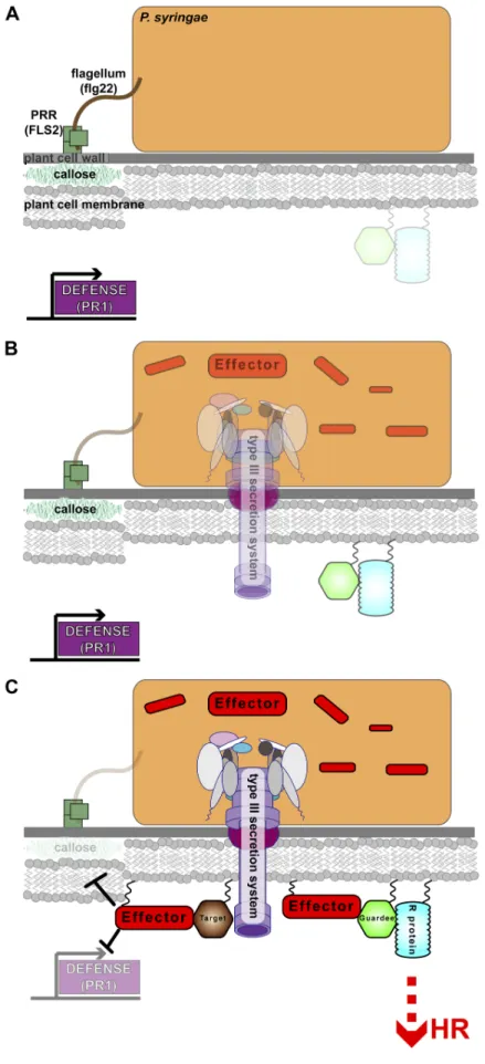
Figure 1.1. The complex
host-pathogen interactions involved in plant immune response. (A) When P. syringae first comes in contact with a host it is perceived by host PRRs such as FLS2. These PRRs recognize MAMPs such as the flagellar tail, and in specific the conserved peptide flg22. This initiates MTI responses inside the host such as callose deposition and transcription of early defense response genes like PR1. (B) In the next stage of infection P. syringae undergoes a
transcriptional and
translational switch where it stops making some PAMPs like the flagellar tail and begins transcription of the hrp/hrc controlled genes which are responsible for maintence and assembly of the TTSS and associated effector proteins. (C) In the final stage of infection, the TTSS effectors are injected into the host and begin to manipulate host targets thus blocking MTI and leading to an increase in pathogen growth on susceptible hosts
(left side). These same beneficial manipulations can betray the pathogen on resistant hosts (right side)
Figure 1.4. Structural conservation exists among SopE and the WXXXE family of TTSS effectors with target specificity for RhoGTPases arising on the α4/α6 helical
CHAPTER TWO
AvrRpm1 missense mutations weakly activate RPS2-mediated immune response in Arabidopsis thaliana
Introduction
This chapter was submitted to PLoS One on 5/15/2012. We completed all
reviewer recommended revisions, resubmitted on 6/19/2012 and the manuscript was
accepted on 7/9/2012. An additional figure showing the purification of AvrRpm1 and
analysis of the resulting protein has been included for this dissertation. I completed
all experiments in this chapter with the exception of the western blot analysis shown
in Figures 2.7 and 2.8. Brenda R.S. Temple provided guidance for the creation of
the homology model and identification of ADP-ribosyl transferase motifs in AvrRpm1
shown in Figure 2.1.
Pseudomonas syringae is a Gram-negative phytopathogen that utilizes
various biochemical means, including analogous enzymatic activity or molecular
mimicry of host proteins, to block or bypass the plant immune system. To achieve
this, each P. syringae strain injects a suite of effector proteins into host cells using a
type III secretion system. The type III secretion system is shared by many
Gram-negative pathogens of plants and animals that use effector proteins to subvert host
cell physiology and bypass defenses [9,20,89]. Plants have evolved an elaborate
MAMP-triggered immunity (MTI), and reinitiate the blocked immune response [29].
Several well-studied nucleotide binding leucine-rich repeat (NB-LRR)-dependent
responses to effectors are mediated by indirect recognition of effector action on a
host target, as described by the Guard Hypothesis [27,29]. In this model effector
targets function as a molecular lure or ‘guardee’, and a specific NB-LRR protein
functions as a ‘guard’ [80,84,90,91]. Upon biochemical manipulation of the guardee
by an effector protein, the NB-LRR protein is activated [27,29,92], leading to a
successful immune response. In the absence of the corresponding NB-LRR,
manipulation of the guardee can contribute to the virulence activity of the effector
[29,80].
This work focuses on the characterization of Pseudomonas syringae type III
effector protein AvrRpm1. AvrRpm1 function requires consensus fatty acid acylation
sites including the myristoylation site of Gly2, likely followed by a subsequent
palmitoylation site at Cys3 [6]. Once localized at the plasma membrane, AvrRpm1
associates with RIN4, and, by an unknown mechanism, triggers its phosphorylation
[80]. RIN4 phosphorylation is presumed to activate RPM1, and consequent
downstream disease resistance responses. This model has been experimentally
validated for a second, sequence diverse type III effector, AvrB, which targets the
same RIN4 sub-domain targeted by AvrRpm1 to activate RPM1 [81]. In the absence
of RPM1, AvrRpm1 [5] and AvrB [93] can contribute to overall pathogen virulence.
Moreover, in the absence of both RPM1 and RIN4, AvrRpm1 still contributes to
virulence [82], strongly suggesting that additional targets for AvrRpm1 exist in
[94-96] and HopF2 [91] suggest that RIN4 is a point of convergence in the arms race
between pathogen effectors and critical host defense machinery [97].
As type III effectors are the main contributors to the overall virulence of a
phytopathogen, their myriad biochemical functions in the host cell have only recently
started to be dissected; these include E3 protein ligase, phosphothreonine lyase,
and ADP-ribosyl transferase activities [45,75,76,98]. Determination of molecular
functions for type III effectors is complicated by their relatively low conservation at
the primary amino acid sequence level to proteins of known biochemical function,
suggesting convergent evolution onto structures that modulate eukaryotic signaling
pathways [55,99]. Therefore, we used tertiary structure prediction in order to gain
insight into AvrRpm1 function. We found that AvrRpm1 can assume the fold from the
catalytic domain of poly(ADP-ribosyl)polymerase-1 (PARP-1).
PARPs belong to a large family of proteins that contain additional domains beyond
the canonical catalytic domain [100]. PARPs undergo self-modification by addition of
ADP-ribose moiety(s) from NAD or function analogously on other targets. The
addition of poly(ADP-ribose) (PAR) is reversible by
poly(ADP-ribose)glycohydrolases (PARGs) [101]. Poly(ADP-ribose) (PAR) can be toxic, often
leading to inflammation, ischemia, and eventually cell death in mammalian systems
[102]. Nudix O-acetyl-ADP-ribose hydrolases are responsible for the breakdown of
free PAR within the cell [103]. The Arabidopsis genome encodes both PARGs and
Nudix hydrolases, and both have been implicated in immune responses [73,104].
results in the manipulation of host signaling and defense machinery in both plant and
animals, as evidenced by the structurally related proteins Diphtheria toxin from
Corynebacterium diphtheriae, ExoS from Pseudomonas aeruginosa, and HopF2
from P. syringae, and the structurally unrelated HopU1 [1,75,105-107].
We demonstrate that the AvrRpm1 family of type III effectors shares the
PARP catalytic fold, including key catalytic and structural components of PARP such
as the catalytic triad H862-Y907-E998, which typically facilitates the ribosylation
reaction. We use mutagenesis and functional tests to demonstrate that the
conserved putative catalytic residues are required for AvrRpm1 to either elicit an
RPM1-dependent immune response or contribute to virulence on a susceptible host.
Furthermore, and quite intriguingly, we show that putative catalytically inactive
AvrRpm1 inhibits the growth of P. syringae pathovar (pv.) maculicola on disease
susceptible plants. This growth inhibition is dependent on activation of the NB-LRR
protein RPS2. These findings support previous work suggesting that over-expressed
AvrRpm1 has an ‘off target’ ability to trigger an RPS2-mediated defense response,
and that RIN4 is not the only target for AvrRpm1 [82,108,109].
Despite our inability to demonstrate enzymatic activity, due to inherent
instability of purified AvrRpm1, our results collectively support the hypothesis that
AvrRpm1 is a PARP-type ADP-ribosylating protein. Our data provide a starting point
for identification of its substrate and for the definition of how that substrate
contributes to RIN4 phosphorylation and inhibition of host defense. Our results also
RPM1, RPS2, RIN4 and RIN-like proteins that may also be functionally relevant in
this system.
Materials and Methods
Creation of the Homology Models. The models were generated by querying the BioInfoBank Institute’s metaserver where we initially were able to detect homology
with the catalytic domain of PARP-1. We compared sequence alignments generated
with ClustalX, using the programs InSIGHTII, Accelrys Software Inc., and
MODELLER [110-113]. We used PDB IDs: IUK0, IGS0, 1A26, and 3GJW as
templates to generate a structural map for which we could align the AvrRpm1
sequences. The model for the Psm allele was then evaluated for fitness using the
Verify 3D application in InSIGHTII.
Generation of AvrRpm1 mutants and P. syringae strains. Missense mutations for AvrRpm1 were generated by gene splicing [114]. The external PCR primers are
Gateway™ compatible so that a common entry vector product could be used for the
generation of multiple destination vectors. Pto DC3000, Psm CR299 carried the
engineered missense mutations in trans on the pDLTrp plasmid, a Gateway™
compatible derivative of the pBBR1MCS vector [115] that uses a constitutively active
tryptophan promoter. Missense alleles of AvrRpm1 were expressed in Pto DC3000
as fusions to Δ79avrRpt2 as previously described [116]. An avirulent P. fluroescens
(Pf0) strain that has been engineered to carry a stable integration of the hrp/hrc
the plasmids pVSP61 carrying avrRpt2 [93,117] or the pDLTrp plasmid mentioned
above carrying either wild type avrRpm1 or the missense mutations.
Electrolyte leakage and bacterial growth assay conditions. Electrolyte leakage assay has been described [84], modified to include 4 leaf discs in 6 mL of water.
Bacterial growth in leaves was measured by inoculating 106cfu/mL into the leaves of
4-5 week old plants. Leaf discs were extracted and ground in 10mM MgCl2 and
serially diluted to measure bacterial numbers on the day of infiltration as well as 3
dpi. ANOVA and a Tukey’s post-hoc analysis were performed on the 3pi data using
the JMP ® Genomics software suite, SAS Institute Incorporated © 2012 to determine
if there was a statistically significant difference among the growth levels of the
various strains. Bacterial growth in seedlings was measured by dip inoculation as
previously described [118]. Briefly, an inoculum of 105cfu/mL was made for Pto
DC3000 carrying either an empty vector or avrRpm1 with missense mutations.
Bacterial growth was measured on the day of inoculation as well as 3pi.
Translocation assays were performed by inoculation of 4-5 week old plants with 5 x
107 cfu/mL on one side of the leaf. Leafs were collected and photographed 20 hpi.
Protein accumulation and immunoblot assay. For accumulation of proteins in plant tissue, leaf samples were ground in extraction buffer containing 20mM Tris pH
8.0, 150mM NaCl,1% Triton X-100, 1mM EDTA pH 8.0, 0.1% SDS, 10 mM DTT
for 20 minutes at 20,000 X g. Supernatant was quantified by Bradford analysis,
subjected to SDS-PAGE and immunoblot analysis.
AvrRpm1 / AvrRpt2 RIN4 competition assay. Pfo strains described in the methods section for generation of AvrRpm1 mutants were infiltrated at 108 cfu/mL into
4-week-old plants. Two leaves were collected for each time point and tissue was
harvest as described above. Extracts were subjected to immunoblot analysis and
probed with an α-RIN4 antibody generated from a highly specific and antigenic
peptide of RIN4.
Ribosylation Assay. Seedlings of either rpm1 or Dex::AvrRpm1-HA in rpm1 genotypes were sparsely sown and grown on MS plates for 14 days [119]. The
seedlings were then sprayed with a solution of 25mM dexamethasone (Sigma) and
25nM biotinylated NAD (Trevigen). The protein was extracted using the protocol
described in the protein accumulation and immunoblot assay methods. Duplicate
preparations were made and one set was treated with phosphodiesterase type I
(Sigma) in 110mM Tris pH 9.0, 110mM NaCl and 15mM MgCl2 [120]. The extracted
protein was subjected to immunoblot analysis and probed using pre-conjugated α
-streptavidin (Thermo). For agrobacterium-mediated transient ribosylation assay we
followed the protocol established in [81]. We then followed the protocol outlined
Purification of AvrRpm1. We generated ligation independent cloning constructs
for full length AvrRpm1 as both 6X-His-TEV and GST-TEV fusion proteins and a Δ30
AvrRpm1 (ΔTTSS) as a GST-TEV fusion protein for expression in E. coli BL21 (DE3)
Rosetta ® cells. An overnight culture of 50mL was grown with 50µL of 100mg/mL
ampicillin and 17µL of 100mg/mL chloramphenicol. One liter of Terrific Broth (TB)
was inculated with 10mL of overnight culture and allowed to grown to and optical
density of 0.6, λ=600nm. The cultures were chilled in an ice water bath for 10
minutes. Induction of protein production was achieved with 100µL/L of 100mM IPTG
followed by overnight incubation at 19°C. Cell pellet was collected and lysed in
20mM Tris, 200mM NaCl, 2mM DTT, and 5% glycerol using Avestin® Emulsiflex-C5.
Cellular debris and insoluble material was centrifuged for 1 hour at 60,000 rpm in
Beckman Culter® ultrafuge. Soluble material was loaded onto a GST column in Tris
pH 8.0, 200mM NaCl, 2mM DTT, and 5% glycerol and eluted in the same buffer with
the addition of 40mM glutathione. Protein was dialyzed in Tris pH 8.0, 200mM NaCl,
2mM DTT, and 5% glycerol and TEV protease was added (at least 6 hours). Salt
concentration was titrated to 50mM and the protein was loaded onto a cation
ammonia-group matrix (Q) column (high initial pH) for ion exchange
chromatography. Differential elution occurred upon the addition of a salt gradient
over 20 column volumes with a buffer composition of Tris pH 8.0, 1M NaCl, 2mM
DTT, and 5% glycerol. The final eluted protein was concentrated using CetriPrep®
concentrators (MW cutoff= 10kDa) and was brought to a final volume of 200µL at a
concentration of 11mg/mL. Ligation independent cloning constructs using
alleles of AvrRpm1 for use as a baculovirus expression system in Spodoptera
frugiperda (Fall Armyworm) cells (Sf-9). Constructs for each allele included including
the missense mutation G2A, double missense mutation G2A D185A, ΔTTSS and
ΔTTSS D185A. Upon successful cloning these constructs were used to transform
DH10Bac™ cells and subjected to blue/white screening using disruption of the lacZα
gene as incorporation of the pFastBac™ clone. Bacmid purification products (high
molecular weight DNA prep) was used to transfect SF-9 cells and generate primary
virus. Virus was collected and stored at °4C and cells were tested for relative
expression of recombinant protein. Constructs yielding highest levels of expression
were used to generate secondary virus to be used to infect cell cultures for protein
production. 1L cultures of SF-9 cells were grown to a final concentration of ~ 2 x 10⁶
and then inoculated with the secondary virus prep. Cultures were allowed to grow
for ~48 additional hours and then harvested immediately. Protein purification from
this point forward followed the protocol previously described for prokaryotic
constructs.
Native gel analysis of AvrRpm1. Purified AvrRpm1 or RIN4 (see [121] for
purification protocol) was diluted to a final concentration of 1mg/mL in Tris pH 8.0,
200mM NaCl, 2mM DTT, and 5% glycerol. A total volume of 6µL was prepared for
each sample (1µL of each protein alone, 1µL of each protein in combination, and
then 1µL of RIN4 + increasing concentrations of RIN4. A 12.5% native gel was run
Circular dichroism (CD) of AvrRpm1 and RIN4. AvrRpm1 and RIN4 were purified as previously described except Phosphate buffer was substituted for Tris and NaF
was substituted for NaCl to comply with accepted protocols for CD. Molar elipticty
(θ) measurements were collected from 185nm to 260nm for AvrRpm1 alone and
AvrRpm1 in combination with RIN4.
Results
Identification of conserved structural homology and a putative PARP catalytic triad in AvrRpm1.
We generated a computational homology model for AvrRpm1 to identify
conserved structural domains shared with proteins of known function. After removing
the first 30 residues, which are predicted to be disordered, we input the remaining
AvrRpm1 amino acid sequence into the BioInfoBank Institute’s metaserver [122].
The highest-ranking outputs for predicted homologous folds from the aggregated
databases were to various catalytic domains of poly(ADP-ribosyl)polymerase
(PARP) [67,100]. PARP is a member of the larger family of Diphtheria toxin-like
ADP-ribosyl transferases [1,66,70]. The catalytic domain of these proteins can be
broken down into three regions (Figure 2.1A). The N-terminal region 1 is a span of
primarily conserved residues highlighted by an aromatic residue (Figure 2.1A,
denoted with φ) followed by the first catalytic triad member H63 (in AvrRpm1; all
residues noted refer to the allele from Psm M6, GEN BANK ID AF359557.1 unless
stated otherwise) and a glycine (G64). We also noted the presence of a conserved
leucine (L62) preceding this region and a serine or threonine (T64) at its end in the