FEEDING THE MEDICALLY FRAGILE INFANT:
EFFECTS OF FEEDING METHOD AND MILK FLOW ON PHYSIOLOGY AND BEHAVIOR
Britt Frisk Pados
A dissertation submitted to the faculty of the University of North Carolina at Chapel Hill in partial fulfillment of the requirements for the degree of Doctor of Philosophy in the School of Nursing.
Chapel Hill 2015
Approved by: Suzanne M. Thoyre Katherine E. Gregory George Knafl
iii ABSTRACT
Britt Frisk Pados: Feeding the Medically Fragile Infant: Effects of Feeding Method and Milk Flow on Physiology and Behavior
(Under the direction of Dr. Suzanne M. Thoyre)
Background: Oral feeding is a challenge for medically fragile infants, particularly those born preterm and with hypoplastic left heart syndrome (HLHS). Rate of milk flow from the bottle nipple affects physiologic stability during feeding in preterm infants, but little data is available on the flow rates of nipples used for feeding hospitalized infants. Changes in milk flow rate likely also affect physiologic stability of infants with HLHS during feeding, however no studies have evaluated responses of infant with HLHS to different feeding methods. Feeding interventions aim to reduce feeding stress in fragile infants to promote growth. Outcome measures that sensitively measure stress are needed.
Purpose: This dissertation is composed of three studies. Chapter two presents milk flow rates of nipples used for feeding hospitalized infants. Chapter three examines the physiologic and behavioral responses of an infant with HLHS to variations in milk flow rate. Chapter four evaluates heart rate variability (HRV) as a feeding intervention outcome measure in the preterm infant.
Methods: In chapter two, milk flow rates of ten each of 29 nipple types (n=290) were tested using a breast pump. In chapter three, a single-subject with HLHS was evaluated during feeding with either a slow-flow or standard-flow nipple. In chapter four, a secondary analysis of heart rate variability indices was conducted from a test of a co-regulated approach to feeding preterm infants (n=14).
Results: In chapter two, flow rates varied widely between nipple types. Chapter three found that oral feeding was distressing for an infant with HLHS, regardless of flow condition. In chapter four, only SD12, a non-linear index of HRV, was found to significantly differentiate between feeding methods.
iv
To my husband, Matt, and my children, Frank, Lily, and Caroline. Matt, thank you for your unending support throughout this journey. Through the challenges of being full-time students, to moving to Boston,
and then becoming parents, you have always valued my work and supported me in every way possible. Frank, Lily, and Caroline, thank you for helping me to find balance through this process – reminding me to
v
ACKNOWLEDGMENTS
I would like to express my sincere gratitude to the countless people who have supported me throughout this journey. Thank you to my parents, Eric and Gwen, to Frank and Barbara Pados, and the rest of our family members (Kristen, Carl, Kelly, Kristin, Kaitlyn, and Patrick) for helping to care for our children when I needed to travel to UNC or to work on my dissertation. I could not have done this without all of your support.
Thank you to my dissertation chair and mentor, Suzanne Thoyre. You have believed in me, brought me in to your family, housed me and fed me on my many visits to UNC. You have taught me so much and you have challenged me to do my best work, but you have also reminded me to find balance between my work and my family and allowed me the flexibility to do all of these things in a way that have made these seven years a true blessing.
Thank you to my dear friends, Hayley Estrem and Jinhee Park. Your friendship, support, and mutual interest in helping children with feeding difficulty have helped me to stay motivated through challenging days. When the “Feeding Flock” first came to be, I could never have imagined the incredible work we would do as a team and the profound impact that it would have on all of our careers. Cara McComish and Eric Hodges, thank you for your contributions to the “Flock.” We have big dreams!
I also want to thank my other classmates and friends from the first and second year Supper Club: Ashley Leak, Carolyn Jones, Kobie Leiper, and Brandy Mechling. You all made the first two years of this journey a little more fun. Also, thank you Leslie Davis for your support as a fellow mother of twins and your prayers during the scary time of being on bedrest with Frank & Lily.
Thank you also to Brant Nix, Ginny Neelon, and the Biobehavioral Laboratory in the School of Nursing at the University of North Carolina at Chapel Hill for supporting my work. I also want to
vi
O’Brien, and Kim Barbas. To the family of the sweet baby girl who participated in my study – I am so grateful that you allowed me in to your world during such a stressful time – thank you.
Finally, I would like to thank my committee members, Katherine Gregory, George Knafl, Gail McCain, and Marcia Van Riper, for their guidance and thoughtful review of this dissertation.
Scholarship support for my doctoral studies was provided by the Elizabeth Scott Carrington Scholarship at the University of North Carolina School of Nursing (2007 & 2008) and Glaxo Fellowship Fund Scholarship at the University of North Carolina School of Nursing (2009).
The study “Milk Flow Rates from Bottle Nipples used for Feeding Hospitalized Infants” was funded by a Sigma Theta Tau International Honor Society of Nursing Alpha Alpha Chapter Research Award (2013) and was supported by the National Institute of Nursing Research of the National Institutes of Health under Award Number 5F31NR011262 (Pados). This project was supported by the Biobehavioral Laboratory at the University of North Carolina at Chapel Hill School of Nursing. Nipples for testing were donated by Boston Children’s Hospital, Duke University Medical Center, Floating Hospital for Children at Tufts University Medical Center, Groningen University (Netherlands), Handi-Craft Co., Medela Inc., North Carolina Children’s Hospital, Royal Children’s Hospital (Australia), and University of Oklahoma Children’s Hospital.
The study “Effects of Milk Flow on the Physiologic and Behavioral Responses to Feeding in an Infant with Hypoplastic Left Heart Syndrome” was funded by a Sigma Theta Tau International Honor Society of Nursing Alpha Alpha Chapter Research Award (2014), Linda Waring Matthews Research Fund Scholarship (2012 & 2014), James and Patricia Leak Fund for Nursing Research (2013), and was
supported by the National Institute of Nursing Research of the National Institutes of Health under Award Number 5F31NR011262 (Pados).
The study “Heart Rate Variability as a Feeding Intervention Outcome Measure in the Preterm Infant” was supported by the National Institute of Nursing Research of the National Institutes of Health under Award Number T32 NR007091 (PI: Mishel; Pre-doc Fellow: Pados). The original data for this project was collected by Dr. Suzanne M. Thoyre and was funded by the National Institute of Nursing Research of the National Institutes of Health under Award Number K01 NR007668 (Thoyre).
vii
TABLE OF CONTENTS
LIST OF TABLES ... x
LIST OF FIGURES ... xi
LIST OF ABBREVIATIONS... xii
CHAPTER 1: INTRODUCTION ... 1
Background and Significance ... 1
Etiology of Feeding Difficulties ... 2
Milk Flow ... 3
Theoretical Framework ... 4
Conceptualization of Feeding within the Polyvagal Theory ... 6
Aims ... 9
Prepared Manuscripts ... 9
REFERENCES ... 11
CHAPTER 2: MILK FLOW RATES FROM BOTTLE NIPPLES USED FOR FEEDING HOSPITALIZED INFANTS ... 15
Overview ... 15
Introduction... 16
Methods... 17
Statistical Analysis ... 19
Results ... 19
Comparisons Within Brand ... 20
Comparisons Within Category ... 21
viii
Conclusions ... 23
REFERENCES ... 33
CHAPTER 3: EFFECTS OF MILK FLOW ON THE PHYSIOLOGIC AND BEHAVIORAL RESPONSES TO FEEDING IN AN INFANT WITH HYPOPLASTIC LEFT HEART SYNDROME ... 35
Overview ... 35
Introduction... 36
Theoretical Framework ... 37
Methods... 38
Sample and Setting ... 38
Flow Conditions ... 39
Study Feeding Protocol ... 39
Variables & Measures ... 40
Statistical Analysis ... 43
Results ... 44
Infant Characteristics ... 44
Feeding Description ... 46
Behavioral Outcomes ... 46
Physiologic Outcomes ... 47
Discussion ... 49
Conclusion ... 55
APPENDIX 3.1: STUDY FEEDING PROTOCOL ... 66
APPENDIX 3.2: PROTOCOL FOR DATA COLLECTION ... 67
APPENDIX 3.3: OBSERVATIONAL CODING SCHEME ... 75
APPENDIX 3.4: RESPIRATORY DATA MANAGEMENT PROTOCOL ... 84
APPENDIX 3.5: LINEAR MIXED MODELING ANALYSIS OF CHAPTER 3 DATA ... 94
ix
CHAPTER 4: HEART RATE VARIABILITY AS A FEEDING INTERVENTION
OUTCOME MEASURE IN THE PRETERM INFANT ... 115
Overview ... 115
Introduction... 116
Heart Rate Variability ... 116
Theoretical Framework ... 118
State of the Literature ... 119
Methods... 121
Setting and Sample ... 121
Procedure ... 122
Statistical Analysis ... 124
Results ... 124
Sample ... 124
Results ... 125
Discussion ... 126
Conclusion ... 127
APPENDIX 4.1: LINEAR MIXED MODELING ANALYSIS OF CHAPTER 4 DATA ... 131
REFERENCES ... 181
CHAPTER 5: DISCUSSION ... 184
Future Directions For Research ... 184
x
LIST OF TABLES
Table 2.1. Nipples Tested ... 25
Table 3.1. Specific Measures ... 57
Table 3.2. Coding Scheme Descriptions of Behavioral State, Engagement, and Organization ... 58
Table 3.3. Feeding Experience ... 58
Table 3.4. Feeding Description ... 59
Table 3.5. Feeder Actions ... 59
Table 3.6. Behavioral Outcomes During Feeding ... 60
Table 3.7. Heart Rate Indices ... 60
Table 3.8. Physiologic Changes During Feeding by Flow Condition ... 61
Table 3.9. Respiratory Indices ... 62
Table 4.1. Demographic and Clinical Data ... 128
Table 4.2. Feeder Actions ... 129
Table 4.3. Standard Physiologic Measures ... 129
xi
LIST OF FIGURES
Figure 2.1. Hydrostatic pressure measured ... 26
Figure 2.2. Nipple testing equipment ... 26
Figure 2.3. Milk flow rates of all nipples tested ... 27
Figure 2.4. Coefficient of variation of milk flow of all nipples ... 28
Figure 2.5. Mean coefficient of variation of milk flow rates by nipple brand ... 29
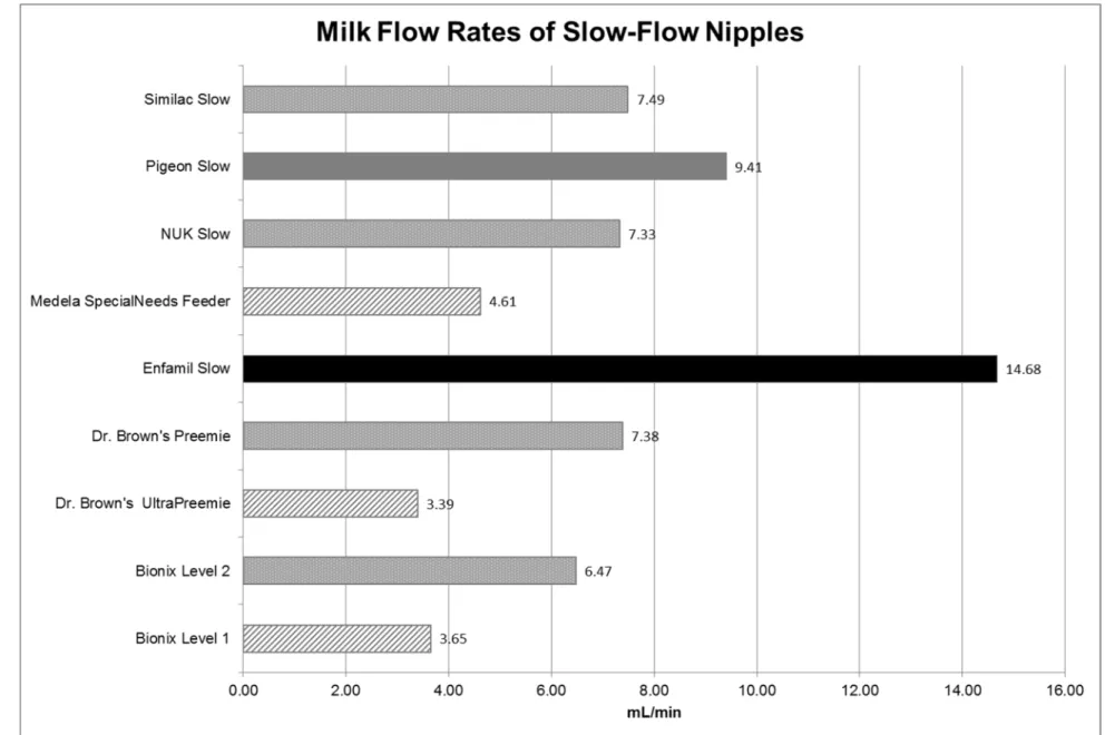
Figure 2.6. Milk flow rates of slow-flow nipples ... 30
Figure 2.7. Milk flow rates of standard-flow nipples ... 31
Figure 2.8. Milk flow rates of premature nipples ... 32
Figure 3.1. Illustration of post-surgical cardiac anatomy of Baby G ... 63
Figure 3.2. Heart Rate plotted every 1 minute ... 64
Figure 3.3. Oxygen saturation plotted every 1 minute ... 64
Figure 3.4. Respiratory rate plotted every 1 minute ... 65
Figure 4.1. Poincaré plot with SD1 (red) and SD2 (green) ... 130
xii
LIST OF ABBREVIATIONS
ANS Autonomic nervous system
Ao Aorta
Base Baseline (this abbreviation was only used in tables 3.7 and 3.9)
bpm Beats per minute
C-CHEWS Cardiac Children's Hospital Early Warning Score
CHD Congenital heart disease
ChOMPS Child Oral and Motor Proficiency Scale
CLD Chronic lung disease
cm centimeters
CoReg Coregulated approach to feeding preterm infants; name of the feeding intervention in the study presented in chapter 4
CV Coefficient of variation
DMNX Dorsal motor nucleus
DOL Day of life
ECG Electrocardiogram
FaMM Feed Family Management Measure: Feeding
Feed Feeding (this abbreviation was only used in tables 3.7 and 3.9)
FR Flow restrictor
GI Gastrointestinal
HF High frequency
HLHS Hypoplastic left heart syndrome
HR Heart rate
HRV Heart rate variability
Hz Hertz
IBI Interbeat interval
IVH Intraventricular hemmorhage
LA Left atrium
LF Low frequency
xiii
LV Left ventricle
min minute
mins minutes
mL milliliters
mmHg millimeters of mercury
MPA Main pulmonary artery
ms2 milliseconds squared
NA Nucleus ambiguous
NBRS Neurobiological Risk Score
NG Nasogastric
No Number
02 Oxygen
Pedi-EAT Pediatric Eating Assessment Tool
PMA Post-menstrual age
PNS Parasympathetic nervous system
Qp:Qs Pulmonary to systemic blood flow ratio
RA Right atrium
RDS Respiratory distress syndrome
Recover Recovery (this abbreviation was only used in tables 3.7 and 3.9)
RIP Respiratory inductance plethysmography
RR Respiratory rate
rri R wave to R wave interval
RSA Respiratory sinus arrhythmia
RV Right ventricle
RV-PA Right ventricle to pulmonary artery
S1P Stage 1 palliation
S2P Stage 2 palliation
SA Sinoatrial
SD Standard deviation
xiv
SD12 Ratio of the length of the transverse axis to the length of the longitudinal axis of the ellipse in a Poincaré plot
SD2 Length of the longitudinal axis of the ellipse in a Poincaré plot
secs seconds
SNS Sympathetic nervous system
1
CHAPTER 1: INTRODUCTION Background and Significance
Feeding is one of the most basic tasks for sustaining life once an infant is no longer being nourished in utero. While in utero, the infant rehearses sucking, swallowing, and breathing behaviors but nutrition and oxygenation are supported by the placenta. As the infant transitions to the extra-uterine environment, survival is dependent on effective coordination of fluid management (sucking and swallowing) with respiration in order to take in enough milk for adequate growth while also sustaining oxygenation. Feeding is essential for survival, but it is not simple. It is extremely complex and its success is dependent on a variety of factors both internal and external to the infant. The infant must have the anatomic structure and neurologic capacity to perform the physical act of feeding as well as the
physiologic support to maintain stability during this act. All of this must happen within an environment that is safe and supportive of the task of feeding.
2 Etiology of Feeding Difficulties
The etiology of feeding difficulties in infants born preterm and those with CHD is slightly different, although there are overlapping features. In this dissertation, chapter 3 focuses on a subset of infants with CHD who have hypoplastic left heart syndrome (HLHS) and chapter 4 focuses on infants born preterm. HLHS is a defect of the left side of the heart that results in a hypoplastic left ventricle. Survival is dependent on either heart transplantation or a series of three palliative reconstructive surgeries of the cardiac anatomy that results in a single right ventricle (RV) providing blood flow to both the pulmonary and systemic circulations (Feinstein, et al., 2012); the latter is the more common course of action. The first surgical procedure (stage 1 palliation (S1P)) typically occurs in the first week of life and the second procedure occurs around 4-6 months of age. The inter-stage period between S1P and stage 2 palliation (S2P) is a time associated with high mortality (Hehir, Cooper, Walters, & Ghanayem, 2011) and feeding difficulties have been implicated in contributing to inter-stage death (Hehir, et al., 2011). Infants with HLHS were chosen at the focus of chapter 3 because this particular group of infants is at high risk for feeding difficulty.
Common risk factors for oral feeding difficulty among preterm infants and those with HLHS include prolonged periods of intubation or respiratory support and prolonged nasogastric tube feedings in conjunction with periods of time without oral feeding (Barlow, 2009; Dodrill, et al., 2004; Einarson & Arthur, 2003). Both groups also frequently have elevated respiratory rates at rest; preterms as a result of respiratory distress syndrome and infants with HLHS as a result of pulmonary overcirculation. Studies of nutritive sucking in infants have shown that ventilation is markedly reduced during the sucking phase of feeding, then stabilizes when the infant pauses to breathe (Mathew, 1991b). Physiologically normal infants are able to increase ventilation during these pauses by increasing respiratory rate and/or tidal volume (Mathew, 1991b). However, in physiologically compromised infants, the change in ventilation during the initial continuous sucking phase may be too great to recover from and/or the challenge of increasing ventilation to recover may interfere with their ability to continue nutritive sucking. Increased ventilation needs at rest results in limited capacity for ventilatory interruption and also increases the risk of aspiration associated with mistiming of the swallow (Barlow, 2009). Finally, both groups frequently
3
1994), and are at risk for swallowing dysfunction. Infants with HLHS are at risk for swallowing dysfunction resulting from manipulation of the left recurrent laryngeal nerve during aortic arch reconstruction
(Sachdeva, et al., 2007). Preterm infants who have undergone surgical closure of a patent ductus arteriosis are also at high risk for swallowing dysfunction (Benjamin, et al., 2010).
In addition to these common risk factors for oral feeding difficulty, infants who are born preterm often encounter difficulty sucking and creating a latch to the bottle or breast as a result of immature oral musculature. They also experience difficulty coordinating sucking, swallowing, and breathing as a result of immature neurologic function (Barlow, 2009). Infants with HLHS have reduced oxygen levels as a result of mixing of oxygenated and deoxygenated blood in the common atrium, which limits their capacity for managing further decline in oxygen level resulting from ventilatory interruptions with oral feeding. Infants with HLHS also typically have some cardiac dysfunction, which limits their ability to respond to the activity of feeding, and both congenital and acquired neurologic abnormalities (Glauser, Rorke, Weinberg, & Clancy, 1990a, 1990b), which influence the coordination of sucking, swallowing, and breathing.
Milk Flow
When medically fragile infants experience difficulty with bottle-feeding in the hospital, a common strategy employed by nurses is to change the bottle nipple. This strategy is used across populations of infants and results in changes in milk flow rate, as flow rates have been found to vary considerably between different nipple types (Jackman, 2013; Mathew, 1988).
Milk flow is the rate at which milk transfers from the bottle to the mouth during feeding (Mathew, 1991b). Milk flow rate is one variable external to the infant that can affect the infants’ ability to safely coordinate swallowing and breathing, and therefore the degree of stress associated with oral feeding (al-Sayed, Schrank, & Thach, 1994; Mathew, 1991a). Given the common anatomical structures for
4
When milk flow is high, the infant is forced to either swallow at a frequency adequate to clear the oropharynx from fluid to prevent aspiration (at the expense of breathing) (al-Sayed, et al., 1994) or divert the milk away by allowing it to drool out his mouth or stop feeding altogether. Healthy, full-term infants have some capacity for self-regulating the flow of milk by changing sucking rate (Schrank, Al-Sayed, Beahm, & Thach, 1998) or pressure (Colley & Creamer, 1958; Mathew, Belan, & Thoppil, 1992). On the other hand, premature infants, with immature neurologic and respiratory systems, have limited ability to self-regulate flow (Mathew, et al., 1992). Unable to self-regulate milk flow, the premature infant exposed to higher flow during bottle-feeding exhibits greater reduction in ventilation than full-term infants (Mathew, 1991a). Premature infants have also been found to drool more with high flow rates than full-term infants (Kao, Lin, & Chang, 2010; Schrank, et al., 1998).
Jackman’s (2013) study is the only study of milk flow rates of currently available nipples. Her findings were limited in the number of nipples tested and the methods used. Additional data on milk flow rates between different types of nipples and variation in flow rate within a given type of nipple is needed to guide clinicians in making decisions about nipple selection for supporting physiological stability during oral feeding of medically fragile infants. The study presented in chapter 2 describes milk flow rates of bottle nipples used for feeding hospitalized infants.
Although there is fairly good evidence that slower milk flow is more appropriate for infants who are preterm (Kao, et al., 2010; Mathew, 1991a), it remains unknown how infants with HLHS respond to changes in milk flow and what capacity they have to self-regulate flow as they attempt to integrate fluid management and respiration despite both cardiac and respiratory compromise. No studies have examined the physiologic or behavioral responses of infants with HLHS to the challenge of oral feeding. More information is needed about this particularly fragile population of infants to identify strategies to support them during oral feeding. Chapter 3 presents a study of the effects of differing milk flow rates on an infant with HLHS.
Theoretical Framework
5
cardiac indices are expected. Physiologic changes can also manifest as behavioral changes during oral feeding (Thoyre & Carlson, 2003). Polyvagal theory (Porges, 1995) provides a theoretical basis for understanding the relationship between physiologic responses to stress and emotional, cognitive, and behavioral regulation as an infant faces the dynamic challenge of oral feeding. An overview of the key concepts of Polyvagal Theory will be presented as well as a conceptualization of feeding within the theory.
Polyvagal Theory describes the physiologic response of mammals to stress as a function of the two pathways of the vagus nerve (Porges, 1995). Stress is defined as a disruption in homeostasis, where homeostasis is the stable state of an organism when internal needs are met (Porges, 1992). The
Polyvagal Theory states that the evolutionary development of the vagus nerve resulted in two pathways: the myelinated nucleus ambiguous (NA) and the unmyelinated dorsal motor nucleus (DMNX) (Porges, 1995, 2009). While the NA controls the muscles of the supradiaphragmatic structures, such as the larynx, pharynx, esophagus, soft palate, heart, head, and face, the DMNX controls the structures below the diaphragm, particularly regulating the digestive functions of the gastrointestinal (GI) tract (Porges, 1995; Rinaman, 2006). In addition to controlling the supradiaphragmatic structures, the NA is also responsible for the coordination of sucking, swallowing, and breathing during feeding (Porges, 1995).
Polyvagal Theory explains the mammalian response to both low stress and high stress states. Mammals are distinguished from reptiles by high baseline vagal tone with temporary decreases in response to stress (Porges, 1995). During times of low stress, there is high vagal tone via the NA, resulting in low heart rate (HR); variability of the heart rate around baseline; increased tone in the inner ear for differentiation of human voices; preservation of metabolic resources for growth and restoration; coordination of sucking, swallowing, and breathing for feeding; and increased tone of the muscles of the head and face for social communication (Porges, 2007). Simultaneously, minimal input from the DMNX during times of low stress encourages digestion and absorption of nutrients from the GI tract (Porges, 2001).
6
activation of the stress response system of the hypothalamic pituitary axis (e.g., release of cortisol), stimulation of the immune system (e.g., release of cytokines), and diversion of blood away from the GI tract to the more vital organs such as the heart, brain, and lungs (Porges, 1992, 2009). HRV is the fluctuation in the interval between consecutive normal heart beats and reflects the balance of input from the sympathetic and parasympathetic divisions of the ANS (Schroeder, et al., 2004). High HRV, or a wide range around baseline, indicates a well-functioning and adaptable ANS, while low HRV signifies inability to adapt to increased physiologic demands (Verklan & Padhye, 2004).
If the SNS response is not able to reestablish homeostasis, the DMNX, the secondary system, is activated resulting in disengagement, hypotonia, apnea, and bradycardia (Porges, 2003). The
unmyelinated DMNX is the portion of the vagus that is common to both mammals and reptiles and its purpose is to conserve resources during stressful events (Porges, 1995). In reptiles, this response is functional, allowing them to freeze in response to predators (Porges, 1995). Unfortunately, in mammals who have relatively high oxygen needs, activation of the DMNX response and the resulting apnea and bradycardia can result in life-threatening oxygen depletion (Porges, 2007). The three different functions of the vagus allow mammals to not only thrive in safe environments, but survive in dangerous and life-threatening ones as well (Porges, 2009).
Conceptualization of Feeding within the Polyvagal Theory
Polyvagal Theory states that the perception of an event as stressful is subjective and dependent on the vulnerability of the individual at the time of the event (Porges, 1992; Porges, Doussard-Roosevelt, Stifter, McClenny, & Riniolo, 1999). An event may be perceived as stressful if it is environmentally or metabolically demanding or if it requires mental effort, attention, or social interaction (Porges, et al., 1999). Feeding has the potential for being perceived as stressful by an infant for a number of reasons, especially if the infant is physiologically compromised at rest. If the infant enters the feeding with unstable physiology, this is compounded by the environmental, metabolic, and social interactional stresses of feeding.
7
combined with the respiratory needs of the infant at baseline, contribute to the degree of physiologic stress associated with feeding. Finally, feeding may be stressful because it requires a great deal of mental effort, attention, and social interaction, particularly if the infant is inexperienced with feeding, is immature, or if the feeder does not adequately or appropriately respond to the infant’s needs (Porges, 2003).
When stressed during feeding, Polyvagal Theory suggests that the infant would respond by withdrawal of vagal input from the NA, which would inhibit their ability to effectively coordinate sucking, swallowing, and breathing and to accurately give the feeder facial cues about their hunger and/or satiety, their level of fatigue, or their need to pause for respiration. Unknowingly, this may lead the feeder to either end a feeding before the infant is satiated or to push the infant to continue to feed despite the infant’s exhaustion or respiratory instability, which may further compromise an already physiologically vulnerable infant. These theoretical changes are consistent with evidence of behavioral disorganization seen during feeding of preterm infants (Pickler, Frankel, Walsh, & Thompson, 1996; Thoyre & Carlson, 2003) and infants with CHD (Lobo & Michel, 1995).
Simultaneous with withdrawal of input from the NA, activation of the SNS would result in increased HR and decreased HRV (Verklan & Padhye, 2004). Although an increase in HR is expected with an activity such as feeding, the higher the HR, the more energy is expended to maintain physiologic homeostasis and the less energy is available for growth. Additionally, as the HR rises above
approximately 180 beats per minute, ventricular filling time is diminished and oxygen consumption by the myocardium is increased (Gupta, 2014). This may be tolerated in a healthy heart, but is extremely problematic for infants with HLHS who are recovering from cardiac surgery and have reduced cardiac function at rest.
Diversion of blood away from the GI tract inhibits the infant’s digestion and possibly places them at risk for developing necrotizing enterocolitis, a disease of the bowel that is initiated by damage to the intestinal mucosa from a hypoxic event and results in bacterial invasion, bowel necrosis, sepsis, and possibly death (Giannone, Luce, Nankervis, Hoffman, & Wold, 2008; McElhinney, et al., 2000).
8
sucking, swallowing, and breathing for feeding, and to digest and absorb what they have eaten. At the same time, a reduction in stress during feeding minimizes the risk of activation of the SNS and DMNX stress pathways and therefore conserves energy and minimizes oxygen-depleting events such as apnea and bradycardia. Since feeding is a frequent event, usually occurring approximately every three hours during early infancy, and because the first several years of life are a critical period in the development of the nervous system, the potential effects of the level of stress experienced during feeding go beyond each individual feeding and may have long-term effects (Beauchaine, Gatzke-Kopp, & Mead, 2007).
If feeding is persistently stressful and accompanied by activation of the SNS and/or DMNX, the developing nervous system may be trained to remain in a state appropriate for dangerous situations even when the conditions are safe (Beauchaine, et al., 2007). This persistent activation may result in immune system dysfunction, respiratory inefficiency, and psychosocial disorder (Porges, 2003). In medically fragile children, immune system impairment and respiratory dysfunction may be further compromising and even life-threatening.
Persistent activation of the SNS and/or DMNX and inability to appropriately alter vagal tone may be potentially damaging to the developing nervous system and contribute to development of psychiatric disorders associated with difficulties in social behavior, such as autism spectrum disorder (Porges, 2003). An over-responsive nervous system has been found to be associated with high trait anxiety, which when combined with poor vagal adjustment has been linked to anxiety and panic disorders (Beauchaine, et al., 2007). Additionally, evaluation of a situation as being safe or dangerous is learned and if feeding is consistently determined to be dangerous, the child may identify feeding as unsafe long after their physiologic state has improved to make feeding safe, resulting in long-term difficulty with eating. On the other hand, if feeding is consistently a non-stressful event and the environment is perceived as safe, the nervous system is exercised to support social behavior, growth, and restoration (Porges, 2003). Infants with appropriate vagal tone responses have been found to have less
9
absorption hormones and has been correlated with shorter hospital stay (DiPietro & Porges, 1991; Field & Diego, 2008).
Polyvagal Theory offers a theoretical framework for conceptualizing the complexity of infant feeding and the relationships between stress and emotional, cognitive, and behavioral regulation. The degree of stress experienced during oral feeding has the potential for profound short-term and long-term effects. As more research is done to evaluate feeding interventions to reduce feeding-related stress in medically fragile infants, outcome measures are needed that will measure stress sensitively and, ideally, provide early indicators of distress. Polyvagal Theory introduces HRV as a potential outcome measure of stress. While several studies have evaluated HRV during feeding (Brown, 2007; Cohen, Brown, & Myers, 2009; Harrison, 2011; Harrison & Brown, 2012; Lappi, et al., 2007; McCain, Fuller, & Gartside, 2005; McCain, Knupp, Fontaine, Pino, & Vasquez, 2010; Portales, et al., 1997; Suess, et al., 2000), only McCain’s (2005) study has used HRV to evaluate the degree of stress experienced by infants during feeding. Further research is needed to determine whether HRV is a sensitive enough measure to detect alterations in the degree of stress related to different feeding strategies. The study presented in chapter 4 explores the use of HRV as a feeding intervention outcome measure in preterm infants.
Aims
This dissertation is composed of three studies that each contribute to the literature with regards to feeding medically fragile infants. Specifically, the aims were to:
1. Present data on the milk flow rates and variability in flow of bottle nipples used for feeding hospitalized infants.
2. Examine the physiologic and behavioral responses of an infant with HLHS to variations in milk flow rate.
3. Evaluate the usefulness of HRV as a feeding intervention outcome measure in the preterm infant. Prepared Manuscripts
10
appendices will be removed prior to submission for publication. Chapter five provides a discussion of the manuscripts, clinical implications of the findings, and presents plans for future study.
Chapter two is titled “Milk flow rates from bottle nipples used for feeding hospitalized infants.” The purpose of this study was to evaluate milk flow rates from bottle nipples commonly used in hospitals for feeding medically fragile infants in order to provide clinicians with evidence with which to base decisions about nipple selection.
Chapter three is a presentation of a single-case experiment of the effects of milk flow on the response to feeding in an infant with HLHS. This manuscript is titled “Effects of milk flow on the physiologic and behavioral response to feeding in an infant with hypoplastic left heart syndrome.” The purpose of this study was to examine the physiologic changes and observational indicators of distress that occur when an infant with HLHS is bottle-fed with either a standard-flow nipple or a slow-flow nipple.
11 REFERENCES
al-Sayed, L. E., Schrank, W. I., & Thach, B. T. (1994). Ventilatory sparing strategies and swallowing pattern during bottle feeding in human infants. Journal of Applied Physiology, 77(1), 78-83. Retrieved from http://jap.physiology.org/
Barlow, S. M. (2009). Oral and respiratory control for preterm feeding. Current Opinions in Otolaryngology & Head and Neck Surgery, 17(3), 179-186. doi: 10.1097/MOO.0b013e32832b36fe
Beauchaine, T. P., Gatzke-Kopp, L., & Mead, H. K. (2007). Polyvagal theory and developmental
psychopathology: Emotion dysregulation and conduct problems from preschool to adolescence. Biological Psychology, 74(2), 174-184. doi: 10.1016/j.biopsycho.2005.08.008
Benjamin, J. R., Smith, P. B., Cotten, C. M., Jaggers, J., Goldstein, R. F., & Malcolm, W. F. (2010). Long-term morbidities associated with vocal cord paralysis after surgical closure of a patent ductus arteriosus in extremely low birth weight infants. Journal of Perinatology, 30(6), 408-413. doi: 10.1038/jp.2009.124
Brown, L. (2007). Heart rate variability in premature infants during feeding. Biological Research for Nursing, 8(4), 283-293. doi: 10.1177/1099800406298542
Cohen, M., Brown, D. R., & Myers, M. M. (2009). Cardiorespiratory measures before and after feeding challenge in term infants are related to birth weight. Acta Paediatrica, 98(7), 1183-1188. doi: 10.1111/j.1651-2227.2009.01284.x
Colley, J. R., & Creamer, B. (1958). Sucking and swallowing in infants. British Medical Journal, 2(5093), 422-423. Retrieved from www.bmj.com
DiPietro, J. A., & Porges, S. W. (1991). Vagal responsiveness to gavage feeding as an index of preterm status. Pediatric Research, 29(3), 231-236. doi: 10.1203/00006450-199103000-00003
Dodrill, P., McMahon, S., Ward, E., Weir, K., Donovan, T., & Riddle, B. (2004). Long-term oral sensitivity and feeding skills of low-risk pre-term infants. Early Human Development, 76(1), 23-37. doi: 10.1016/j.earlhumdev.2003.10.001
Einarson, K. D., & Arthur, H. M. (2003). Predictors of oral feeding difficulty in cardiac surgical infants. Pediatric Nursing, 29(4), 315-319. Retrieved from: http://www.pediatricnursing.net
Eisenberg, N., Fabes, R. A., Murphy, B., Maszk, P., Smith, M., & Karbon, M. (1995). The role of emotionality and regulation in children's social functioning: A longitudinal study. Child Development, 66(5), 1360-1384. doi: 10.1111/1467-8624.ep9510075268
Fabes, R. A., Eisenberg, N., & Eisenbud, L. (1993). Behavioral and physiological correlates of children's reactions to others in distress. Developmental Psychology, 29, 655-663. doi: 10.1037/0012-1649.29.4.655
Feinstein, J. A., Benson, D. W., Dubin, A. M., Cohen, M. S., Maxey, D. M., Mahle, W. T., et al. (2012). Hypoplastic left heart syndrome: Current considerations and expectations. Journal of the American College of Cardiology, 59(1 Suppl), S1-42. doi: 10.1016/j.jacc.2011.09.022
Field, T., & Diego, M. (2008). Vagal activity, early growth and emotional development. Infant Behavior and Development, 31(3), 361-373. doi: 10.1016/j.infbeh.2007.12.008
12
Glauser, T. A., Rorke, L. B., Weinberg, P. M., & Clancy, R. R. (1990a). Acquired neuropathologic lesions associated with the hypoplastic left heart syndrome. Pediatrics, 85(6), 991-1000. Retrieved from http://pediatrics.aappublications.org/
Glauser, T. A., Rorke, L. B., Weinberg, P. M., & Clancy, R. R. (1990b). Congenital brain anomalies associated with the hypoplastic left heart syndrome. Pediatrics, 85(6), 984-990. Retrieved from http://pediatrics.aappublications.org/
Gupta, S., & Sinha, S.K. (2014). Shock and hypotension in the newborn. Retrieved October 25, 2014, from http://emedicine.medscape.com/article/979128-overview
Harrison, T. M. (2011). Trajectories of parasympathetic nervous system function before, during, and after feeding in infants with transposition of the great arteries. Nursing Research, 60(3 Suppl), S15-27. doi: 10.1097/NNR.0b013e31821600b1
Harrison, T. M., & Brown, R. L. (2012). Autonomic nervous system function in infants with transposition of the great arteries. Biological Research for Nursing, 14(3), 257-268. doi:
10.1177/1099800411407687
Hehir, D. A., Cooper, D. S., Walters, E. M., & Ghanayem, N. S. (2011). Feeding, growth, nutrition, and optimal interstage surveillance for infants with hypoplastic left heart syndrome. Cardiology in the Young, 21 Suppl 2, 59-64. doi: 10.1017/S1047951111001600
Hyman, P. E. (1994). Gastroesophageal reflux: One reason why baby won't eat. Journal of Pediatrics, 125(6 Pt 2), S103-109. doi: 10.1016/S0022-3476(05)82933-6
Izard, C. E., Porges, S. W., Simons, R. F., Haynes, O. M., Hyde, C., Parisi, M., et al. (1991). Infant cardiac activity: Developmental changes and relations with attachment. Developmental Psychology, 27(3), 432-439. doi: 10.1037/0012-1649.27.3.432.
Jackman, K. T. (2013). Go with the flow: Choosing a feeding system for infants in the neonatal intensive care unit and beyond based on flow performance. Newborn & Infant Nursing Reviews, 13, 31-34. doi: 10.1053/j.nainr.2012.12.003
Kao, H. M., Lin, C. H., & Chang, Y. J. (2010). Feeding with cross-cut teats has better sucking effects and oxygenation in preterm infants with chronic lung disease. Journal of Clinical Nursing, 19(21-22), 3016-3022. doi: 10.1111/j.1365-2702.2010.03290.x
Lappi, H., Valkonen-Korhonen, M., Georgiadis, S., Tarvainen, M. P., Tarkka, I. M., Karjalainen, P. A., et al. (2007). Effects of nutritive and non-nutritive sucking on infant heart rate variability during the first 6 months of life. Infant Behavior and Development, 30(4), 546-556. doi:
10.1016/j.infbeh.2007.04.005
Lobo, M. L., & Michel, Y. (1995). Behavioral and physiological response during feeding in infants with congenital heart disease: A naturalistic study. Progress in Cardiovascular Nursing, 10(3), 26-34. Retrieved from http://onlinelibrary.wiley.com/journal/10.1111/(ISSN)1751-7117
Mathew, O. P. (1988). Nipple units for newborn infants: A functional comparison. Pediatrics, 81(5), 688-691. Retrieved from http://pediatrics.aappublications.org/
Mathew, O. P. (1991a). Breathing patterns of preterm infants during bottle feeding: Role of milk flow. Journal of Pediatrics, 119(6), 960-965. doi: 10.1016/S0022-3476(05)83056-2
13
Mathew, O. P., Belan, M., & Thoppil, C. K. (1992). Sucking patterns of neonates during bottle feeding: Comparison of different nipple units. American Journal of Perinatology, 9(4), 265-269. doi: 10.1055/s-2007-994786
McCain, G. C., Fuller, E. O., & Gartside, P. S. (2005). Heart rate variability and feeding bradycardia in healthy preterm infants during transition from gavage to oral feeding. Newborn & Infant Nursing Reviews, 5(3), 124-132. doi: 10.1053/j.nainr.2005.04.005
McCain, G. C., Knupp, A. M., Fontaine, J. L., Pino, L. D., & Vasquez, E. P. (2010). Heart rate variability responses to nipple feeding for preterm infants with bronchopulmonary dysplasia: Three case studies. Journal of Pediatric Nursing, 25(3), 215-220. doi: 10.1016/j.pedn.2009.01.009 McElhinney, D. B., Hedrick, H. L., Bush, D. M., Pereira, G. R., Stafford, P. W., Gaynor, J. W., et al.
(2000). Necrotizing enterocolitis in neonates with congenital heart disease: Risk factors and outcomes. Pediatrics, 106(5), 1080-1087. Retrieved from http://pediatrics.aappublications.org/ Pickler, R. H., Frankel, H. B., Walsh, K. M., & Thompson, N. M. (1996). Effects of nonnutritive sucking on
behavioral organization and feeding performance in preterm infants. Nursing Research, 45(3), 132-135. Retrieved from http://journals.lww.com/nursingresearchonline/pages/default.aspx Porges, S. W. (1992). Vagal tone: A physiologic marker of stress vulnerability. Pediatrics, 90(3), 498-504.
Retrieved from http://pediatrics.aappublications.org/
Porges, S. W. (1995). Orienting in a defensive world: Mammalian modifications of our evolutionary heritage. A polyvagal theory. Psychophysiology, 32(4), 301-318. Retrieved from
http://www.blackwellpublishing.com/journal.asp?ref=0048-5772.
Porges, S. W. (2001). The polyvagal theory: Phylogenetic substrates of a social nervous system. International Journal of Psychophysiology, 42(2), 123-146. doi: S0167-8760(01)00162-3 Porges, S. W. (2003). The polyvagal theory: Phylogenetic contributions to social behavior. Physiology &
Behavior, 79(3), 503-513. doi: S0031938403001562
Porges, S. W. (2007). The polyvagal perspective. Biological Psychology, 74(2), 116-143. doi: 10.1016/j.biopsycho.2006.06.009
Porges, S. W. (2009). The polyvagal theory: New insights into adaptive reactions of the autonomic nervous system. Cleveland Clinic Journal of Medicine, 76 Suppl 2, S86-90. doi:
10.3949/ccjm.76.s2.17
Porges, S. W., Doussard-Roosevelt, J. A., Stifter, C. A., McClenny, B. D., & Riniolo, T. C. (1999). Sleep state and vagal regulation of heart period patterns in the human newborn: An extension of the polyvagal theory. Psychophysiology, 36(1), 14-21. Retrieved from
http://onlinelibrary.wiley.com/journal/10.1111/(ISSN)1469-8986
Portales, A. L., Porges, S. W., Doussard-Roosevelt, J. A., Abedin, M., Lopez, R., Young, M. A., et al. (1997). Vagal regulation during bottle feeding in low-birthweight neonates: Support for the gustatory-vagal hypothesis. Developmental Psychobiology, 30(3), 225-233. doi:
10.1002/(SICI)1098-2302(199704)30:3<225::AID-DEV5>3.0.CO;2-R
Porter, C. L., Wouden-Miller, M., Silva, S. S., & Porter, A. E. (2003). Marital harmony and conflict: Links to infants' emotional regulation and cardiac vagal tone. Infancy, 4(2), 297-307. doi:
10.1207/S15327078IN0402_09
14
Sachdeva, R., Hussain, E., Moss, M. M., Schmitz, M. L., Ray, R. M., Imamura, M., et al. (2007). Vocal cord dysfunction and feeding difficulties after pediatric cardiovascular surgery. Journal of Pediatrics, 151(3), 312-315. doi: 10.1016/j.jpeds.2007.03.014
Schrank, W., Al-Sayed, L. E., Beahm, P. H., & Thach, B. T. (1998). Feeding responses to free-flow formula in term and preterm infants. Journal of Pediatrics, 132(3 Pt 1), 426-430. doi: 10.1016/S0022-3476(98)70014-9
Schroeder, E. B., Whitsel, E. A., Evans, G. W., Prineas, R. J., Chambless, L. E., & Heiss, G. (2004). Repeatability of heart rate variability measures. Journal of Electrocardiology, 37(3), 163-172. doi: S0022073604000421
Stifter, C. A., & Fox, N. A. (1990). Infant reactivity: Physiological correlates of newborn and 5-month temperament. Developmental Psychology, 26(4), 582-588. doi: 10.1037/0012-1649.26.4.582. Suess, P. E., Alpan, G., Dulkerian, S. J., Doussard-Roosevelt, J., Porges, S. W., & Gewolb, I. H. (2000).
Respiratory sinus arrhythmia during feeding: A measure of vagal regulation of metabolism, ingestion, and digestion in preterm infants. Developmental Medicine & Child Neurology, 42(3), 169-173. doi: 10.1111/j.1469-8749.2000.tb00065.x
Thoyre, S., & Carlson, J. (2003). Preterm infants' behavioural indicators of oxygen decline during bottle feeding. Journal of Advanced Nursing, 43(6), 631-641. doi: 10.1046/j.1365-2648.2003.02762.x. Verklan, M. T., & Padhye, N. S. (2004). Spectral analysis of heart rate variability: An emerging tool for
15
CHAPTER 2: MILK FLOW RATES FROM BOTTLE NIPPLES USED FOR FEEDING HOSPITALIZED INFANTS
Overview
Medically fragile infants often experience physiologic compromise during oral feeding. Milk flow is an easily manipulated variable that may contribute to the degree of physiologic instability experienced. Very little evidence is currently available to guide the selection of a bottle nipple for these infants. This study tested the milk flow rates and the variability in flow of currently available nipples used for bottle-feeding hospitalized infants. Clinicians in three countries were informally surveyed regarding nipples used for feeding hospitalized infants. Twenty-nine nipple types were identified and 10 nipples of each type were tested by measuring the amount of infant formula expressed in one minute using a breast pump. Mean milk flow rate (mL/min) and coefficient of variation (CV) were used to compare nipples within brand and within category (i.e., Slow, Standard, Premature). Flow rates varied widely between nipples, ranging from 2.10 for the Enfamil Cross-cut to 85.34 mL/min for the Dr. Brown’s Y-cut. Variability of flow rates among nipples of the same type ranged from a CV of 0.05 for Dr. Brown’s Level 1 Standard- and Wide-Neck to 0.42 for the Enfamil Cross-cut. Mean CV by brand ranged from 0.08 for Dr. Brown’s to 0.36 for Bionix. Given the wide range of flow rates and variability of nipples used for feeding hospitalized infants, nipple selection is an important decision in supporting the medically fragile infant during feeding. This study provides clinicians with information for choosing the best available nipple to support oral feeding in fragile infants.
16 Introduction
Feeding can be physiologically challenging for premature and medically fragile infants who are learning to orally feed. While breast-feeding may be the ultimate goal, most hospitalized infants will receive some bottle-feedings. Many variables contribute to the infant’s ability to bottle-feed safely and effectively, but one easily manipulated variable is the rate of milk flow from the bottle nipple. Milk flow is defined as the rate of transfer of milk from the bottle into the mouth during sucking. The rate of milk flow can affect infants’ ability to integrate fluid management with respiration and the degree of physiologic instability associated with feeding (al-Sayed, Schrank, & Thach, 1994; Mathew, 1991a). When an infant swallows, the airway is closed for about one second to prevent aspiration of milk (Mathew, 1991b). As milk flow increases and requires increased swallowing frequency, ventilation is increasingly interrupted and respiratory rate decreases (al-Sayed, et al., 1994). When milk flow slows, the swallow is delayed until a critical volume is accumulated (al-Sayed, et al., 1994), allowing the infant to breathe more and better maintain physiologic stability during feeding.
Rate of milk flow varies considerably between different brands and types of nipples (Jackman, 2013; Mathew, 1988). Healthy, full-term infants are typically resilient feeders and are able to alter sucking rate (Schrank, Al-Sayed, Beahm, & Thach, 1998) and pressure (Colley & Creamer, 1958; Mathew, Belan, & Thoppil, 1992)in order to regulate milk flow. On the other hand, medically fragile infants, such as those born preterm, have a limited ability to self-regulate flow (Mathew, 1991a). When milk flow is too high, the infant must either swallow at a frequency adequate to clear the oropharynx from fluid to prevent aspiration (at the expense of ventilation) (al-Sayed, et al., 1994); allow the milk to pool in the oropharynx (and risk aspiration); divert the milk away by allowing it to drool out their mouth (Schrank, et al., 1998); or stop feeding.
17
some having three times the flow as others (Jackman, 2013). Finally, significant variation was reported between nipples of the same type (Jackman, 2013). Given the variability between nipples of the same type, Jackman’s study was limited in that only one nipple per type was tested for nipples intended for multiple use and three nipples per type were tested for single-use nipples. To account for the variability between nipples and determine an accurate mean flow rate of each nipple type, more tests were needed. No statistical analysis was presented in the report of this study.
More information is needed to support clinicians in decision-making regarding nipple selection for feeding hospitalized infants. Without this information, infants are often exposed to multiple types of nipples in an effort to find a good match. The variability in nipples during early oral feeding may contribute to the length of time required to successfully feed and ultimately, to length of stay. This comparative, descriptive study tested the milk flow rates and variability of nipples used for bottle-feeding hospitalized infants.
Methods
Clinicians from the United States, Netherlands, and Australia were informally surveyed regarding nipples available to them for feeding infants in the hospital. Twenty-nine nipples were identified and tested (Table 2.1). A power analysis revealed that ten of each type of nipple was sufficient to compare flow rates between the nipple types with 80% power at an alpha of 0.05.
All of the nipples except the Dr. Brown’s Level 1 Wide-Neck fit on a 60 mL Grad-U-Feed Nurser (Mead Johnson & Co, Glenview, IL) and were tested with this bottle. Bottles were filled with Similac Advance Stage 1 (20 calories/ounce) ready-to-feed formula (Abbott Laboratories, Abbott Park, IL). To ensure equal levels of hydrostatic pressure, the height from the level of the liquid surface to the tip of the nipple was maintained at 2.5 cm (Figure 2.1), requiring 50 mL of formula for nipples tested with the Grad-U-Feed Nurser and 70 mL for the Dr. Brown’s Wide-Neck bottle. The formula was changed every ten tests to prevent increased viscosity as a result of denaturation of proteins from prolonged exposure to air.
18
suction pressure of 180 mmHg was used for all tests. Given the opportunities for loss of suction from the pump to the nipple, negative pressure within the bottle was tested every 50 tests using the Samba 201 Micro Pressure Measurement System (BIOPAC Systems, Inc., Goleta, CA). Mean suction rate was 110 cycles per minute and mean negative pressure within the bottle was 14 mmHg.
Formula was expressed for one minute into a 500 mL beaker situated on a calibrated platform scale (Thermo Fisher Scientific, Inc, Waltham, MA), accurate to 0.01 grams. At the conclusion of one minute, the weight of formula expressed was recorded. Outliers were re-tested to ensure accuracy of the measurement. Tests were video-recorded and measurements were confirmed by video review. Milk flow rates (mL/min) were calculated using the density of Similac Advance formula of 0.97 mL/gram (AVCalc, 2014).
The Bionix Controlled Flow Baby Feeder consists of two parts that may contribute to variability in milk flow: the nipple with the silicone inner channel and the flow restrictor (FR) system, consisting of the yellow flow restrictor, purple seal, and green flow adjuster (Bionix Medical Technologies, 2014).Since the nipple and FR system may contribute to flow in different ways, ten nipples were tested using the same FR system and separately ten FR systems were tested using the same nipple. For both the nipple and FR tests, the Bionix was tested on each of the five flow levels, resulting in a total of 100 tests. Also of note, the Dr. Brown’s nipples were tested with the venting system in place, which is how the nipple is intended to be used. The venting system comprises the cream colored vent insert and the blue vent resevoir (Handi-Craft Company, 2014a). The Medela SpecialNeeds Feeder was tested without the white circular valve membrane or the yellow circular disc (Medela Inc., 2014). The method used in this study for applying negative pressure to the nipple could not work with the valve membrane in place. The
SpecialNeeds Feeder is intended to have three flow levels: zero flow, medium flow, and maximum flow, depending on the position of the slit opening in the infant’s mouth when positive pressure (i.e.,
compression) is applied by the infant’s mouth (Medela, Inc., 2014). In this study, no positive pressure was applied. In the presence of negative pressure only, the slit opening should, theoretically, respond similarly regardless of positioning, but nipples were tested in the same position for consistency.
19
feeding. Infants feed with varying sucking rates and pressures and will achieve different flow rates within and between feedings. Thus, this data should be interpreted only as a means to compare flow rates between nipples.
Statistical Analysis
Mean milk flow rate (mL/min) and SD were calculated for each nipple type. Variability within nipple types was assessed using the coefficient of variation (CV; SD/mean). To compare variability between nipple types, CV was categorized into three levels: low (< 0.1), moderate (0.1 – 0.2), and high (> 0.2).
The Shapiro-Wilk statistic was used to assess nipples for normality with an alpha of 0.05 considered significant. Comparisons between nipple types were made within brand and within category (Slow, Standard, and Premature) using way ANOVA when normally distributed; non-parametric one-way ANOVA was used otherwise. Multiple comparison tests for the post-hoc analysis of one-one-way ANOVA utilized Duncan’s multiple range test, with an alpha of 0.05 being significant. When non-parametric one-way ANOVA was utilized, pairwise comparisons were made using the Wilcoxon Rank Sum Test and the alpha was adjusted using a Bonferroni adjustment.
For the purpose of comparing nipples within the categories of “Slow,” “Standard,” and
“Premature,” nipples were categorized by name, with a few exceptions. The Bionix Level 1 is intended to “introduce taste” and the Level 2 is intended to deliver a slow flow (Bionix Medical Technologies, 2014); these two levels were categorized as “Slow.” The Bionix Level 5 is intended to deliver flow “at or near a flow rate of a Stage 1 nipple” (Bionix Medical Technologies, 2014) so this was categorized as “Standard.” For comparisons within category, the Bionix nipple and flow restrictor tests were combined for each level. Dr. Brown’s Preemie and Ultra-Preemie were included in the categories of both “Slow” and “Premature.” Dr. Brown’s Level 1 Wide- and Standard-Neck were categorized as “Standard.” The Medela
SpecialNeeds Feeder was categorized as “Slow.” Results
20
ranged from a CV of 0.05 for Dr. Brown’s Level 1 Standard- and Wide-Neck to 0.42 for the Enfamil Cross-cut (Figure 2.4). Mean CV by brand ranged from 0.08 for Dr. Brown’s to 0.36 for Bionix (Figure 2.5). Comparisons Within Brand
Bionix Controlled Flow Baby Feeder. This system was tested to evaluate the flow and
variability of the nipples (indicated by an “N” after the level in the text) and the flow restrictor (FR) systems (indicated by a “FR” after the level in text and figures) separately. For both the nipple and FR tests, milk flow increased overall in the direction intended (level 1 being the slowest and level 5 being the fastest). Within the nipple tests, each level provided a significantly different flow rate (p<0.001), with the exception of levels 2N and 3N, which were not significantly different. Within the FR tests, 2FR and 3FR were not significantly different and 4FR and 5FR were not significantly different.
Comparing the FR tests to the nipple tests, level 1N and 1FR were comparable and levels 2N and 2FR were comparable. For all levels above 2, the nipple tests were significantly (p<0.001) slower than the FR tests. For all levels above 1, there was overlap between levels. Levels 2N, 2FR, and 3N were all similar to one another. Level 3FR was comparable to Levels 4N and 5N. At each level, the CV was higher for the FR tests than the nipple tests. Bionix Levels 3N and 5N were the only levels with CV < 0.1.
Dr. Brown’s. All levels of Dr. Brown’s nipples were found to be significantly different (p<0.005), with the exception of the Preemie and Level 1 Wide-Neck, which were found to be comparable. Dr. Brown’s Ultra-Preemie performed as intended with the lowest flow (3.39 mL/min) of all the nipples by this brand; this nipple was the second lowest flow of the 29 nipple types tested in this study. Dr. Brown’s Y-cut had the highest flow of all the nipples tested (85.34 mL/min) and was moderately variable with a CV of 0.13. All of the other Dr. Brown’s nipples had a CV < 0.1.
Enfamil. All levels of Enfamil nipples were found to be significantly different (p<0.05), with the Cross-Cut being the slowest and the Preemie nipple being the fastest. Enfamil Standard was the only nipple with a CV < 0.1.
21
Pigeon. All levels of Pigeon nipples were significantly different (p<0.05). The No-Drip was the slowest, but also the most variable (CV > 0.2).
Comparisons Within Category
Slow Flow Nipples. Nine of the 29 nipples tested were categorized as “Slow” and flow ranged from 3.39 to 14.68 mL/min (Figure 2.6). Dr. Brown’s Ultra-Preemie, Bionix Level 1, and Medela
SpecialNeeds Feeder were comparable to one another; these were all significantly slower (p<0.001) than the other “Slow” nipples. Bionix Level 2, Dr. Brown’s Preemie, NUK Slow, and Similac Slow all delivered comparable flow. Enfamil Slow was significantly faster (p<0.001) than all other “Slow” nipples. Pigeon Slow was significantly slower than Enfamil Slow (p<0.001) but significantly faster (p<0.05) than all other “Slow” nipples.
Standard Flow Nipples. Seven nipples were categorized as “Standard” and flow ranged from 6.61 to 25.07 mL/min (Figure 2.7). Similac Standard, Difrax, and Dr. Brown’s Level 1 Wide-Neck and Standard-Neck nipples were comparable to one another; these were all significantly slower than the other “Standard” nipples (p<0.05).
Premature Nipples. Four nipples were categorized as “Premature” and flow rates ranged from 3.39 to 22.68 mL/min (Figure 2.8). All four “Premature” nipples delivered significantly different flow rates (p<0.05).
Discussion
Choosing a nipple for feeding a medically fragile infant is an important decision given the wide range of flow rates found in this study. The name of a nipple (e.g., “Slow”) is not always an accurate indicator of the flow rate. Additionally, variability in flow rate between and within nipple types is an added challenge that may contribute to feeding difficulty.
22
Bionix does provide a Flow Rate Comparison chart on their website (Bionix Medical
Technologies, 2014). According to Bionix, a similar, but not completely transparent, method was used to test flow rates for 50 seconds using a Medela Classic Breast Pump (S. Herzig, e-mail communication, March 2014). Our results were consistent with theirs for the increase in flow between levels 1 and 2; they found a 75% increase while we found 77%. Both our tests and theirs found the greatest increase in flow to be between levels 3 and 4. Bionix also tested nipples made by other companies, but it is difficult to make comparisons because the names of the nipples have changed and because the methods may have been different. In the current study, Bionix Level 1 was among the slowest of the nipples tested and may be useful for feeding infants who require a very slow flow.
Dr. Brown’s markets the Ultra-Preemie nipple as being 35% slower than their Preemie nipple (Handi-Craft Company, 2014b). In our tests, the Ultra-Preemie was 54% slower than the Preemie nipple. Dr. Brown’s brand was the most consistent brand, with the lowest mean CV of all brands (Figure 2.5).
The Enfamil Cross-cut was the slowest of all nipples tested. The cross-cut has two slits that form a cross at the tip of the nipple. Enfamil advertises this nipple as having a faster flow than their standard nipple (Amazon.com, 2014), which is not consistent with our findings. Cross-cut nipples are described as varying in flow, with increasingly faster flow as the infant applies suction and opens the cross wider (Start & St James-Roberts, 2000). Two clinical studies have evaluated the physiologic effects of feeding with either a cross-cut or a single-hole nipple and found that, at sucking pressures established by preterm infants, the cross-cut yielded slower flow than the single hole (Chang, Lin, Lin, & Lin, 2007; Kao, Lin, & Chang, 2010). These studies did not use Enfamil nipples, but may support further investigation of our findings and how the cross-cut performs in practice.
23
fatigued. Certain premature nipples may have been designed based on these assumptions. More current evidence supports slower flow for maintaining physiologic stability during feeding for these infants, allowing them to breathe more (al-Sayed, et al., 1994; Mathew, 1991a; Park, Thoyre, Knafl, Hodges, & Nix, 2014), maintain better oxygenation, and endure oral feeding longer.
Another clinically significant finding was that the Similac Slow and Standard nipples do not deliver significantly different flow rates. Compared to the Enfamil products, both the Similac Slow and Standard were slower than the Enfamil Slow. Anecdotally, we have heard from clinicians that the Enfamil Slow delivers a slower flow than the Similac Slow nipple. There may be other qualities of nipples, such as the mechanical stiffness of the nipple material, that affect flow from the nipple or the infant’s sucking during feeding that could not be detected using our methods (Barlow, 2009). Our findings, however, are consistent with Jackman’s (2013) findings for these nipples.
This study had some limitations. The method used in this study applied only negative pressure to nipples. Nipples with a slit opening as opposed to a hole opening likely perform differently when positive pressure is applied during feeding, changing the shape of the opening. The two nipples in this study with slit openings were the Enfamil Cross-Cut and the Medela SpecialNeeds Feeder; caution should be used when interpreting these results as the flow rates may be different in practice. Additionally, it should be noted that the Dr. Brown’s Y-cut nipple was tested with standard thickness formula. In clinical practice, this nipple is typically used with thickened milk in medically fragile infants.
Conclusions
Milk flow is an important variable in the complex task of oral feeding for the medically fragile infant. This study confirmed results of previous studies (Jackman, 2013; Mathew, 1988), which found a wide range in milk flow rates from different nipple types. This study has built on previous work by testing additional nipples that are currently available for feeding hospitalized infants, further exploring variability within nipple types, and by improving upon the testing and analysis methods.
24
nipples should consider providing information on nipple packaging that reflects flow rate and variability of each nipple type; this would help to reduce any confusion related to the naming of nipples (i.e, premature, slow, standard), which may not accurately reflect flow rate. Manufacturers could also use the information from this study to improve upon nipple construction to reduce variability, particularly in nipples intended for fragile infants.
Researchers should use this data to make decisions about nipples used in tests of feeding interventions and select nipples with low variability in order to ensure consistency of flow. The specific nipple(s) used, flow rate, and variability of nipples should be documented in reports of feeding
intervention studies.
25 Table 2.1.
Nipples Tested
Brand Name Company & Location Nipples Tested Bionix Bionix Medical Technologies
Toledo, OH
Controlled Flow Baby Feeder Levels 1-5; nipples and flow restrictor (FR) systems tested separately
Difrax Difrax BV
Bilthoven, Netherlands
Teat Natural Standard-Neck Small (0+ months)
Dr. Brown’s Handi-Craft Co. St. Louis, MO
Level 1 Standard-Neck Level 1 Wide-Neck Ultra Preemie Preemie Y-cut Enfamil Mead Johnson & Co. Glenview,
IL
Standard-Flow (royal blue collar) Slow-Flow (turquoise collar) Preemie (light blue collar) Cross-Cut (yellow collar) Medela Medela Inc.
McHenry, IL
SpecialNeeds Feeder (formerly Haberman Feeder)
NUK NUK USA LLC
Hackensack, NJ
Orthodontic Silicone Slow-Flow Standard-Neck
Pigeon Pigeon
Tokyo, Japan
Standard-Flow Slow-Flow No-Drip Similac Abbott Nutrition
Lake Forest, IL
26
Figure 2.1. Hydrostatic pressure measured as the height from the level of the nipple opening to the height of the level of fluid.
Figure 2.3. Milk flow rates of all nipples tested (mL/min). FR ‒ flow restrictor.
2
Figure 2.4. Coefficient of variation (CV) of milk flow of all nipples. Nipples are color coded by category of CV. Diagonal pattern indicates CV < 0.1, gray indicates CV 0.1 – 0.2, and black indicates CV > 0.2. CV = mean/SD. FR ‒ flow restrictor.
0.38 0.10 0.22 0.12 0.19 0.09 0.25 0.10 0.28 0.07 0.33 0.09 0.09 0.05 0.05 0.13 0.42 0.16 0.16 0.07 0.15 0.11 0.39 0.14 0.14 0.35 0.14 0.14 0.39
0.00 0.05 0.10 0.15 0.20 0.25 0.30 0.35 0.40 0.45
Bionix Level 1 FR Bionix Level 1 Nipple Bionix Level 2 FR Bionix Level 2 Nipple Bionix Level 3 FR Bionix Level 3 Nipple Bionix Level 4 FR Bionix Level 4 Nipple Bionix Level 5 FR Bionix Level 5 Nipple Difrax Dr. Brown's UltraPreemie Dr. Brown's Preemie Dr. Brown's Level 1 Standard-Neck Dr. Brown's Level 1 Wide-Neck Dr. Brown's Y-cut Enfamil Cross-cut Enfamil Preemie Enfamil Slow Enfamil Standard Medela SpecialNeeds Feeder NUK Slow Pigeon No-Drip Pigeon Slow Pigeon Standard Similac Orthodontic Similac Premature Similac Slow Similac Standard
Coefficient of Variation of Milk Flow of All Nipples
3
Figure 2.5. Mean coefficient of variation (CV) of milk flow rates by nipple brand. Calculated as the mean of the CV of each nipple type by each brand. Brands are color coded by category of CV. Diagonal pattern indicates mean CV < 0.1, gray indicates CV 0.1 – 0.2, and black indicates CV > 0.2.
3
Figure 2.6. Milk flow rates of slow-flow nipples (mL/min). Nipples of the same color/pattern indicate that they are comparable in flow rate.
3
Figure 2.7. Milk flow rates of standard-flow nipples (mL/min). Nipples of the same color/pattern indicate that they are comparable in flow rate.
3
Figure 2.8. Milk flow rates of premature nipples (mL/min). Nipples of the same color/pattern indicate that they are comparable in flow rate.
3
33 REFERENCES
al-Sayed, L. E., Schrank, W. I., & Thach, B. T. (1994). Ventilatory sparing strategies and swallowing pattern during bottle feeding in human infants. Journal of Applied Physiology, 77(1), 78-83. Retrieved from http://jap.physiology.org/
Amazon.com. (2014). Enfamil Cross-cut nipple. Retrieved March 31, 2014, from
http://www.amazon.com/Enfamil-Cross-Cut-Nipple-12- Count/dp/B004TON9FQ/ref=sr_1_1?s=hpc&ie=UTF8&qid=1396278510&sr=1-1&keywords=enfamil+crosscut
AVCalc. (2014). Food weight to volume conversions. Retrieved May 18, 2014, from http://www.aqua-calc.com/calculate/food-weight-to-volume
Barlow, S. M. (2009). Central pattern generation involved in oral and respiratory control for feeding in the term infant. Current Opinions in Otolaryngology & Head and Neck Surgery, 17(3), 187-193. doi: 10.1097/MOO.0b013e32832b312a
Bionix Medical Technologies. (2014). Controlled Flow Baby Feeder. Retrieved July 8, 2014, from http://www.bionixmed.com/MED_Pages/ControlledFlow.html
Chang, Y. J., Lin, C. P., Lin, Y. J., & Lin, C. H. (2007). Effects of single-hole and cross-cut nipple units on feeding efficiency and physiological parameters in premature infants. Journal of Nursing
Research, 15(3), 215-223. doi: 10.1097/01.JNR.0000387617.72435.c6
Colley, J. R., & Creamer, B. (1958). Sucking and swallowing in infants. British Medical Journal, 2(5093), 422-423. Retrieved from www.bmj.com
Handi-Craft Company. (2014a). How They Work: Bottles. Retrieved July 8, 2014, from http://www.drbrownsbaby.com/bottles-accessories/how-they-work
Handi-Craft Company. (2014b). Ultra-Preemie Nipple. Retrieved June 9, 2014, from http://www.drbrownsbaby.com/medical/products/ultra-preemie-nipples
Jackman, K. T. (2013). Go with the flow: Choosing a feeding system for infants in the neonatal intensive care unit and beyond based on flow performance. Newborn & Infant Nursing Reviews, 13, 31-34. doi: 10.1053/j.nainr.2012.12.003
Kao, H. M., Lin, C. H., & Chang, Y. J. (2010). Feeding with cross-cut teats has better sucking effects and oxygenation in preterm infants with chronic lung disease. Journal of Clinical Nursing, 19(21-22), 3016-3022. doi: 10.1111/j.1365-2702.2010.03290.x
Mathew, O. P. (1988). Nipple units for newborn infants: A functional comparison. Pediatrics, 81(5), 688-691. Retrieved from http://pediatrics.aappublications.org/
Mathew, O. P. (1990). Determinants of milk flow through nipple units. Role of hole size and nipple thickness. American Journal of Diseases of Children, 144(2), 222-224. Retrieved from www.archpediatrics.com
Mathew, O. P. (1991a). Breathing patterns of preterm infants during bottle feeding: Role of milk flow. Journal of Pediatrics, 119(6), 960-965. doi: 10.1016/S0022-3476(05)83056-2