INVESTIGATING THE ROLE OF POST-TRANSLATIONAL MODIFICATIONS IN THE CORE RAS GTPASE DOMAIN
Samantha Kathleen Kistler
A dissertation submitted to the faculty at the University of North Carolina at Chapel Hill in partial fulfillment of the requirements for the degree of Doctor of Philosophy in Chemical
Biology and Medicinal Chemistry in the Eshelman School of Pharmacy.
Chapel Hill 2019
Approved by: Sharon Campbell Michael Jarstfer
Stephen Frye David Williams
© 2019
ABSTRACT
Samantha Kathleen Kistler: Investigating the Role of Post-Translational Modifications in the Core Ras GTPase Domain
(Under the direction of Sharon Campbell)
Ras proteins are the most commonly mutated oncoproteins in cancer (~30%). Oncogenic, activating Ras mutations are known drivers of the deadliest human cancers, including lung, pancreatic and colorectal cancers. Ras proteins function as critical regulators of cellular growth by acting as molecular switches, cycling between active, GTP- and inactive, GDP-bound states. In their active form, Ras proteins signal through downstream pathways that regulate cellular growth, differentiation and apoptosis. Early attempts to target Ras proteins (farnesyltransferase inhibitors) were directed toward inhibiting key carboxyl (C)-terminal lipid post-translational modifications (PTMs), which are crucial for proper Ras localization and function at the cellular membrane. Despite their failure, FTIs represent the first direct targeting efforts of Ras proteins.
Promising new classes of anti-cancer drugs directed at targeting the dysregulation of PTM status in cancers (kinase inhibitors, histone deacetylase inhibitors, HDACi and
signaling and tumorigenesis, suggesting that PTMs in this region are capable of regulating Ras behavior. Further, aberrant dysregulation in the balance of PTMs has been characterized in several cancer types, including the Ras-driven pancreatic cancer. It is therefore reasonable that Ras PTMs may present a novel avenue for therapeutic targeting in cancer. Despite more than three decades of research, Ras has remained an elusive target for cancer therapy.
TABLE OF CONTENTS
LIST OF TABLES ... vii
LIST OF FIGURES ... viii
LIST OF ABBREVIATIONS ... xi
Chapter 1 – Introduction to the Ras superfamily of GTPases and Ras proteins ... 1
Ras Proteins as GTPases ... 1
Ras History and Signaling in Cancer ... 2
Ras Structure and Dynamics ... 6
Ras and Post-Translational Modifications ... 11
Strategies to Therapeutically Target Ras Proteins: A Broad Overview ... 16
Chapter 2. – HDACi treatment causes Ras acetylation, directing signaling through the MAPK pathway through a reordering of the Ras:Raf binding interface ... 22
Introduction ... 22
Results ... 25
Discussion ... 54
Materials and Methods ... 57
Chapter 3. The ‘Ras-opathy’ mutant KRas K5N potentiates protein activation through destabilization of the GDP-bound state ... 73
Introduction ... 73
Results ... 76
Discussion ... 95
Chapter 4. A KRas GTPase K104Q Mutant Retains Downstream Signaling by
Offsetting Defects in Regulation ... 105
Introduction ... 105
Results ... 108
Discussion ... 123
Materials and Methods ... 126
Chapter 5. A Tool for Site-Specific Methyl-Lysine Generation and Selective Enrichment in Intact Proteins... 135
Introduction ... 135
Site-specific methyl-lysine analogue alkylation reaction ... 137
Methyl-binding domain enrichment of methylated Ras ... 138
Results ... 140
Discussion ... 143
Materials and Methods ... 146
Chapter 6. Conclusions and Future Directions ... 149
Final Conclusions ... 154
LIST OF TABLES
Table 2.1. KRas WT and KRas K5AcK rates of nucleotide exchange and
hydrolysis ... 37
Table 2.2. Thermal melting temperature for KRas WT and KRas acetylated protein ... 38
Table 2.3. Calculated binding affinities of KRas WT, KRas G12V and KRas G12V-K5AcK to BRaf RBD, CRaf RBD and PI3Kα RBD ... 43
Table 3.1. Melting temperature of KRas K5N and wild-type protein. ... 79
Table 3.2. Nucleotide exchange properties of KRas K5N and KRas WT proteins. ... 80
Table 3.3. Nucleotide association rates for KRas WT and KRas K5N ... 81
Table 3.4. Calculated binding affinities of KRas WT and KRas K5N to BRaf and CRaf RBDs ... 84
LIST OF FIGURES
Figure 1.1. Ras Regulation and Effector Binding ... 2
Figure 1.2 Ras Mutations in Cancer. ... 5
Figure 1.3. Ras Domain Architecture and Structure. ... 7
Figure 1.4. Ras switch dynamics. ... 9
Figure 1.5. Ras interactions with Raf and PI3K RBDs... 10
Figure 1.6. Ras proteins are extensively regulated by post-translational modifications... 15
Figure 2.1. Oncogenic Ras is an effective predictor of resistance to HDAC inhibitors. ... 27
Figure 2.2. HDAC inhibition promotes KRas acetylation ... 29
Figure 2.3. HDAC inhibition increases the steady-state levels of KRas-GTP complexed with Raf-RBD... 31
Figure 2.4. HDAC inhibitor treatment potentiates MAPK-mediated signaling ... 32
Figure 2.5. Mass spectrometry identification and characterization of acetylated KRas ... 35
Figure 2.6. GEF and GAP-mediated defects identified in acetylated KRas ... 36
Figure 2.7. Minor alterations in thermal melting temperature due to KRas acetylation ... 38
Figure 2.8. KRas G12V-K5AcK displays altered affinity to Raf-RBD ... 40
Figure 2.9. ITC binding analysis of KRas WT, KRas G12V and KRas G12V-K5AcK to BRaf RBD displays altered affinities ... 41
Figure 2.10. ITC binding analysis of KRas WT, KRas G12V and KRas G12V-K5AcK to CRaf RBD displays altered affinities ... 42
Figure 2.11. ITC binding analysis of KRas WT, KRas G12V and KRas G12V-K5AcK displays no difference in affinity to PI3Kα RBD ... 43
Figure 2.13 Cα backbone fluctuations calculated throughout the MD simulation demonstrate that acetylation stabilizes the Ras WT GDP- and GTP-bound
structures ... 47
Figure 2.14. Representative models of MD simulations of Ras WT ,Ras G12Vand K5 acetylated Ras proteins in complex with Raf RBD demonstrating overall reorientation of the K5 sidechain in response to acetylation. ... 50
Figure 2.15 Reordering and strengthening of the electrostatic network of the Ras:Raf binding interface due to lysine 5 acetylation. ... 51
Figure 2.16 Reorientation of critical α-helical and -strand pairing binding interfaces due to mutation or acetylation are identified in Molecular Dynamic simulations ... 52
Figure 2.17. Altered conformational dynamics in the Ras:Raf RBD complex due to mutation or acetylation identified in molecular dynamics simulations. ... 53
Figure 3.1. KRas K5N mutant is less thermostable in both active and inactive forms. ... 79
Figure 3.2. KRas K5N has a GEF defect in the GDP-bound form. ... 80
Figure 3.3. KRas K5N mutant displays alterations in the ability to associate GDP ... 81
Figure 3.4. KRas K5N does not alter binding to the Raf RBDs... 82
Figure 3.5. KRas K5N mutation does not disrupt binding to Raf RBDs ... 83
Figure 3.6. KRas K5N mutation significantly disrupts the Ras effector lobe in the GDP-bound state. ... 86
Figure 3.7. K5N mutation displays modest impact in GMPPCP-bound KRas. ... 87
Figure 3.8. Molecular dynamics simulations identify altered contacts in SWI and SWII due to K5N mutation. ... 90
Figure 3.9. K5N mutation causes structural changes in the effector lobe of Ras. ... 93
Figure 3.10 K5N mutant causes significant structural perturbations in GDP-bound form ... 94
Figure 4.1. The KRAS K104Q mutation impairs regulation by GEFs and GAPs yet retains effector binding interactions with RAF and PI3K RAS binding domains. ... 110
Figure 4.3. 2D 1H-15N HSQC NMR spectral overlay of 15N-enriched KRAS
K104Q (red) and WT (blue). ... 114 Figure 4.4. K104Q causes structural and dynamic perturbations primarily in helix
2 and helix 3. ... 115 Figure 4.5. The side chain of Lys104 in helix 3 interacts with helix 2 in switch II. ... 117
Figure 4.6. Backbone 15N relaxation parameters for K104Q KRAS (red) and WT
KRAS (blue). ... 119 Figure 4.7. Exogenous KRAS K104Q expression supports the growth of Rasless
MEFs. ... 121 Figure 4.8. The K104Q mutation does not alter the levels of GTP-bound KRAS. ... 122 Figure 4.9. The K104Q mutation does not alter KRAS effector signaling in NIH
3T3 cells. ... 122 Figure 4.10. The K104Q mutation does not alter wild type or activated KRAS
morphologic transforming activity. ... 123 Figure 5.1 Ras methylation has been identified at several sites within the core
GTPase domain ... 140 Figure 5.2 Methyl-lysine analogues are able to be installed in a site-specific
manner in intact Ras protein ... 141 Figure 5.3 Dimethyl-lysine reaction optimization at K147 and K117 in intact
LIST OF ABBREVIATIONS
BEZ235 Dactolisib, an ATP-competitive PI3K (phosphoinositide 3-kinase) and mTOR (mammalian target of rapamycin) inhibitor
BME 2-Mercaptoethanol C-terminus carboxyl-terminus
CD circular dichroism
CFC cardio-facio cutaneous syndrome CRC colorectal cancer
CS Costello Syndrome
CSP chemical shift perturbations DTPA diethylenetriaminepentaacetic acid
EGFR Epidermal growth factor receptor FTase farnesyltransferase
FTI farnesyltransferase inhibitor GAP GTPase activating protein GDP guanosine diphosphate
GEF guanine nucleotide exchange factor
GMPPCP Guanosine-5'-[(β,γ)-methyleno]triphosphate
GSK1120212 Trametinib, a reversible, selective MEK1/MEK2 kinase inhibitor
GTP guanosine triphosphate
GTPS guanosine 5'-O-(gamma-thio)triphosphate
GTPase domain G-domain, guanine nucleotide binding domain
H2 Helix 2 in Ras
HDACi Histone deacetylase inhibitor HEK-HT cells Human embryonic kidney cells HNSCC Head and neck small cell carcinoma HVR Hypervariable region
KD Equilibrium dissociation constant KDM Histone lysine demethylase KMT Histone lysine methyltransferase
MANT-GDP 2'-(or-3')-O-(N-Methylanthraniloyl) Guanosine 5'-Diphosphate
MANT-GMPPCP 2'/3'-O-(N-Methyl-anthraniloyl)-guanosine-5'-((β,γ)-methyleno)triphosphate
MANT-GTPS 2'/3'-O-(N-Methyl-anthraniloyl)-guanosine-5'-(γ-thio)-triphosphate
MAP3K2 (MEKK2) Mitogen-activated protein kinase kinase kinase 2 MAPK Mitogen-activated protein kinase
MD Simulations Molecular dynamic simulations MEFs Rasless mouse embryo fibroblasts MLA Methyl-lysine analogues
N-terminus Amino-terminal
NS Noonan’s Syndrome
P-loop Phosphoryl binding loop
PDAC Pancreatic ductal adenocarcinoma
PDX101 Belinostat, pan- (class I/II) histone deacetylase (HDAC) inhibitor
PI3K Phosphoinositide 3-kinase PLCε Phospholipase C epsilon PTM Post-translational modification
Raf A family of serine/threonine-specific protein kinases RalGDS Ral guanine nucleotide dissociation stimulator RBD Ras binding domain
Rce1 Ras and a-factor converting enzyme-1 RTK Receptor tyrosine kinase
SAHA Vorinostat, suberoyl+anilide+hydrozamic acid, pan- (class I/II) histone deacetylase (HDAC) inhibitor
SMYD3 SET and MYND domain containing protein 3 SN2 Nucleophilic substitution reaction mechanism SOS Son of Sevenless exchange factor
SWI Switch I
SWII Switch II
TCEP (tris(2-carboxyethyl)phosphine hydrochloride) TEV Tobacco Etch Virus
Tiam1 T-cell lymphoma invasion and metastasis-inducing protein 1
TM Melting temperature
Chapter 1 – Introduction to the Ras superfamily of GTPases and Ras proteins
Ras Proteins as GTPases
Ras proteins are predominately GDP-bound and inactive. After proper membrane localization, Ras proteins are capable of being activated. One mechanism of Ras activation involves upstream receptor tyrosine kinases (RTKs). Receptor activation via an external stimuli triggers
phosphorylation of the internal portion of the receptor and subsequent recruitment of scaffolding and modulatory proteins to the cellular membrane, which in turn serves to activate Ras proteins at the membrane (14). When bound to GTP, Ras proteins assume a conformation that confers high affinity binding to downstream effector proteins (15),(16), initiating signaling through downstream pathways to regulate multiple aspects of cellular growth, differentiation and apoptosis (4),(17) (Figure 1.1).
Figure 1.1. Ras Regulation and Effector Binding
Ras proteins cycle between “off” and “on” states with the aid of the modulatory factors, GEFs (guanine nucleotide exchange factors) and GAPs (GTPase activating proteins), A. In their active form, Ras proteins display significantly higher affinity to their downstream effector proteins, which promotes binding and signaling through Ras-mediated signaling cascades (16), B.
Ras History and Signaling in Cancer
1970s were the first glimpses into what we now know as Ras oncoproteins (18),(19). In hallmark discoveries in 1980s, Ras genes were discovered in human cancers and their role in driving oncogenic cancer cell transformation was first described (20)–(23). This has triggered decades of extensive research, aimed at therapeutically targeting and understanding Ras oncoproteins as drivers of human cancers. To date, no clinically effective anti-Ras therapies have been developed (24).
Ras proteins are some of the most commonly mutated oncoproteins in human cancers (25). Oncogenic, gain-of-function mutations in Ras genes promote Ras protein hyper-activation and are present in approximately 30% of the most deadly human cancers, including melanoma, lung, pancreatic and colorectal cancers (5),(17),(26). While the ability of wild-type Ras proteins to serve as tumor suppressors is still in debate (27), ‘hotspot’ mutations in Ras proteins are
is the least frequently mutated Ras isoform in human cancers (~4%). This is followed by NRas, which is mutated in approximately 11% of human cancers. KRas proteins are by far the most mutated of the isoforms, revealing an almost 85% mutation rate in Ras-driven cancers (4),(5),(17). Ras-driven cancers also exhibit isoform preferences. Pancreatic ductal
adenocarcinoma (PDAC) and colorectal cancers (CRC) are driven by oncogenic KRas proteins (nearly 100% of cases and ~86% of cases, respectively), whereas melanoma is driven by mutant NRas (~94% of cases) (4),(5),(26),(28). While HRas proteins are the least mutated of the Ras isoforms, they are known drivers of bladder urothelial carcinoma and head and neck squamous cell carcinoma (HNSCC) (4),(5),(26). Taken together, these data suggest a functionally unique role for each Ras isoform and mutation in cancer. Even though each of these mutations have a distinct mechanistic role in modulating Ras activity, the details of which are still under
Figure 1.2 Ras Mutations in Cancer.
Ras proteins are oncogenic drivers in several of the deadliest cancers, including melanoma, lung (LAC), pancreatic (PDAC) and colorectal cancers (CRC). Strikingly, these cancers display tissue, mutation and isoform-specific preferences.
Reprinted (adapted) with permission from Hobbs, G. A., Der, C. J., & Rossman, K. L. (2016). RAS isoforms and mutations in cancer at a glance. Journal of Cell Science, 129(7), 1287–1292. Copyright 2016 Journal of Cell Science.
Ras Structure and Dynamics
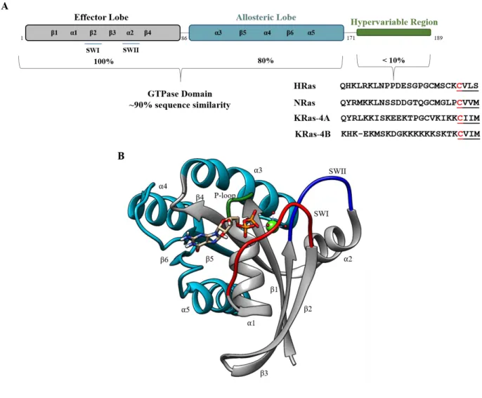
Figure 1.3. Ras Domain Architecture and Structure.
A. Ras isoforms share a highly similar core GTPase domain but a highly dissimilar C-terminal hypervariable region (HVR). The effector lobe is strictly conserved between Ras isoforms and plays roles in
effector/modulatory protein binding. The allosteric lobe houses isoform-specific sequence differences between Ras proteins, the roles of which are currently unknown. The c-terminal HVR is highly post-translationally modified, allowing for proper Ras location and activity at the cellular membrane. B. The Ras core GTPase domain (PDB 4LPK) is shown in cartoon representation. Core structural elements are labeled. The effector lobe is represented in grey and is the primary location of effector and regulatory protein interactions. The role of the allosteric lobe (teal) is more poorly understood and may contribute to Ras isoform-specificity. The highly dynamic switch regions are colored in red and blue for SWI and SWII, respectively.
on a Rossmann-type fold of alternating -helices and -sheets (Figure 1.3B) (1). It is further divided into five different structural motifs (G1-G5). G1 contains the phosphate binding loop (P-loop) that aids in coordinating the nucleotide within the Ras binding pocket. G2, more commonly termed ‘switch I’ (SWI, residues 32-38) is a highly dynamic region of Ras proteins that distinctly changes conformation upon nucleotide binding and exchange (41) (Figure 1.4). This region is also termed the ‘effector region’ as Ras effector proteins (such as the Raf kinases) are known to recognize and bind to Ras through this highly conserved region (Figure 1.5) (1),(41)–(43). G3 comprises another highly dynamic region in Ras proteins, termed ‘switch II’ (SWII, residues 59-67). Together with switch I, these regions describe the nucleotide-bound state of Ras (1). In an ‘open’ conformation, Ras is GDP-bound and inactive. However, in a closed conformation, SWI
Figure 1.4. Ras switch dynamics.
Left, cartoon representation of the NMR structures of bound HRas (PDB 1CRP (48)). In the GDP-bound form, Ras proteins exist in a more ‘open’ conformation, as seen by the increased conformational dynamics of SWII. Right, cartoon representation of the lowest energy NMR structures of GppNHp (GTP analogue)-bound HRasT35S (49). In the active, GTP-bound form the switch regions exist in a closed
conformation, facilitating contacts with the - and -phosphates. Reprinted (adapted) with permission from Lu, S., Jang, H., Muratcioglu, S., Gursoy, A., Keskin, O.,
Nussinov, R., & Zhang, J. (2016). Ras Conformational Ensembles, Allostery, and Signaling. Chemical Reviews. https://doi.org/10.1021/acs.chemrev.5b00542. Copyright 2016 Chemical Reviews ACS.
The ability to bind downstream effector proteins is also dependent upon the nucleotide-bound state of Ras proteins. In the active, GTP-nucleotide-bound form, Ras proteins exhibit a significantly higher affinity to downstream effector proteins (15). Effector proteins such as the Raf kinases and PI3-kinases (phosphoinositide 3-kinase) bind Ras proteins within their Ras binding domain (RBD) through differential SWI and SWII residue engagement (Figure 1.5). These binding interfaces are often highly electrostatic in nature (42),(50),(51). Binding will also be a result of
-strand pairing between Ras and the effector RBD (52). Effector binding and engagement will
and SWII and the surrounding regions to engage with Ras proteins (13),(53),(54).
Figure 1.5. Ras interactions with Raf and PI3K RBDs.
Ras proteins interact with effector proteins primarily through the switch regions. Raf RBD (top figure, teal) interacts with Ras proteins (grey) through SWI (red) and β-strand pairing at the interaction interface (PDB 4G0N (55)). Interactions shown are < 4.0 Å (56). PI3Kγ RBD (teal, bottom figure) also displays an interaction interface with β-strand pairing but engages both SWI (red) and SWII (blue) of Ras (grey) for binding (PDB 1HE8 (50)). Binding interfaces in both models are shown as a surface. The switch regions are labeled accordingly.
implicated in an allosteric modulation mechanism. Several groups of inter-connected residues have been identified through computational simulations that propagate structural and dynamic changes upon effector binding from the N-terminus to the C-terminus of Ras (39). Results of these computational simulations indicate that the binding of effector proteins locked Ras in the active state, primarily due to the conformational restriction of SWI. Effector binding then stimulated conformational changes in unique residue groupings that stretch from the N- to C-terminus of Ras (39),(40). While the dynamic switch regions are critical determinants of Ras activation state, their engagement in effector and regulatory protein binding can lead to larger global conformational and dynamic changes. These inside-out, allosteric structural changes are only beginning to be described in recent years and may serve to provide a more comprehensive understanding of Ras conformational and dynamic relationships alone and in complex with effector or regulatory proteins.
Ras and Post-Translational Modifications
be palmitoylated at two other sites, while NRas can be palmitoylated at one other site. The KRas-4B HVR contains a polybasic lysine tail that is not further modified. This differential processing is thought to lead to isoform-specific trafficking at the cellular membrane (57)–(59). Early attempts to target Ras proteins (farnesyltransferase inhibitors, FTIs) were directed toward inhibiting this key carboxyl (C)-terminal lipid modification, crucial for proper Ras localization and function at the cellular membrane. FTIs ultimately failed as NRas and KRas can undergo an alternative type of lipid modification (geranyl-geranylation) (60).
Ras proteins are also post-translationally modified within their core GTPase domains (61) (Figure 1.6). However, the role of these PTMs has not been intensively studied. Importantly, PTMs in this region can directly regulate Ras activity. Monoubiquitylation has been identified at three sites within the core GTPase domain of Ras proteins: K104, K117 and K147 (62),(63). K147 is located in the conserved G5 box, which plays a role in the stabilization of the guanine nucleotide (1). Monoubiquitination at K147 has been demonstrated to up-regulate protein
activity primarily through an insensitivity to GAP proteins, leading to persistent GTP-bound Ras (63). This was further verified in cellular studies where an amplified population of GTP-bound Ras was identified in RBD pulldown experiments (62). However, monoubiquitination at K147 significantly impaired binding of activated Ras to the downstream effectors PI3Kγ, CRaf and RalGDS RBDs (64), which would seem to contradict the cellular findings. One possible explanation for increased RBD binding in cells could be due to an increased affinity of CRaf RBD to the GDP-bound from of monoubiquitinated Ras (64). Monoubiquitination of K117 in Ras also led to an activated phenotype in cells; however, this occurred through a unique
increase rates of nucleotide exchange, in turn activating Ras proteins (46),(47),(66). These mutations have been demonstrated to promote tumorigenesis in human cancers and have also been identified in ‘Ras-opathies’ (developmental disorders characterized by Ras germline mutations). It was determined that monoubiquitination of K117 lead to increased guanine nucleotide dissociation rates, which served to activate Ras (65).
Beyond ubiquitination, Ras proteins are also capable of being acetylated in the core GTPase domain at K104 (67)–(69). Lysine (K) 104 is a highly conserved residue in the Ras superfamily. Structurally, K104 is located in loop 7, following α-helix 3, in the Ras core G-domain. In molecular dynamic simulations using an acetylation mimetic, glutamine (Q), Yang et al. identified that KQ mutation destabilized the α2 helix of SWII (67). They determined that
the destabilization was primarily due to a disruption in the electrostatic interactions resulting from the KQ mutation (67). Given that these regions are critical for GEF-mediated nucleotide exchange, it was not surprising that K104Q mutation disrupted SOS-mediated exchange (67). Subsequent NMR studies using the same K104Q mutant indicated that the disruption of α2 was not as severe as predicted computationally, but partial helix disruption was able to be identified (69). In addition to the GEF defect, K104Q mutation in Ras also demonstrated a GAP defect (69). However, in cellular studies K104Q mutation in Ras did not significantly alter steady-state GTP levels, cellular growth or proliferation, leading to the conclusion that the GEF and GAP defects were compensatory in nature, and acetylation at K104 likely did not impact overall Ras activity (69). However, the validity of using canonical amino acids as a mimetic of PTMs is still under debate (70). Our lab and others were able to use a genetic approach to site specifically install Nε-acetyl-L-lysine into Ras proteins, generating natively acetylated lysine, and
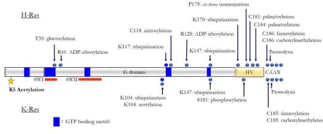
Figure 1.6. Ras proteins are extensively regulated by post-translational modifications.
Here, a schematic of the Ras core G-domain is shown with Ras PTMs reported. On the top, HRas PTMs are identified, where KRas is on the bottom. We can see that Ras proteins are highly post-translationally modified both within their core G-domain and also their carboxy-terminal hypervariable regions. Several of these PTMs are known to regulate Ras activity.
Reprinted (adapted) with permission from Ahearn, I. M., Haigis, K., Bar-Sagi, D., & Philips, M. R. (2012). Regulating the regulator: post-translational modification of RAS. Nature Reviews Molecular Cell Biology, 13(1), 39–51. https://doi.org/10.1038/nrm3255. Copyright 2011, Springer Nature.
Research in our lab and others has eluded to the identification of novel methylation sites within the core Ras G-domain. Several methylation sites have been identified, but their roles in regulating Ras activity are not clear (manuscripts submitted, data not shown). Aberrant
methylation patterns have been described in several Ras-mediated signaling pathways and of Ras effector or modulatory proteins (71)–(75). Further, the Ras-driven cancer, pancreatic ductal adenocarcinoma (PDAC), displays strong dysregulation of crucial histone lysine
identified as a direct target of SMYD3 and its inhibition resulted in decreased MAPK signaling (73),(77). Taken together this data suggests that lysine methylation may be a novel therapeutic target in Ras-driven cancers. While the functional role of Ras methylation remains unclear, the role of methylation regulating other cancer-related proteins is well established. Lysine
methylation (or acetylation) of the tumor suppressor gene p53, fine-tunes its overall activity in cancer (78),(79). Inhibitors of KMTs or KDMs present viable therapeutic opportunities in several cancer types (80). If Ras methylation contributes to aberrant growth control, methyltransferase inhibitors may represent a potential targeting mechanism. As most lysine PTMs in Ras occur at conserved sites involved in structural integrity or nucleotide binding (81), it is likely that these PTMs will alter the intrinsic function of the protein. In Chapter 5 I present novel methods to generate site-specifically methylated intact Ras proteins. This could be a crucial first step in understanding how methylation is capable of regulating Ras protein activity.
Taken together, these data demonstrate the complexity by which Ras proteins are regulated by post-translational modifications. While our lab and others have demonstrated that Ras protein activity can be modulated by PTMs, namely acetylation and monoubiquitination, the mechanisms behind this regulation are complicated in nature. PTMs in the C-terminal HVR presented therapeutic opportunities in Ras proteins with farnesyltransferase inhibitors (60). This may also be the case with PTMs that occur in the core G-domain of Ras proteins. By
understanding the distinct mechanisms by which PTMs elicit their activity in Ras proteins, we may develop novel therapeutic opportunities in Ras-driven cancers.
Strategies to Therapeutically Target Ras Proteins: A Broad Overview
Farnesylation of the C-terminal cysteine and subsequent proteolysis of -AAX leads to
carboxymethylation of the terminal cysteine and facilitates proper Ras membrane localization, which is crucial for Ras activity (4),(57). FTIs were designed to inhibit the farnesyltransferase (FTase) responsible for acting on Ras proteins, thereby rendering them cytosolic and inactive (60). However, while FTIs were one of the first examples of rational drug design targeting Ras proteins, they were designed to be specific for the FTase and not for Ras itself. FTIs were
successful at blocking the prenylation of HRas proteins, but this was not the case for N- or KRas proteins (60). It was soon discovered that in the presence of FTIs, N- and KRas could undergo alternative prenylation in the form of geranylgeranylation, allowing them to be effectively trafficked to the cellular membrane where they could be activated (4),(60),(82),(83). In recent years, the idea of blocking Ras membrane association has again become a topic of conversation. A salicylic acid derivative, Salirasib has been reported to dislodge prenylated Ras proteins from the cellular membrane. As a mimetic of farnesyl-cysteine, Salirasib has been reported to compete with farnesylated Ras binding sites at the cellular membrane (84)–(86). In cellular studies using human hepatocellular carcinoma cell lines, Salirasib was demonstrated to reduce Ras expression and activation and decreased phosphorylation of Akt, a readout of PI3-kinase pathway activation (85). In early preclinical trials in patient derived PDAC mouse xenograft studies, Salirasib in combination therapy demonstrated low overall toxicity, increased overall survival and decreased levels of signaling through both PI3-kinase and MAPK pathways as determined through western blotting (87). Unfortunately, in human phase 2 clinical trials of Salirasib in non-small cell lung cancer, low drug toxicity and good tolerance were noted but no increase in progression free survival was demonstrated (88). However, a more recent phase I clinical trial in Japanese
upon salirasib treatment (89). While these are preliminary studies, they bring to light the previous strategy of targeting Ras membrane localization as a potentially effective anti-Ras therapy.
Since the failure of traditional FTIs, targeted Ras therapies are being pursued using both direct and indirect strategies. As Papke & Der outline, there are five general strategies for the development of anti-Ras therapeutics (83). These strategies include: 1) small molecules that bind directly to Ras proteins, disrupting interactions with regulatory/effector proteins, 2) inhibition of Ras membrane association/localization, 3) inhibition of Ras downstream effector signaling cascades, 4) inhibition of genes whose functions are crucial for mutant Ras (synthetic lethal interactions) and 5) inhibition of Ras-mediated metabolic processes (83),(86). Direct strategies to target Ras proteins have proven challenging as Ras proteins lack easily discernable druggable pockets on their surface, which greatly limits the efficacy of these approaches (24),(82). In recent years, potentially druggable novel pockets have been identified in Ras, and the hunt is on for specific and selective Ras therapeutics (24),(90),(91). One direct Ras targeting strategy that has garnered some initial successes has been the efforts to develop G12C-selective inhibitors. KRas G12C mutations have been identified in non-small cell lung cancer (NSCLC) and are associated with poor prognosis (92). Using the thiol of the cysteine mutation, G12C-selective inhibitors form covalent adducts with small molecules, thereby inhibiting GTP binding and rendering Ras inactive (83),(90),(93),(94). Currently there are 2 drugs that target Ras G12C in clinical trials for the treatment of G12C-specific Ras solid tumors. MRTX894 (clinical trial identifier
drugs represent the first efforts for mutation-specific targeting of Ras proteins for the treatment of cancer.
The most clinically successful inhibitors to date have been those that target downstream Ras effector signaling pathways. Arguably, the most important downstream Ras signaling cascade is the Ras/Raf/MEK/ERK (MAPK) cascade, responsible for regulating cellular proliferation (4),(82),(83),(95). However, given the multitude of Ras-regulated signaling pathways, targeting a specific pathway presents challenges due to significant pathway crosstalk and paradoxical activation (83). Initial efforts focused on developing direct inhibitors of Raf and MEK as a means to inhibit downstream ERK activation. However, this approach was not
successful, as targeting BRaf led to paradoxical MAPK pathway activation due to compensatory CRaf activity (83). Since these initial findings, several generations of Raf, MEK and ERK-specific inhibitors have been developed and have demonstrated varying levels of clinical success (83),(86). Pathway-specific inhibitors have also been developed for the PI3-kinase signaling cascade. However, there is contradicting evidence as to whether PI3K is a potent Ras effector or is its importance is situationally dependent (82). As monotherapies, inhibitors of the PI3K-AKT-mTOR pathway have not demonstrated success in Ras-driven cancers (82),(83). While dual therapies targeting both MAPK and PI3K pathways seems promising, they have demonstrated limited clinical efficacy due to toxicity and drug-resistance concerns (83),(86).
However, these drugs have not demonstrated successes as monotherapies in Ras-driven cancers. Ras proteins are known to be regulated by PTMs, and these PTMs has been demonstrated to modulate Ras activity (67)–(69). Recently, several labs have been able to demonstrate that HDACi are successful in Ras-driven cancers when administered as combination therapies (100)– (102). In particular, the combination of a MEK inhibitor (trametinib, GSK1120212)+ PI3K inhibitor (belinostat, BEZ-235)+ HDAC inhibitor (TSA,SAHA or PDX101) resulted in >99% inhibition of cellular proliferation and dramatic induction of cellular apoptosis in pancreatic cancer cells (101). Additionally, a pan-HDAC inhibitor, Belinostat when combined with the MEK inhibitor, Trametinib functioned to synergistically decrease tumor formation in a mouse lung cancer xenograft model (102). This data suggests that HDACi may represent an untapped therapeutic potential in Ras-driven cancers. In addition to acetylation, methylation may also represent a therapeutically targetable PTM in Ras. It was recently discovered that there are significant alterations in methylation patterns and signaling in the Ras-driven cancer, PDAC (76). Additionally, the Ras-mediated MAPK signaling cascade has been demonstrated to be regulated by methylation. Methylation of MAP3K2 (MEKK2) by the methyltransferase SMYD3 is linked to increases in MAPK signaling and promotes the formation of Ras-driven carcinomas in mouse models of PDAC and lung cancer (77). This effect was reversed in SMYD3 knock-out studies. This may suggest that lysine methylation is a tractable therapeutic target in Ras-driven cancers. HDACi and methyltransferase inhibitors have not been extensively studied in Ras-driven cancers. This is due in part to the lack of clinical knowledge surrounding the exact
Chapter 2. – HDACi treatment causes Ras acetylation, directing signaling through the
MAPK pathway through a reordering of the Ras:Raf binding interface1
Introduction
Ras proteins are the most commonly mutated oncoproteins in cancer. They function as critical regulators of cellular growth by acting as molecular switches, cycling between active and inactive states (4),(17),(33). In their active form, two highly dynamic regions of Ras proteins, termed switch I and switch II, assume a conformation that confers high affinity binding to downstream effectors (15)–(17). Effector engagement then stimulates signaling through downstream pathways that regulate cellular growth, differentiation and apoptosis (17),(33). Oncogenic, gain-of-function mutations in Ras genes promote Ras protein hyper-activation and are present in approximately 30% of the most deadly human cancers, including melanoma, lung, pancreatic and colorectal cancers (5),(17). Mutationally activated Ras proteins have a well-validated role in driving oncogenic cancer cell transformation (107), and mutations in Ras at position 12, 13 or 61 are particularly oncogenic, and are widely recognized as critical
determinants of therapeutic response (107)–(109). Despite more than three decades of research, Ras has remained an elusive target for cancer therapy and is commonly considered undruggable (24). This has stimulated the search for comprehensive approaches to develop efficient
therapeutic strategies to target mutant Ras proteins for cancer treatment. Early attempts to target Ras proteins (farnesyltransferase inhibitors, FTIs) were directed toward inhibiting a key carboxyl
(C)-terminal lipid modification, crucial for proper Ras localization and function at the cellular membrane (4),(34),(60). Unfortunately, as NRas and KRas can undergo an alternative type of lipid modification (geranyl-geranylation), the use of FTIs as an anti-Ras targeted therapy was unsuccessful (60). Current approaches to target oncogenic Ras proteins are more focused on indirect strategies, including disruption of regulator or effector protein interactions and inhibiting downstream effector signaling pathways (83).
Histone deacetylase inhibitors (HDACi) are a very promising emerging class of anti-cancer drugs directed at targeting the frequent dysregulation of PTMs in anti-cancers. Aberrant dysregulation of acetylation due to altered expression of HDACs or histone acetyl transferases (HATs) has been observed in several cancer types (110)–(112). Additionally, HDACi have shown multiple clinical successes in the treatment of a myriad of primarily non-solid tumor cancers (96). Historically, the effects of PTMs have been most extensively studied in histone regulation (113); however, acetylation of non-histone proteins is known to alter protein stability and localization as well as protein-protein interactions (105). While the role of acetylation in modulating protein activity in several cancer-related proteins such as p53 has been well
established (79),(105),(114), the role of acetylation has not been thoroughly investigated in Ras-driven cancers. Despite the early promise of HDACi, they have not proven to be a clinically viable monotherapy treatment option for Ras-driven solid tumors (106),(115). The rationale for this ineffectiveness is currently unknown. However, the use of an HDACi as part of a
proliferation was inhibited and dramatic cellular apoptosis was induced (101). Further, belinostat (HDACi) combination therapy with a MEK inhibitor (trametinib) synergistically acted to
decrease tumor formation in a mouse lung cancer xenograft model (102).
Ras proteins have been reported to be acetylated within their core GTPase domain, but it is unclear exactly how acetylation modulates Ras activity (67)–(69),(116),(117). Acetylation of a receptor tyrosine kinase upstream of Ras, EGFR (epithelial growth factor receptor), causes enhanced signaling and sustained downstream activation, leading to resistance of tumor cells to HDACi treatment (118). Also, HDAC2 overexpression has also been identified in colorectal cancer (CRC) (96),(119)–(122) and it is correlated with poor survival (121). CRC is one of the leading causes of cancer deaths in the United States (123), and Ras proteins are mutated in approximately 52% of CRCs (5). These findings suggest that acetylation likely plays a role in regulating Ras-driven CRC, and therefore, HDACi may be an important and novel therapeutic option for Ras-driven cancers. Given the lack of clinical knowledge surrounding HDACi therapy in Ras-driven cancers, we have used cellular, biophysical and computational approaches to characterize the mechanism by which HDAC inhibitors display limited clinical utility as a monotherapy in Ras-driven CRC. This may lead to novel therapeutic approaches for the treatment of Ras-driven cancers.
the Raf RBD and an overall restructuring of the highly electrostatic binding network, consistent with the increased affinity for Raf and subsequent increased signaling.
Results
Oncogenic Ras is an effective predictor of resistance to HDACi treatment.
To gain insight into the limited clinical utilities of HDAC inhibitors as therapeutics in Ras-driven solid tumors the Sers lab conducted a drug sensitivity screen using a selection of HDACi on a panel of CRC cell lines that differ in their KRas or BRaf mutational status. Inhibitors that were chosen target either class I HDACs or both classes I and II, serving as pan-HDAC inhibitors. Three of the selected inhibitors in the include the pan-pan-HDAC inhibitors panobinostat (LBH589), belinostat (PXD101) and vorinostat (SAHA), all of which are US Food and Drug Administration (FDA)-approved for the treatment of multiple myeloma (MM),
being apoptotic (Figures 2.1A, B). They were further able to demonstrate that in the presence of the oncogenic BRaf V600E mutant, cells harboring KRas WTdemonstrated no alterations in their sensitivity profiles. This suggested a mechanism dependent upon KRas and not the oncogenic BRaf V600E-mediated dysregulation of the downstream MAPK-signaling cascade. To confirm that oncogenic Ras is an adequate predictor of resistance to HDAC inhibitor treatment, in particular the class I HDACi MS-275 (Entinostat), an isogenic system using CaCO-2 cells transduced with either KRas G12Vor KRas WT was employed to allow for conditional expression of the respective proteins. Upon expression of the oncogenic KRas G12V, a
Figure 2.1. Oncogenic Ras is an effective predictor of resistance to HDAC inhibitors.
A. Heatmap representing the degree of sensitivity to HDAC inhibitors. 15 CRC cell lines were treated with either DMSO (0.1% v/v), MS-275 (5 μM), FK228 (5 nM), SAHA (5 μM), PXD101 (1 μM) or LBH589 (750 nM) for 72 hrs. The level of apoptosis was determined based on the percentage of cleaved capspase-3 positive cells as detected by flow cytometry. The white end of the spectrum denotes no detectable apoptosis (< 5%), while the blue end of the spectrum represents increasing to complete activation of apoptosis (100%). KRas and BRaf mutational status is indicated in left column. Data were compiled as mean ± standard deviation B. The mutational status of KRas determines the degree of MS-275-induced apoptosis as based on the level of cleaved PARP (poly ADP-ribose polymerase). 11 CRC cell lines from A, were treated with either DMSO (0.1% v/v) or MS-275 (5 μM) for a total of 72 hrs. Samples were immunoblotted with the indicated
antibodies. C. A KRAS G12V-dependent decrease in sensitivity to MS-275 treatment. CaCO-2 KRAS WT and CaCO-2 KRAS G12V cells were treated with either DMSO (0.1% v/v), MS-275 (5 μM) (left) or increasing concentration of MS-275 (right) for 72 hrs. The ectopic expression of KRAS WT and KRAS G12V was initiated with doxycycline (2 μg/ml) 72 hrs prior and maintained for the full duration of the experiment. The level of apoptosis (left) was determined as in A, while growth inhibition was measured using XTT
KRas is acetylated at position K5 in response to MS-275 treatment.
KRas has been reported to be regulated via post-translational acetylation at lysine 104 (K104) and lysine 147 (K147), with HDAC6 and SIRT2 serving as the lysine deacetylases modulating these processes (67)–(69),(125),(126). In order to investigate whether the resistance to the HDACi MS-275 observed upon expression of oncogenic KRas G12V protein is a
consequence of modulation of the acetylation state of Ras, the Sers lab assessed the overall acetyl-lysine levels of immunoprecipitated Ras from CaCO-2 cells ectopically expressing either KRas WT or KRas G12V. Immunoblotting data demonstrated that irrespective of the mutational status of KRas, treatment with MS-275 resulted in elevated levels of detected acetyl-lysine (Figure 2.2A). This data suggests that MS-275 induced therapeutic resistance via modulation of acetylation state in oncogenic KRas G12V proceeds via a mechanism unique or perpetuated by oncogenic Ras that is distinct from KRas WT. In order to determine which residue was being acetylated in Ras proteins independent immunoprecipitation followed by protein shotgun LC-MS/MS analysis was conducted. Intriguingly, only one lysine residue, K5 where acetylation was detected as a consequence of treatment with MS-275 was identified (Figure 2.2B). To verify that acetylation of K5 was solely responsible for the observed increase in detectable levels of
acetylated lysine identified in response to MS-275 treatment, substitution mutants were made at K5. K5 was substituted for either alanine (K5A) or arginine (K5R). This functioned to eliminate the detectable overall lysine acetylation, permanently locking KRas in a constitutively
deacetylated state upon MS-275 treatment. This indicated that K5 is likely to be the only
previously unidentified acetylation site in Ras proteins, the functional consequence of acetylation at K5 are unknown.
Figure 2.2. HDAC inhibition promotes KRas acetylation
A. Detection of acetyl-lysine of immunoprecipitated KRAS. CaCO-2 KRas WT and CaCO-2 KRas G12V cells, with an induced ectopic expression of the respective proteins, were treated with either DMSO (0.1% v/v) or MS-275 (5 μM) for 72 hrs. Total Ras was immunoprecipitated and acetyl-lysines (AcK) on KRas were detected by immunoblotting. B. Identification of the acetylated lysine residue by LC/MS/MS analysis of immunoprecipitated KRas G12V. C. Effect of K5A and K5R substitution mutations on overall lysine acetylation of KRas G12V. Ectopically expressed KRas G12V, KRas G12V-K5A and KRas G12V-K5R were immunoprecipitated form CaCO-2 cells treated as in A. Acetylated lysine on KRas was detected by immunoblotting. *Data collected by the Sers lab.
MS-275 induced acetylation of Ras increases the steady-state Ras-GTP levels in cells.
evaluated whether MS-275 treatment and Ras acetylation were capable of modulating the overall activation status of Ras in cells. As Ras proteins bind to their downstream effectors in a GTP-dependent manner (15), Raf-RBD (Ras binding domain) pulldown assays can be used to evaluate the GTP-bound population of Ras proteins in cells (127). The Raf-RBD pull-down assay
revealed an immediate and persistent increase in the steady-state GTP-bound levels of KRas following treatment with MS-275. This effect was observed independent of KRas mutational status (Figure 2.3A). Although the steady-state GTP-bound level of KRas is expectedly
Figure 2.3. HDAC inhibition increases the steady-state levels of KRas-GTP complexed with Raf-RBD A. MS-275-induced changes in steady-state GTP-bound KRas levels. Cells were treated with either DMSO (0.1% v/v) or MS-275 (5 μM) for 72 hrs. Changes in the levels of KRas G12V-GTP and KRas WT-GTP pulled-down with Raf1-RBD agarose beads were assayed in the absence and presence of an induced ectopic expression of the respective proteins and detected by immunoblotting analysis. B. Effect of K5A and K5R mutations on MS-275-induced changes in steady-state GTP-bound KRas levels. Changes in the levels of KRas G12V-GTP, KRas G12V-K5A-GTP and KRas G12V-K5R-GTP were assessed as described in A.
MS-275 induced acetylation of Ras results in preferential signaling through the downstream Raf-MAPK signaling cascade.
the Sers lab assessed whether MS-275 treatment and acetylation of Ras at K5 led to altered MAPK-mediated downstream signaling. They uncovered that only in the setting of an oncogenic KRas G12V were they able to observe hyper-activation of the downstream MAPK signaling cascade due to MS-275 treatment. This is marked by an increase in the phosphorylation of
MEK1/2 and ERK1/2 (Figure 2.4A). In case of KRas WT, this remained largely unchanged. This identified disparity is not entirely unexpected as oncogenic KRas G12V is known to populate a constitutively activated phenotype primarily due to the lack of GAP-stimulated GTP hydrolysis (4),(5),(27),(33). In response to MS-275 treatment, acetylation of K5 in KRas G12V resulted in further increase in MAPK pathway activation. No observed defect was identified in PI3K signaling (data not shown).
Figure 2.4. HDAC inhibitor treatment potentiates MAPK-mediated signaling
MS-275-induced hyperactivation of MAPK-signaling in KRas G12V expressing cells. CaCO-2 KRas WT and CaCO-2 KRas G12V were treated with either DMSO (0.1% v/v) or MS-275 (5 μM) for 24, 48 and 72 hrs. Changes in the levels of MAPK-signaling components were assayed in the absence and presence of an induced ectopic expression of the respective proteins. Samples were immunoblotted with the indicated antibodies.
located in the 1 sheet of Ras and extends into a region known to be important for GEF, GAP and effector protein recognition and binding (16),(53),(131). To explore the possibility that acetylation of K5 is capable of altering Ras activity in vitro, we generated KRas protein
containing acetyl-lysine at position K5 using an unnatural amino acid approach. Here, a cognate pair of tRNACUA/tRNA synthase was used to direct the installation of Nε-acetyl-lysine into KRas in response to an amber codon at the genetic level, in a manner similar to as described previously (68),(132),(133). Incorporation of acetyl-lysine was verified by mass spectrometry to be greater than 95% (Figure 2.5A,B).
Cellular studies conducted by the Sers lab demonstrate an equal increase in Ras-GTP levels as identified by Raf RBD pulldowns independent of mutational status due to MS-275 treatment and therefore Ras acetylation (Figure 2.3A). This suggests that regulation of Ras activation due to K5 acetylation likely occurs via a similar mechanism in wild-type and
oncogenic KRas G12V. This would hold true despite the propensity of oncogenic KRas G12V to remain in an activated phenotype due to significantly impaired GAP-mediated hydrolysis
2.85±0.326 x 10⁻⁴ s-1 for KRas WT and acetylated protein, respectively) (Figure 2.7, A-D). Interestingly, we were able to identify mild defects in the ability of acetylated KRas protein to undergo regulator-mediated nucleotide exchange and hydrolysis, with larger defects observed in the activated form of Ras proteins. Slower GMPPCP nucleotide exchange (348±6.97 x 10⁻⁴ s-1 vs. 182±1.83 x 10⁻⁴ s-1 for KRas WT and acetylated protein, respectively) and GTP hydrolysis rates (24.3±0.277 x 10⁻⁴ s-1 vs. 9.96±0.178 x 10⁻⁴ s-1 for KRas WT and acetylated protein, respectively) in the presence of GEFs and GAPs may demonstrate a propensity for acetylated Ras proteins to remain in the active, GTP-bound form (Figure 2.6, A-D), although the noted defects are small in nature. Compiled nucleotide exchange and hydrolysis rates can be seen in
Table 2.1. SOS is known to coordinate nearly every sidechain of the SWII region of Ras proteins. At the core of these interactions is a hydrophobic network in Ras proteins containing Y71 (54). K5 is noted to pack against Y71 particularly in the GDP-bound form (128).
Figure 2.5. Mass spectrometry identification and characterization of acetylated KRas
Full-MS spectra of intact KRas protein verifies acetylation. Unmodified wild-type Ras (top panel, 19230.74 Da), acetylated wild-type Ras (middle panel, 19272.743 Da) and acetylated G12V Ras (bottom panel,
Figure 2.6. GEF and GAP-mediated defects identified in acetylated KRas
Table 2.1. KRas WT and KRas K5AcK rates of nucleotide exchange and hydrolysis
Figure 2.7. Minor alterations in thermal melting temperature due to KRas acetylation
A. Thermal melting temperature was determined using circular dichroism measurements for GDP- and GMPPCP-bound KRas WT and acetylated proteins. Protein unfolding was measured as a function of increasing temperature over time (20–95°C, 2°C per minute) at 222 nm of 30 M Ras protein. The thermal melting temperature was determined by fitting the curve to a Boltzmann’s sigmoidal equation in GraphPad Prism 5, where the V50 is indicative of the protein melting temperature. Results are reported as the mean ± S.E. (n = 3). Statistical analysis was conducted using the built-in one-way ANOVA analysis in GraphPad Prism 5 (p<0.0001) followed by a post hoc Tukey comparison to determine statistical significance. Results of GDP- and GMPPCP-bound thermal melts can be seen in B and C, respectively.
Table 2.2. Thermal melting temperature for KRas WT and KRas acetylated protein Acetylation of KRas at K5 alters the binding affinity of Ras to the Raf RBDs
Ras proteins are highly dynamic and display distinct conformations in both their GDP- and GTP-bound forms dictated by the positioning of two highly flexible regions, namely
switches I and II (SWI, SWII) (9),(41). In their GTP-bound form, SWI and SWII form additional contacts with the guanine nucleotide, stabilizing the active form of Ras proteins (41). Ras
and biochemical analyses of Ras proteins demonstrate that K5 acetylation leads to an increased GTP-bound population of protein as identified in Raf RBD pulldowns and enhanced signaling through the MAPK cascade. As the structural role of K5 in regulating Ras activity is unclear, we sought to determine if enhanced MAPK signaling was due to changes in the affinity of acetylated Ras protein to the Raf kinase RBD (Ras binding domain) or due solely to the increase in the GTP-bound population of the acetylated protein. Raf RBDs primarily interact with Ras proteins through SWI, whereas other effector proteins such as PI3K interact with Ras proteins using both SWI and SWII (16),(50),(55). To assess whether K5 acetylation is capable of altering
interactions with downstream effector proteins, we used isothermal titration calorimetry (ITC) to determine the binding affinity of Ras proteins to the RBDs of Raf and PI3K. Consistent with literature (17), we were able to observe a weakened binding affinity of KRas G12V to both BRaf and CRaf RBD. Interestingly, acetylation at K5 was able to restore the weakened binding affinity of KRas G12V to wild-type values for both BRaf and CRaf (Figures 2.8, 2.9, 2.10). This would suggest that the increased MAPK signaling observed in cells as a result of MS-275 treatment and Ras acetylation is at least due in part to altered interactions with the Raf RBDs. Cellular analysis also indicated no change in PI3K-mediated signaling as a result of mutation or acetylation (data not shown). This was verified in further binding studies, where ITC analysis indicated no statistical differences in the binding affinities for KRas WT, KRas G12V or K5 acetylated KRas G12V for the PI3K RBD (Figures 2.8, 2.11). Combined results of ITC analyses are provided in
Figure 2.8. KRas G12V-K5AcK displays altered affinity to Raf-RBD
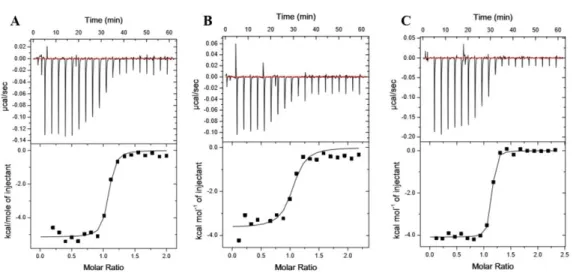
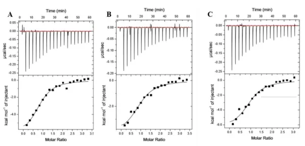
Figure 2.9. ITC binding analysis of KRas WT, KRas G12V and KRas G12V-K5AcK to BRaf RBD displays altered affinities
Representative isotherms of Ras:BRaf RBD binding experiments conducted using isothermal titration calorimetry (ITC). Either 150 μM or 200 μM Ras proteins were titrated into Raf RBDs at a starting molar ratio of 1:10. Isotherms are shown for A. KRas WT, B. KRas G12V and C. K5 acetylated KRas G12V binding to BRaf RBD. Experiments were conducted as described in Figure 2.8. Calculated affinities of 37.3 ± 5.7 nM, 223 ± 21.94 nM and 49.2 ± 12.62 nM correspond to A. KRas WT, B. KRas G12V and C. K5 acetylated KRas G12V binding to BRaf RBD, respectively. Compiled data can be seen in Table 2.3 where data is represented
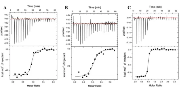
Figure 2.10. ITC binding analysis of KRas WT, KRas G12V and KRas G12V-K5AcK to CRaf RBD displays altered affinities
Representative isotherms of Ras:CRaf RBD binding experiments conducted using isothermal titration calorimetry (ITC). Either 150 μM or 200 μM Ras proteins were titrated into Raf RBDs at a starting molar ratio of 1:10. Isotherms are shown for A. KRas WT, B. KRas G12V and C. K5 acetylated KRas G12V binding to CRaf RBD. Experiments were conducted as described in Figure 2.8. Calculated affinities of 60.6 ± 6 nM, 344 ± 38.58 nM and 21.75 ± 10.71 nM correspond to A. KRas WT, B. KRas G12V and C. K5 acetylated KRas G12V binding to CRaf RBD, respectively. Compiled data can be seen in Table 2.3 where data is represented
Figure 2.11. ITC binding analysis of KRas WT, KRas G12V and KRas G12V-K5AcK displays no difference in affinity to PI3Kα RBD
Representative isotherms of Ras:PI3K RBD binding experiments conducted using isothermal titration calorimetry (ITC). Either 150 μM or 200 μM Ras proteins were titrated into Raf RBDs at a starting molar ratio of 1:15. Isotherms are shown for A. KRas WT, B. KRas G12V and C. K5 acetylated KRas G12V binding to PI3K RBD. Experiments were conducted as described in Figure 2.8. Calculated affinities of 2.39 ± 0.010
M, 2.0 ± 0.271 M and 2.6 M correspond to A. KRas WT, B. KRas G12V and C. K5 acetylated KRas G12V
binding to PI3K RBD, respectively. Compiled data can be seen in Table 2.3 where data is represented in replicate ± standard error (N=2 for Ras WT and Ras G12V, acetylated protein is N=1).
Acetylation of K5 causes sidechain reorientation and altered dynamics in GDP- and GTP-bound protein
While K5 of Ras extends into the binding interface for regulatory and effector proteins, it is not noted to make any direct contacts (128). In the GDP-bound form, the K5 sidechain has been suggested to interact with SWII residues T74 and Y71 (128). Acetylation of K5 would likely disrupt these contacts, as is indicated by the mild change in thermal stability identified in
Figure 2.7. In order to understand how acetylation alters intrinsic Ras protein activity, we
conducted 200 ns Molecular Dynamic (MD) simulations of Mg+2-GDP bound Ras proteins and
Mg+2-GTP bound Ras WT, Ras G12V and K5 acetylated (K5AcK) Ras G12V. Trajectories were subjected to clustering analysis using Gromacs (137), and the centroids of the most populated, lowest energy clusters for each was examined using PyMol 2.1 (Schrödinger, LLC). Results from this analysis (Figure 2.12) indicate that acetylation causes reorientation of K5, altering contacts with residues in SWII and β2. Throughout the simulations, the lysine 5 backbone carboxyl oxygen and amide can be seen forming polar contacts with the backbone amides of Gly77 and Glu76 and the C oxygen of Glu76. Acetylation of Ras wild type results in the formation of a polar contact with Asp54 in both the GDP- and GTP- bound forms (Figure 2.12B,F), where the Asp54 contact is only identified for Ras WT bound to GTP (Figure 2.12E). Similar to Ras wild type protein, lysine 5 in Ras G12V forms backbone contacts with the
the lysine 5 sidechain and Thr74 in any models (polar contact distance cutoff of 3.6Å). Since the backbone of lysine 5 forms several contacts with the highly dynamic switch II region, it is possible that acetylation may result in altered protein dynamics.
Figure 2.12 Lysine 5 acetylation causes reorientation of switch II and β2 contacts.
Lysine 5 contacts as identified from the lowest energy structure resulting from MD simulations and clustering analysis. Sidechains are shown as sticks. Ras wild type K5 (grey), wild type acetylated K5 (green), Ras G12V K5 (blue) and acetylated Ras G12V K5 (orange) models are shown in both GDP-bound (A-D) and GTP-bound (E-H) states. The backbone carboxyl oxygen and amide from backbone contacts with the switch II residues Gly77 and Glu76. Sidechain contacts are also note with Asp54 of β2. Mutation or acetylation results in reorientation of these contacts.
Figure 2.13 Cα backbone fluctuations calculated throughout the MD simulation demonstrate that acetylation stabilizes the Ras WT GDP- and GTP-bound structures
Cα backbone fluctuations throughout the course of the MD trajectory are mapped onto the structure of GDP- or GTP-bound Ras proteins. Fluctuations are labeled from least to most severe by color and size. Ras wild type and Ras wild type acetylated K5 GDP-bound are show in A and B respectively. Ras G12V and Ras G12V acetylated K5 GDP-bound are show in C and D respectively. Ras wild type and Ras wild type acetylated K5 GTP-bound are show in E and F respectively. Ras G12V and Ras G12V acetylated K5 GTP-bound are show in G and H respectively. In wild type Ras, acetylation (B and D vs A and C) appears to minimize dynamic fluctuations of SWII in particular. RasG12V proteins display less dynamic fluctuations as a whole and changes due to acetylation are minimal (E-H).
Acetylation of K5 in KRas fosters enhancement of the electrostatic network between Ras and Raf RBD
proteins to interact with the Raf RBD. As Raf RBDs interact with Ras proteins primarily through SWI (16),(56), it is not immediately clear how acetylation could impact Raf RBD binding. The interaction interface between Ras and Raf is highly electrostatic in nature (16),(42),(51), and mutation to any of the critical binding residues significantly alters or ablates binding (43). To understand how acetylation at K5 is capable of altering the affinity of Ras proteins to Raf RBDs,
we conducted 500 ns Molecular Dynamic (MD) simulations of Mg+2-GTP bound Ras WT, Ras G12V, K5 acetylated (K5AcK) Ras WT and K5 acetylated (K5AcK) Ras G12Vin complex with CRaf RBD. Analysis of these simulations was completed as described previously. Initial findings demonstrated that the acetyl sidechain of K5 reorients toward the effector interface (Figure 2.14), forming a novel electrostatic contact between the sidechain acetyl oxygen of K5AcK and the CRaf R67 guanidino NH sidechain. Mutation to either alanine or leucine at this site (R67) in the CRaf RBD dramatically alters binding to Ras proteins, indicating the importance of these electrostatic interactions in Ras:Raf RBD binding (43). Based on the analysis of all centroids, the distance (CRaf R67 NH to Ras K5 acetyl oxygen) is in the range of ~3 Å, which is indicative of an energetically favorable electrostatic interaction (138)–(140). The relative distance for Ras wild type is longer (~9 Å), possibly resulting from the repulsive nature of the positively charged lysine and arginine sidechains. This finding led us to further investigate the electrostatic network of binding interactions between Ras and the Raf RBD. Strikingly, we were able to identify a reordering and increased electrostatic network formation in Ras due to acetylation at K5 (Figure 2.15A-D). Several new electrostatic contacts were able to be identified due to acetylation
involving Ras residues AcK5, Glu31, Glu37 and Asp54, which appear to be largely
similar trend is also observed (data not shown). Taken together, the formation of novel
Figure 2.15 Reordering and strengthening of the electrostatic network of the Ras:Raf binding interface due to lysine 5 acetylation.
Novel electrostatic contacts and networks are identified due to acetylation at K5. Identified residues for Ras WT and acetylated Ras WT are noted in A and B, respectively. Distances noted are the minimum distance
Figure 2.16 Reorientation of critical α-helical and -strand pairing binding interfaces due to mutation or acetylation are identified in Molecular Dynamic simulations
In final structural models of Ras:Raf RBD complexes, large structural rearrangements can be identified due to K5 acetylation. Relative to Ras WT protein (A, grey), K5 acetylation alters critical -helical and -strand pairing networks for Ras binding to the Raf RBD (B).
Dramatic rearrangement of the binding interface between Ras and the Raf RBD have been identified in our MD simulations. However, as these are static structures and Ras proteins are known to be highly dynamic in nature, we calculated the RMSF fluctuations of each Cα backbone carbon throughout each trajectory. This will provide insight into how protein dynamics may play a role in Raf RBD binding. Consistent with the structural perturbations identified in
between Ras and Raf RBD. This is consistent with the formation of a very stable Ras:Raf RBD complex and is reflected in the increased binding affinity of acetylated Ras G12V to the Raf RBDs (Figure 2.8). Here, we have identified the formation of a larger and more interconnected electrostatic binding network due to protein acetylation, which would support the increased affinity observed for acetylated protein to the Raf RBDs and increased MAPK cellular signaling.
Figure 2.17. Altered conformational dynamics in the Ras:Raf RBD complex due to mutation or acetylation identified in molecular dynamics simulations.
Discussion
Ras proteins have remained elusive drug targets for more than 30 years (24). However, small molecules that target the mutational status of Ras proteins are currently in clinical trials for the treatment of Ras G12C specific cancers (clinical trial identifier: NCT03785249 and
NCT03600883). As direct targeting strategies have been largely ineffective, new approaches to target Ras in cancers focus on using indirect strategies including inhibiting Ras association with effector proteins and inhibiting the ability of Ras proteins to associate with the cellular
membrane (83). Drugs that target post-translational modifications are gaining much interest as novel anti-cancer therapeutics. Histone deacetylase inhibitors (HDACi) have demonstrated success in the treatment of many non-solid tumors (96),(110)–(112). However, as the class I HDACi, Entinostat and other pan-HDAC inhibitors have been primarily studied in the context of hematologic (blood) cancers (142), it is unclear why HDACi have not proven to be clinically viable monotherapies in Ras-driven solid tumors (106),(115).
therapeutic strategy in Ras-driven CRC. Surprisingly, we were able to determine that Ras is a direct target of Entinostat treatment. In CRC cells Entinostat caused Ras acetylation and fostered protein hyper-activation and preferential signaling through the Ras/MAPK mediated pathway.
We were able to discover that Ras proteins are acetylated in colorectal cancer (CRC) cells at a novel location, K5 due to treatment with the class I HDACi, Entinostat (Figure 2.2). This data represents a novel finding that Ras is acetylated at a never-before described location, K5, due to HDACi treatment. Using a genetic code expansion technique to generate acetylated Ras proteins, we were able to determine that acetylation does not severely impact the innate functions of Ras proteins. The ability of Ras proteins to bind and cycle nucleotides intrinsically or in the presence of modulatory proteins was not significantly altered due to acetylation (Figure 2.6). This is not surprising as K5 is not noted to play any role in binding or coordinating nucleotides, nor is it noted to interact with the Ras modulatory proteins, GEFs and GAPs. However, in Raf RBD pulldown assays using colorectal cancer (CRC) cells, HDACi treatment led to an increase in the GTP-bound population of cells (Figure 2.3). Consistent with this, HDACi treatment in wild type and G12V oncogenic CRC cells caused increased MAPK signaling (Figure 2.4), while no changes in PI3K signaling were observed. This was supported by our findings of increased binding affinity of acetylated proteins to the Raf RBDs most striking in the case of oncogenic G12V, where complete restoration to wild-type affinity was observed (Figure 2.8). Mutation nor acetylation resulted in alterations in binding affinity to the PI3Kα RBD (Figure 2.8).