The Association Between Quadriceps Activity and Loading Variables During Walking Gait in Individuals with ACL-R
By Andrew Allen
Senior Honors Thesis
Department of Exercise and Sport Science University of North Carolina at Chapel Hill
April 18, 2016
Chapter 1: Introduction
The anterior cruciate ligament (ACL) is the most frequently disrupted knee ligament,
injured at the highest incidence among physically active individuals under the age of 30.1 Surgical reconstruction of the ACL aims to increase knee stability, reduce the chance of further
injury, and facilitate the return to normal activities and sports. However, ACL reconstruction
(ACL-R) does not restore the knee to its pre-injury state,2 and is associated with a substantial increase in the risk of early onset knee osteoarthritis (OA).3-8 Among a cohort of 122 male soccer players with ACL-R, von Porat et al.8 found that 95 (78%) showed radiographic signs of knee OA 14 years after the initial injury. Culvenor et al.9 reported considerable joint disease in one-third of individuals within one year following ACL-R. Similarly, recent findings suggest
cartilage changes indicative of OA during the first two years after ACL injury.10 In young adults, joint injury is the most common risk factor for OA and can lead to its development prior to
patients reaching 50 years of age.11 Approximately 12% of symptomatic OA of the hip, knee, and ankle arises from joint injury and is thus characterized as post-traumatic osteoarthritis
(PTOA).12 PTOA affects 5.6 million individuals in the United States and creates a financial burden of more than $3 billion in direct healthcare costs.11 With incidences of ACL-R historically rising,13 identifying factors that contribute to knee OA development that can be targeted via intervention is critical.
Knee OA progression is provoked by biomechanical forces that cause structural joint
deterioration.14 Ground reaction forces are the primary external contributors to knee joint loading during human movement and can reach magnitudes up to three times body weight.15,16 During gait, muscles and trabecular bone have been assumed to counteract most of the external forces at
a small fraction of the loading.17 Healthy articular cartilage provides a low-friction surface at the articulating surfaces of bones to allow for free movement of diarthrodial joints, while also
distributing loads to minimize bony stress.18 Eccentric quadriceps activity is the primary
mechanism of force attenuation at the knee joint. Thus, when the quadriceps are impaired, more
force attenuation is required from the other connective tissues. Chronic quadriceps dysfunction
following ACL injury has been well documented19,20 and can persist for several years, potentially impeding the ability to absorb impact loading and contributing to the development of knee
OA.21,22
Quadriceps dysfunction has been associated with impulsive, or high rate, loading.22,23 Impulsive loading produces microfractures in the trabecular and subchondral bone, subsequently
increasing bone stiffness during remodeling.24 The increase in bone stiffness decreases force attenuation capacity, facilitating cartilage break down and joint degeneration.24 Mikesky et al.25 reported that healthy women with weaker quadriceps displayed greater loading rates during gait.
Appropriate quadriceps activation prior to heel strike (HS) is critical for attenuation of impact
loading,17 as it is essential for decelerating the limb. Diminished quadriceps activity is thus characteristic of impulsive loading.23,25 Multiple studies support the notion that diminished quadriceps function leads to impulsive loading during gait,23,26-28 which potentially contributes to OA development and progression. There is further evidence to suggest the existence of an
independent relationship between rate of loading and joint degeneration, regardless of the peak
magnitude of the load.26
Near the end of the swing phase, muscles control the lower limb in preparation for
ground contact. In order to decelerate the lower limb prior to HS, the quadriceps are normally
associated with lesser loading rates.22,23 Healthy individuals exhibiting impulsive loading have delayed muscular activation compared to those with normal gait patterns, and rely on firm
contact with the ground to decelerate the limb rather than muscle activity.22 While timing of this activation is key, there also must be significant activation amplitude of the quadriceps to
effectively slow the lower limb during its decent towards HS. Quadriceps activation deficits
following ACL-R likely contribute to knee OA development, as the alterations of muscle forces
affect natural loading conditions during locomotion.21,29,30 Lower activity of the vastus medialis and rectus femoris prior to HS is linked to impulsive loading in individuals with radiographic,
but asymptomatic, knee OA.17 Furthermore, individuals with quadriceps weakness resulting from ACL-R tend to adopt quadriceps avoidance gait patterns.21 Palmieri-Smith et al.31 showed that lower preparatory activity of the quadriceps resulted in greater loading at the knee by increasing
peak GRF, decreasing knee flexion angles, and decreasing knee extension moments. Thus,
altered quadriceps activation timing and amplitude may contribute to impulsive loading that
could lead to joint degeneration following ACL-R.
Quadriceps dysfunction following ACL-R may contribute to the development of OA due
to the inability of the muscles to attenuate impact forces. Both the timing and amplitude of
quadriceps activity early in the gait cycle appear to be important factors in decreasing loading
rates. Because gait is a repetitive and perpetual task, slight changes in loading rates may have
substantial effects on the articular cartilage of the knee joint. However, the influence of
quadriceps activation characteristics on impulsive loading during gait has yet to be evaluated in
individuals with ACL-R. Therefore, the purpose of this investigation was to evaluate associations
between the timing and amplitude of quadriceps EMG activity and impulsive loading during
Research Questions
• What is the relationship between the onset of quadriceps activity (EMG) relative to heel strike and vertical ground reaction force (vGRF) loading rate and magnitude during the first 50% of the stance phase during walking gait in individuals with ACL-R?
o H1: Earlier onset of quadriceps activity will be associated with lower vGRF loading rate and peak vGRF.
• What is the relationship between the amplitude of quadriceps activity prior to heel strike and vGRF loading rate and magnitude during the first 50% of the stance phase during walking gait in individuals with ACL-R?
o H2: Greater quadriceps activity prior to heel strike will be associated with lower vGRF loading rate and peak vGRF.
• What is the relationship between the amplitude of quadriceps activity during the load acceptance phase of walking gait and vGRF loading rate and magnitude during the first 50% of the stance phase in individuals with ACL-R?
Chapter 2: Literature Review ACL Injury and Epidemiology
Approximately 200,000 ACL injuries occur annually in the United States,32 with over 50% of these traumas sustained by young athletes between the ages of 15 and 25 years.33 ACL reconstruction (ACL-R) is by far the most common treatment strategy, with over 90% of patients
opting for surgical reconstruction as opposed to rehabilitation alone.32 For each patient undergoing ACL-R, Mather et al.32 estimates a lifetime cost to society of $38,000. Therefore, annually, the total cost to society for ACL-R in the US amounts to $7.6 billion. Between the
years 1994 and 2006, the total number of ACL reconstruction procedures increased by 50%,
largely a result of the increase in procedures in patients younger than 20 years of age.13 Published data does not support the idea that ACL-R is able to fully restore the knee to its
preoperative state. In a 15-year follow-up study comparing individuals treated with ACL-R to
those who were treated with a conservative rehabilitation program, nearly 60% of individuals in
both groups were classified as abnormal or severely abnormal based on knee rotational
instability, extension deficits, flexion deficits, and radiological OA grade.4 It has been reported that only 31% of individuals regain normal gait patterns after ACL-R.34 While ACL-R does successfully limit anteroposterior translation of the tibia, it does not adequately limit tibial
rotation.35 Importantly, less knee extension during gait and muscular weakness have been documented in individuals following ACL-R.36 Changes in knee joint kinematics and altered muscle function during gait likely put individuals at risk of developing OA following ACL-R.
Numerous studies have documented a heightened risk of knee OA following ACL-R.3-10 In fact, degenerative changes in cartilage observable via MRI have been reported as early as one year
radiographic knee OA was detected in 71% of individuals, with 24% showing moderate or
severe radiographic OA.37 In addition to the high costs associated with ACL injury, this increased risk of OA development causes an even greater healthcare burden.
Knee Osteoarthritis Epidemiology
In the United States, approximately 27 million people are affected by OA.38 This amounts to an aggregate annual healthcare expenditure of $185.5 billion,39 at an average cost of $5,700 per year for each individual.40 However, while the financial burden placed on those affected by OA is notable, the disabling effects of the disease are even more substantial. As knee OA is a
disease of a weight-bearing joint, it has great impact on function, and often results in difficulty
performing activities of daily living such as walking, stair climbing, bathing, or using the
bathroom.41 In fact, knee OA accounts for difficulty climbing stairs and walking more than any other disease.42 Nearly 45% of individuals with symptomatic knee OA have at least some difficulty walking a quarter mile, and 42% have extreme difficulty or are unable to perform
activities that require kneeling or crouching. Furthermore, 15% of individuals with knee OA use
an assistive device to walk.43 Surgery is a common treatment option, but can be an emotionally stressful event as well as a potentially dangerous form of therapy. In 2009, there were over
620,000 hospital discharges associated with total knee replacements in the United States,44 with OA being the most common cause for treatment.41 The effects of knee OA are multidimensional, and are not limited to physical disability. A study by Salaffi et al.45 showed that the disease significantly affects individuals’ mental and social health. The pervasive effects of this illness
individuals who are known to be at higher risk, such as those suffering injury to the structures of
the knee.
Post-traumatic Osteoarthritis
OA is defined as a multifactorial disease, mainly resulting from the degeneration of
articular cartilage. Under normal, healthy conditions, articular cartilage is smooth and lubricates
the articular surfaces of bones to provide for low-friction joint movements. It additionally acts to
distribute mechanical loading, protecting the bone from damaging levels of stress. Because it is
avascular, there is little the body can do to self-repair or regenerate articular cartilage following
injury. Symptomatic OA usually includes, but is not limited to, joint pain, inflammation, and
limited range of motion.18 The Kellgren-Lawrence scale is the most widely used OA
classification and has been adopted by the World Health Organization as standard for evaluation
of OA for epidemiologic studies. As it is based on the presence of osteophytes and joint space
narrowing, the scale allows for good comparison between different types of joints.
Kellgren-Lawrence scores range from 1 – 4, with “1” representing minute osteophyte of doubtful
significance, “2” representing definite osteophyte, but joint space unimpaired, “3” representing
moderate diminution of joint space, and “4” representing greatly impaired joint space and
subchondral sclerosis.46 In addition to the Kelgren-Lawrence scale, alternative methods of OA diagnosis, such as MRI, have also been utilized and supported as providing reliable and valid
classifications.47
In contrast to idiopathic OA in which the cause is non-specific, post-traumatic
osteoarthritis (PTOA) results from joint trauma associated with impact loading of the synovial
Following ACL-R, the risk for developing OA in the injured knee has been found to be 57%,
compared to only 18% in the contralateral knee.48 In the United States, lower-extremity PTOA generates an economic burden of $11.79 billion annually. Furthermore, approximately 10% of all
incidences of symptomatic knee OA result from joint trauma as opposed to natural means. Of
particular concern, individuals with PTOA of the knee are 10.4 years younger on average than
individuals with idiopathic OA, and must face the debilitating effects of OA for a greater portion
of their lives.12 As the literature reveals a strong correlation between joint injury and OA, effective preventative interventions must be developed and implemented following injury,
regardless of the presence of symptoms. By the time PTOA individuals are symptomatic, there is
already irreversible damage to the articular cartilage.49
Gait Biomechanics Adaptations Following ACL-R
ACL-R following joint injury does not seem to restore the knee to its pre-injury state, as
investigations have revealed adaptations to gait biomechanics that may predispose the articular
cartilage to future degeneration. It has been reported in the literature that ACL-R knees are
deficient in knee extension36 and exhibit less hip and knee flexion during gait.50 In an
investigation of gait kinematics, 85% of subjects displayed more external rotation of the tibia in
the ACL-R knee compared to the contralateral knee at HS and during the midstance, terminal
extension, and toe off phases of gait.51 Papannagari et al.52 found that reconstructed knees showed more anterior tibial translation and external rotation during weight bearing tasks. Other
studies have contradictorily noted more internal rotation in ACL deficient knees during gait.53 Furthermore, some literature reports significantly reduced peak knee extension at HS in the
the femoral and tibial articular cartilage align when the knee is extended, as it is prior to HS. It
has been suggested that the locations of the regions of thick articular cartilage are tibiofemoral
adaptations to help alleviate the impact loading that occurs at HS.55 As such, kinematic changes following ACL-R result in a shift in the areas of contact of the articular cartilage such that there
is increased loading in areas of cartilage not conditioned for extensive weight bearing.56 This shift in areas of cartilage contact provides a potential explanation for the relationship between
ACL-R and OA development.
In addition to the gait kinematic alterations that have been reported following ACL-R, the
literature shows several kinetic changes as well. During a stair climbing exercise, individuals
with ACL-R relied more heavily on hip extension moments, as they displayed a 22% decrease in
peak knee extension moments. Such alterations have been suggested to result from greater
quadriceps and hamstring co-contraction, subjecting the articular cartilage to increased
compressive loading.57 Additionally, greater knee adduction moments during gait have been observed in the injured and contralateral limbs of ACL-R subjects relative to control subjects.58,59 This is cause for concern as greater external knee adduction moment, a measure of loading in the
medial compartment of the knee, has been reported to highly correlate with OA progression.60 Some studies, however, have found ACL-R individuals to display similar or decreased external
knee adduction moment,61,62 demonstrating the variability of individuals’ gait patterns following ACL-R. The discrepancies in these investigations (i.e. some ACL-R subjects display altered gait
kinetics while others do not) may parallel differential development of knee OA.
Ground reaction forces are indicative of lower extremity loading and are useful proxies
for dynamic joint loading during gait. Specifically, the vertical component of the ground reaction
Chen et al.63 provided evidence that the extent of damage to articular cartilage is dependent on peak stress, stress rate, and loading duration by subjecting canine cartilage to repetitive impacts.
Even when peak stress was low (2.5 MPa) and administered for a duration of only 2 minutes,
damage to the cartilage matrix was observed at high stress rates, suggesting that loading rate is
highly correlated with cartilage degeneration. Furthermore, impact stresses resulted in more
damage than did smoothly arising compressions at the same peak stress level. In a study of
bovine knee articular cartilage, higher rates of loading resulted in matrix damage characterized
by longer fissure length and greater fissure depth compared to low loading rates.64 In damaged articular cartilage, cyclic loading has been shown to propagate fissure length in vitro, suggesting
a potential mechanism of OA progression related to repetitive loading in gait.65 In addition to the mechanical damage to articular cartilage associated with high loading rates, Kurz et al.66 found decreases in the biosynthetic activity of cartilage following high rate impact loading. In the
healthy joint, cartilage responds to low-amplitude cyclic loading with an anabolic response.
Deficient anabolic activity following high rate loading may provide further explanation
regarding the pathogenesis of PTOA.
In a cohort of females, Noehren et al.67 found that average loading rates and initial impact forces during walking and running were significantly higher in the ACL-R group compared to
control. Due to the repetitive and perpetual nature of gait, slight increases in impact loading
could result in significant consequences for the articular cartilage. Because higher impact loading
was found during walking, this suggests that ACL-R individuals experience greater cartilage
loading during daily activities, not just during sport or exercise. Of additional concern is the lack
of between-limb differences, suggesting that gait adaptations following ACL-R could increase
individuals with ACL-R, which found higher levels of impulsive loading in both extremities of
ACL-R individuals compared to both extremities of controls.68 Contrary to studies showing no between-limb differences in loading rates, a more recent study by Blackburn et al.69 found
significantly higher instantaneous loading rates in ACL-R limbs. Likewise, instantaneous loading
rates were higher in individuals characterized as “impulsive loaders,” who commonly displayed
high peaks in vertical ground reaction force at HS. Greater loading rate has been identified as a
common gait characteristic in individuals with knee OA, suggesting a link between ACL-R and
OA development.28 Using MR imaging, Morgenroth et al.26 found an independent relationship between dynamic loading rate and degeneration of the medial knee cartilage in vivo. As evidence
suggests that high loading rates can significantly damage articular cartilage even at small
magnitudes, interventions targeted at preventing increased rates of loading following ACL-R
could inhibit cartilage degeneration and the development of PTOA.
Quadriceps Influence on Loading Rate
During gait, the quadriceps play an important role in controlling the limb prior to HS and
managing contact forces during loading. Eccentric action of the quadriceps is responsible for
controlling knee flexion as a way of attenuating the large magnitude of force that is transferred to
the limb during the load acceptance phase of gait.21 Decreased quadriceps activity during gait may result in higher loading rates due to the diminished utilization of eccentric action to absorb
shock. Jefferson et al.23 showed that inhibition of the quadriceps caused by injecting lidocaine in the femoral nerve invokes impulsive loading in individuals with normal gait patterns. When the
quadriceps were inhibited, subjects displayed increased vertical foot velocity prior to ground
prior to HS as a way of reducing high rate loading. In a group of healthy subjects, Verdini et al.22 found that 100% of subjects with delayed activation of the vastus medialis displayed impulsive
loading during gait. Whereas those with normal gait patterns used quadriceps activity at the end
of the swing phase to decelerate the limb and prepare for ground contact, individuals with
delayed activation likely relied on the ground to abruptly decelerate the limb. Repetitive loading
in this manner may accelerate damage to the articular cartilage. Lewek et al.21 found that
individuals with quadriceps strength deficits after ACL-R exhibited movement patterns similar to
ACL deficient patients. The individuals with weak quadriceps exhibited reduced knee flexion,
thus inhibiting force attenuation during the load acceptance phase. In a cohort of women,
Mikesky et al.25 further emphasized the importance of the quadriceps during gait, reporting much higher loading rates in individuals with weaker quadriceps compared to a group of
strength-trained women. Likewise, in individuals with knee OA, lower preparatory activity of the
quadriceps has been associated with impulsive loading.17 As ACL-R limbs have been observed to display greater magnitude loading rates compared to the contralateral limb,69 diminished quadriceps activity after ACL injury may be a viable target for interventions aimed at preventing
PTOA.
Quadriceps EMG after ACL Injury
Surface electromyography (EMG) is an easy, non-invasive method of quantifying muscle
activity during static or dynamic tasks. Through the use of electrodes, surface EMG produces a
signal based on the sum of the nearby motor unit action potentials (MUAP) that are transmitted
during contraction. While the EMG signal is not equivalent to muscle force, it is indicative of the
of muscle activation as well as the patterns and strength of the neuromuscular signaling for a
given muscle.70
Following ACL-R, studies involving EMG have revealed alterations in quadriceps
activity. In an animal model, altered EMG patterns during the loading phase of gait were directly
associated with transection of the ACL.71 Torry et al.72 observed decreases in quadriceps activity during gait with increasing levels of joint effusion, providing evidence that knee injury is
associated with quadriceps dysfunction. At high levels of knee joint effusion (80 cm3), vastus medialis EMG activity decreased by 25% while rectus femoris activity decreased by 50.4 %.
While the literature concerning quadriceps EMG activity during gait is limited in individuals
with ACL-R, Hurd et al.73 reported significant reductions in EMG activity of the quadriceps during the load acceptance phase of gait in a cohort of ACL-R individuals. This is cause for
concern as individuals with knee OA who display impulsive loading have been found to exhibit
43.5% lower pre-activity of the quadriceps.17 The lack of quadriceps activity prior to HS was associated with peak forces at the start of the loading phase of gait, regardless of walking speed.
Furthermore, it has been reported that individuals with knee OA activate their quadriceps
significantly later than controls during other functional tasks as well, such as stair descent.74 Altered EMG activity of the quadriceps during normal activity may contribute to greater loading
rates and the progression of cartilage degeneration. Because ACL-R individuals display
quadriceps EMG patterns similar to those who suffer from knee OA, decreased quadriceps
EMG Processing during Gait
There is no consensus among the literature regarding the best method for processing
EMG signals. In studies involving EMG during gait, it is common for the signal to be full-wave
rectified (taking the absolute value of the EMG signal) and filtered to produce a linear
envelope.75-78 The linear envelope is a processed form of the raw EMG data that reduces noise to provide more useful results for analysis. However, upon obtaining this more useful output,
methods for determining EMG amplitude and muscle onset are varied. It should be noted that
many investigations fail to provide detailed explanations of the methodology used to evaluate
EMG data, further complicating the selection of an ideal protocol.79 In a previous investigation of gait parameters in ACL-R individuals, Alkjaer et al.75 calculated mean amplitude during the weight acceptance period from 100ms prior to HS to the first peak on the vGRF curve. Theoret et
al.76 quantified muscle activation by calculating the area under the curve of the linear envelope for each participant’s average running cycle (from heel strike to heel strike). Tang et al.78 utilized a different method, calculating root mean square (RMS) values in a 20ms smoothing window
throughout the gait cycle. To determine preparatory activity of the quadriceps, peak RMS EMG
values were obtained for the 200ms interval prior to HS. Furthermore, most EMG data in the
literature is normalized for each individual based on a selected reference activity. Multiple
studies have normalized all signals obtained during gait to the maximal EMG amplitude recorded
during a maximal voluntary isometric contraction for the muscle group of interest.17,75,78 Others, however, have normalized signals to the average of the peak EMG signal among multiple gait
time interval prior to HS. All data was normalized to the average EMG values of the quadriceps
during maximal voluntary isometric contraction.
In regards to the determination of muscle onset, investigations have utilized protocols
based on visual determination, computer assistance, and fully automated algorithms.80 Theoret et al.76 first determined a noise level of the signal by selecting a period of the gait cycle when the muscle was not active, then set a threshold for onset of the contraction at two standard deviations
higher than the resting value. Similarly, Tang et al.78 used an algorithm that determined onset when the signal differed by three standard deviations. To determine the validity of different
methods for determining muscle onset, Hodges et al.79 compared the onset times determined by numerous computer-based algorithms to the onset times determined by an experienced examiner.
The study compared twenty-seven different combinations of parameters, with each algorithm
differing in regards to the degree of smoothing (low pass filter), the number of samples assessed
in the sliding window (20, 50, or 100), and the magnitude of the deviation from baseline activity
required to indicate muscle onset (threshold value). Based on the results, multiple algorithms
proved relatively valid, but the most accurate algorithm used a 50 Hz low pass filter, threshold
level of 1 standard deviation above baseline, and a 100-sample sliding window (50ms epoch).
Summary
ACL injury is prevalent in the US and is becoming more frequent among younger
populations. This is of concern as ACL injury, despite reconstructive surgery, is highly
associated with the development of PTOA in the literature. ACL-R does not restore the knee to
its pre-operative state, as the literature reports gait kinematic and kinetic alterations that persist
alterations observed during gait, and dysfunction of the quadriceps specifically may serve as the
mechanism for greater loading rates in ACL-R individuals. Without proper quadriceps activation
timing and activation amplitude both prior to HS and throughout the load acceptance phase of
gait, individuals with ACL-R may be subject to repetitive, impulsive loading. Such loading has
proven to be independently damaging to articular cartilage. Therefore, understanding the
relationship between quadriceps activity and loading rate could divulge a viable target for
Chapter 3: Methods Subjects
Nine individuals were recruited from two local orthopaedic clinics (UNC Department of
Orthopaedics and Triangle Orthopaedic Associates), rehabilitation clinics, the University
population, and the Durham Veterans Affairs Medical Center. Subjects were included if they
were between the ages of 18-35 years and had undergone unilateral ACL-R within 5 years, but
no less than 6 months prior to participation. Exclusion criteria included a history of ACL graft
rupture or revision surgery, a history of neurological disorder, or a history of injury to either leg
within 6 months prior to participation. Furthermore, central activation ratio was assessed to
ensure that subjects possessed deficits in quadriceps function in the ACL-R limb (<95%).
Likewise, the Knee Injury and Osteoarthritis Outcome Score (KOOS) self-report survey was
used to ensure that subjects were functional enough to complete the study requirements. To be
included, subjects were required to score greater than 53.1 on the KOOS Pain subscale, and
greater than 44.9 on the KOOS Symptom subscale. Lastly, subjects had to have been cleared by
a physician for return to physical activity, and currently physically active, participating in at least
20 minutes of physical activity 3 times per week. During the screening session on the first day,
subjects were evaluated for the inclusion criteria and required to read and sign an informed
consent form prior to data collection. (See Table 1 for subject demographics)
Experimental Design
This project was a part of a larger ongoing investigation evaluating the effects of
vibratory stimuli on factors linked to OA in individuals with ACL-R. The overarching
convenience sample of 75 individuals who had undergone ACL-R. It includes assessments of
quadriceps function outcomes (peak voluntary torque, rate of voluntary torque development, and
the central activation ratio), proprioception/sensory outcomes (joint repositioning assessment and
vibratory perception threshold), and gait biomechanics outcomes (kinematics, kinetics, and EMG
variables). Subjects complete the aforementioned assessments prior to and following vibratory
interventions designed to enhance quadriceps activity.
The present investigation was cross-sectional, and evaluated the relationship between
quadriceps activity and loading rates during gait using data obtained from the baseline
assessments. While the larger study required subjects to complete 3 testing visits, the present
study only included data collected during the first 2 visits to the Sports Medicine Research Lab at
the University of North Carolina at Chapel Hill. Data obtained during the screening session on
the first day was used to ensure that subjects met all inclusion criteria. On the second visit,
subjects completed the gait biomechanics assessment.
Gait Biomechanics Assessment
During the screening session, subjects performed 5 walking trials along a 6-meter
walkway. Subjects walked barefoot at a self-selected pace and gait speed was monitored using
infrared timing gates. Using the 5 trials during the screening session, an average preferred gait
speed was recorded for each participant. Subjects completed 5 walking trials for which gait
speed was within +/- 5% of the preferred speed and the entire foot made contact on each force
plate. Trials that did not meet these criteria were repeated.
Vertical ground reaction forces were measured using two force plates (model 4060,
could be recorded for both limbs in a single trial. Loading rate was assessed using the vGRF data
obtained from the force plates. Ground reaction forces were sampled at 1,200 Hz and low-pass
filtered at 75 Hz. Outcome variables included the peak vGRF during the first 50% of the stance
phase, linear loading rate (slope of the line connecting HS and peak vGRF during the first 50%
of the stance phase), and the peak instantaneous loading rate (1st time derivative). The stance phase was defined as the interval from HS (vGRF ≥ 20N) to toe off (vGRF ≤ 20N). All vGRF
magnitudes and loading rates were normalized to body weight (xBW and xBW/s, respectively).
EMG data was sampled during each of the 5 trials at 1,200 Hz using surface EMG
(DelSys Bagnoli-8; Input Impedance: 1.0 MΩ; EMG100C Amplifiers, CMRR: 110 dB min,
gain: 10000). Preamplified EMG electrodes were placed on the vastus medialis and vastus
lateralis of each limb. The electrode sites were shaved if necessary, and skin was abraded and
cleaned with alcohol to improve the quality of the signal. The raw EMG signal was bandpass
(20-350 Hz) and notch (59.5-60.5 Hz) filtered (4th order zero-phase lag Butterworth filter). Signal smoothing was accomplished using a low pass filter at 10Hz to create a linear envelope.
Quadriceps EMG variables of interest included the onset of activity prior to HS,
preparatory activity of the quadriceps prior to HS, and the amplitude of activity during the
loading phase. The onset of quadriceps activity was defined as the point prior to HS at which the
EMG amplitude exceeds the mean amplitude of a selected 100ms quiet period (quiet mean)
during the gait cycle plus 2 SD for at least 120ms. Quadriceps preparatory activity was
quantified in two ways: firstly, by calculating the mean EMG amplitude from muscle onset to
HS, and secondly, by calculating the mean EMG amplitude during the 100ms prior to HS.
EMG amplitude from HS to the peak vGRF during the first 50% of the stance phase. All mean
EMG amplitudes were normalized to the quiet means for each respective trial.
Statistical Analyses
Partial correlations (Pearson r) were used to evaluate the relationships between each
EMG variable (quadriceps onset, pre-activity, and loading phase activity) and loading variable
(peak vGRF, vGRF linear and instantaneous loading rates) after controlling for gait speed. The
strength of the associations will be interpreted according to the r value, with values from 0.00 to
0.25 representing little or no relationship, 0.25 to 0.50 representing a fair relationship, 0.50 to
0.75 representing a moderate to good relationship, and above 0.75 representing a good to
N Age (years) ± SD
Height (cm) ± SD
Weight (N) ± SD
Time Since Injury (months)
± SD
Time Since Surgery (months)
± SD 9
(2 male, 7 female)
19.7 ± 1.7 171.8 ± 13.3 751.4 ± 288.3 33.1 ± 13.0 30.8 ± 12.6 Table 1 Subject Demographics
Chapter 4: Results
Two subjects were excluded from statistical analysis due to the fact that their EMG
signals contained high levels of noise and did not demonstrate cyclical activity representing gait.
One subject was excluded on the basis of inaccurate vGRF data. Therefore, while nine subjects
completed the gait biomechanics assessment, data from six subjects was used for statistical
analysis. Demographic data for the retained subjects is presented in Table 2. Additionally, due to
high levels of noise in the vastus medialis EMG signals, statistical analysis was only performed
using EMG data from the vastus lateralis.
VL EMG Onset
There was no significant correlation between VL EMG onset and any of the three kinetic
variables: peak vGRF (r = -0.090, p = 0.443), vGRF linear loading rate (r = 0.196, p = 0.376),
and vGRF instantaneous loading rate (r = -0.573, p = 0.156). (See Table 4)
VL Preparatory Activity (Onset to HS)
There was no significant correlation between mean VL EMG amplitude from onset to HS
and any of the three kinetic variables: peak vGRF (r = -0.293, p = 0.316), vGRF linear loading
rate (r = -0.071, p = 0.455), and vGRF instantaneous loading rate (r = -0.637, p = 0.124).
VL Preparatory Activity (100ms prior to HS)
There was a significant negative correlation between mean VL EMG amplitude during
the 100ms prior to HS and vGRF instantaneous loading rate (r = -0.819, p = 0.045). This
indicates that subjects with greater VL activity over the 100ms interval prior to HS demonstrated
lower vGRF instantaneous loading rates (See Figure 2). No significant correlation was found
between mean VL EMG amplitude during the 100ms prior to HS and peak vGRF (r = -0.415, p =
0.244) or vGRF linear loading rate (r = -0.052, p = 0.467). (See Table 4)
VL Loading Phase Activity
There was no significant correlation between mean VL EMG amplitude during the
loading phase any of the three kinetic variables: peak vGRF (r = -0.445, p = 0.226), vGRF linear
loading rate (r = -0.379, p = 0.265), and vGRF instantaneous loading rate (r = -0.142, p = 0.410).
(See Table 4)
N Age (years) ± SD
Height (cm) ± SD
Weight (N) ± SD
Time Since Injury (months) ± SD
Time Since Surgery (months) ± SD 6
(1 male, 5 female)
Peak vGRF vGRF Linear LR vGRF Instantaneous LR
VL Onset -0.090 0.196 -0.573
Preparatory VL
Activity (100ms) -0.415 -0.052 -0.819*
Preparatory VL
Activity (onset to HS) -0.293 -0.071 -0.637
Loading Phase VL
Activity -0.455 -0.379 -0.142
VL EMG Onset (ms before HS) VL Pre-activity from
Onset to HS (% quiet
mean)
VL Pre-activity 100ms before
HS (% quiet mean) VL Loading Phase Activity (% quiet mean) Peak vGRF (xBW) vGRF Linear Loading Rate (xBW/s) vGRF Instantaneous Loading Rate (xBW/s)
111.64 ± 40.83 2.98 ± 0.86 2.99 ± 1.13 5.62 ± 2.57 1.12 ± 0.40 7.80 ± 1.73 62.99 ± 29.40
Partial correlations reflect relationships between the respective quadriceps activity
measures and loading variables after accounting for the influence of gait speed. * p < 0.05 Table 4 Partial Correlations between Quadriceps Activity Measures and
Chapter 5: Discussion
The primary finding of this study was that greater activity of the VL during the 100ms
prior to HS was associated with lower vGRF instantaneous loading rates in the ACL-R limb
during gait. This is consistent with previous research that demonstrates the importance of the
quadriceps in attenuating impact forces during gait,23 and builds on these findings by clarifying how muscle activity alters loading during gait. Activity of the VL during the 100ms prior to HS
was not associated with peak vGRF or linear loading rate. Other measures of quadriceps activity,
including timing of EMG onset, preparatory activity from onset to HS, and VL activity during
the loading phase, were not associated with peak vGRF, linear loading rate, or instantaneous
loading rate. Thus, findings from this study are only partially consistent with the experimental
hypotheses. It was hypothesized that earlier onset of the quadriceps, greater quadriceps activity
during the loading phase and greater preparatory activity both during the 100ms preceding HS
and from onset to HS would result in lower peak vGRF, linear loading rate, and instantaneous
loading rate. Our findings suggest that quadriceps activity immediately prior to HS (specifically
during the preceding 100ms) influences vGRF instantaneous loading rate, and may be an
important target for interventions aimed to reduce impulsive loading following ACL-R.
Blackburn et al.69 found that peak vGRF and linear loading rate did not differ between the ACL-R limb and the contralateral limb during gait, but instantaneous loading rate was
significantly higher in the ACL-R limb. Additionally, instantaneous loading rates, but not linear
loading rates or peak vGRF, were significantly greater in individuals classified as impulsive
loaders compared to normal loaders. This suggests that changes in loading associated with
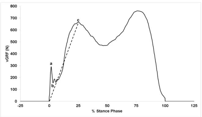
ACL-R may not be detectible via measures such as vGACL-RF linear loading rate and peak vGACL-RF. Linear
calculation is based on data from HS to peak vGRF, and is not influenced by the sharp rise in
vGRF immediately following HS (represented by point “a” in Figure 3). Likewise, peak vGRF is
not influenced by the sharp rise in vGRF immediately following HS. Peak instantaneous loading
rate is a measure of the maximum slope at any point on the vGRF curve, which usually occurs
during the first 20ms following HS.69 Thus, instantaneous loading rate provides a representation of kinetics directly following HS, and is a better indicator of the impulsive loading thought to
damage articular cartilage. As such, our finding that greater preparatory activity of the
quadriceps is associated with lesser instantaneous loading rates supports that notion that
appropriate quadriceps activity is critical for regulating impulsive loading that is hypothesized to
lead to knee OA.
The lack of association between onset of quadriceps activity and the loading variables is
inconsistent with research showing that delayed muscle activation may result in increased impact
forces.22 A potential explanation for this discrepancy could be due to difficulties identifying onset times in the present study. The method for identifying muscle onset utilized a mean EMG
amplitude from a 100ms quiet interval during the gait trial. Onset was identified when EMG
amplitude exceeded the mean plus two standard deviations for a burst duration of 120ms at any
point following the selected start time for the quiet interval. In many trials, the EMG signal was
noisy prior to HS, making it difficult to select an interval of 100ms where the muscle appeared to
be quiet. Therefore, the quiet mean was likely artificially high in some trials, compromising the
validity of the detected onset times. Additionally, some level of subjectivity was introduced
when detecting onset. Depending on which quiet period was selected, the onset time could vary
significantly. The limitation in reliably and accurately detecting quadriceps onset affected the
measure of preparatory quadriceps activity from onset to HS as well. This method of quantifying
preparatory muscle activity was dependent on the onset time, and is likewise limited in its
validity. Future studies should alter onset detection methods, potentially establishing the
threshold level based on a mean EMG amplitude during a quiet stance period instead using a
quiet period from each gait trial.
Another important finding of this study was that quadriceps activity during the loading
phase was not associated with peak vGRF, linear loading rate, or instantaneous loading rate.
Loading phase activity quantifies quadriceps activity from HS to the peak vGRF during the first
50 percent of the stance phase. One possible explanation for the observed results is that
preparatory activity of the quadriceps is more important in attenuating loading rates than activity
following HS. However, loading phase activity is an average EMG amplitude over an
approximately 200ms interval, from HS to peak vGRF.69 Since the majority of loading phase activity occurs after the time point when maximal instantaneous loading rates occur, it is not
surprising that these two variables are not significantly correlated.
Previous research by Liikavainio et al.17 is consistent with the findings of the present study, showing that preparatory activity of the quadriceps during the 100ms preceding HS
influences loading during walking gait. Liikavainio et al. found that lower pre-activity of the
quadriceps was associated with impulsive loading in individuals with osteoarthritis. Calculating
preparatory activity in this way, as opposed to utilizing muscle onset, avoids the bias and
detection errors associated with identifying muscle onset, potentially explaining the correlation
outcomes when using this method. Another potential explanation is that activity within 100ms
prior to HS may have a greater influence on loading immediately following HS compared to
quadriceps activity earlier in the swing phase.
It should be noted that several moderate correlations were observed that did not achieve
significance, potentially due to the small sample size of the present study. Greater instantaneous
loading rate was moderately correlated (i.e. r ≥ 0.50) with later onset of the quadriceps (r =
-0.573, p = 0.156) and lower preparatory activity of the quadriceps from onset to HS (r = -0.637,
p = 0.124).81 A larger sample size and improved methodology for reliably identifying muscle onset could result in significant correlations between these variables. If future observations were
to identify greater instantaneous loading rates to be significantly correlated with later onset and
lower preparatory activity of the quadriceps from onset to HS, this would complement the
As knee osteoarthritis is evident as early as one year following ACL-R,9 it is imperative to identify and address the underlying deficiencies contributing to cartilage breakdown. Evidence
suggests that high rate loading is independently damaging to articular cartilage,26,63 and can inhibit its biosynthetic activity.66 Noehren et al.67 found that individuals with ACL-R had higher average loading rates during gait compared to controls, and Blackburn et al.69 found that
instantaneous loading rates were significantly greater in the ACL-R limb versus the contralateral
limb. The present findings suggest that lower quadriceps activity 100ms prior to HS in ACL-R
limbs is correlated with higher instantaneous loading rates. Therefore, future research should
evaluate interventions aimed to increase preparatory activity of the quadriceps during gait in
individuals with ACL-R. In a previous investigation, Verschueren et al.82 used vibration of the patellar tendon to significantly increase quadriceps EMG activity during gait in healthy
individuals. By attaching a cylindrical vibrator to the tendon using a rubber band, mean EMG
across the complete gait cycle was found to increase by 69% in the rectus femoris and 65% in the
vastus lateralis when compared to mean EMG values during trials with no vibration. Similarly,
Cotey et al.83 observed increased quadriceps activity during gait in healthy individuals and individuals with neurological injury through the use of vibration. By strapping the vibration
device over the anterior thigh, mean EMG area increased by 96% in the rectus femoris and by
211% in the vastus lateralis compared to the control condition (no vibration). Interestingly, phase
specific increases in quadriceps activity could be invoked when the vibration was only applied to
specific periods of the gait cycle. Mean EMG area in the vastus lateralis increased by 123%
during the swing phase and by 100% during the transition into stance phase when phasic
vibration was applied to healthy individuals. Further research should investigate whether similar
References
1. Friel NA, Chu CR. The role of ACL injury in the development of posttraumatic knee osteoarthritis. Clin Sports Med. 2013;32(1):1-12.
2. Dare D, Rodeo S. Mechanisms of post-traumatic osteoarthritis after ACL injury. Curr Rheumatol Rep. 2014;16(10):448.
3. Kessler MA, Behrend H, Henz S, Stutz G, Rukavina A, Kuster MS. Function,
osteoarthritis and activity after ACL-rupture: 11 years follow-up results of conservative versus reconstructive treatment. Knee Surg Sports Traumatol Arthrosc. 2008;16(5):442-448.
4. Streich NA, Zimmermann D, Bode G, Schmitt H. Reconstructive versus non-reconstructive treatment of anterior cruciate ligament insufficiency. A retrospective matched-pair long-term follow-up. Int Orthop. 2011;35(4):607-613.
5. Oiestad BE, Holm I, Aune AK, et al. Knee function and prevalence of knee osteoarthritis after anterior cruciate ligament reconstruction: a prospective study with 10 to 15 years of follow-up. Am J Sports Med. 2010;38(11):2201-2210.
6. Salmon LJ, Russell VJ, Refshauge K, et al. Long-term outcome of endoscopic anterior cruciate ligament reconstruction with patellar tendon autograft: minimum 13-year review.
Am J Sports Med. 2006;34(5):721-732.
7. Lohmander LS, Ostenberg A, Englund M, Roos H. High prevalence of knee
osteoarthritis, pain, and functional limitations in female soccer players twelve years after anterior cruciate ligament injury. Arthritis Rheum. 2004;50(10):3145-3152.
8. von Porat A, Roos EM, Roos H. High prevalence of osteoarthritis 14 years after an anterior cruciate ligament tear in male soccer players: a study of radiographic and patient relevant outcomes. Ann Rheum Dis. 2004;63(3):269-273.
9. Culvenor AG, Collins NJ, Guermazi A, et al. Early knee osteoarthritis is evident one year following anterior cruciate ligament reconstruction: a magnetic resonance imaging evaluation. Arthritis Rheumatol. 2015;67(4):946-955.
10. Eckstein F, Wirth W, Lohmander LS, Hudelmaier MI, Frobell RB. Five-year followup of knee joint cartilage thickness changes after acute rupture of the anterior cruciate
ligament. Arthritis Rheumatol. 2015;67(1):152-161.
11. Roos EM. Joint injury causes knee osteoarthritis in young adults. Curr Opin Rheumatol.
2005;17(2):195-200.
13. Mall NA, Chalmers PN, Moric M, et al. Incidence and trends of anterior cruciate
ligament reconstruction in the United States. Am J Sports Med. 2014;42(10):2363-2370.
14. Englund M. The role of biomechanics in the initiation and progression of OA of the knee.
Best Pract Res Clin Rheumatol. 2010;24(1):39-46.
15. Shelburne KB, Torry MR, Pandy MG. Contributions of muscles, ligaments, and the ground-reaction force to tibiofemoral joint loading during normal gait. J Orthop Res.
2006;24(10):1983-1990.
16. Taylor WR, Heller MO, Bergmann G, Duda GN. Tibio-femoral loading during human gait and stair climbing. J Orthop Res. 2004;22(3):625-632.
17. Liikavainio T, Isolehto J, Helminen HJ, et al. Loading and gait symmetry during level and stair walking in asymptomatic subjects with knee osteoarthritis: importance of quadriceps femoris in reducing impact force during heel strike? Knee. 2007;14(3):231-238.
18. Becerra J, Andrades JA, Guerado E, Zamora-Navas P, Lopez-Puertas JM, Reddi AH. Articular cartilage: structure and regeneration. Tissue Eng Part B Rev. 2010;16(6):617-627.
19. Hopkins JT, Ingersoll CD. Arthrogenic muscle inhibition: A limiting factor in joint rehabilitation. J Sport Rehabil. 2000;9(2):135-159.
20. Ingersoll CD, Grindstaff TL, Pietrosimone BG, Hart JM. Neuromuscular consequences of anterior cruciate ligament injury. Clin Sports Med. 2008;27(3):383-404, vii.
21. Lewek M, Rudolph K, Axe M, Snyder-Mackler L. The effect of insufficient quadriceps strength on gait after anterior cruciate ligament reconstruction. Clin Biomech (Bristol, Avon). 2002;17(1):56-63.
22. Verdini F, Marcucci M, Benedetti MG, Leo T. Identification and characterisation of heel strike transient. Gait Posture. 2006;24(1):77-84.
23. Jefferson RJ, Collins JJ, Whittle MW, Radin EL, O'Connor JJ. The role of the quadriceps in controlling impulsive forces around heel strike. Proc Inst Mech Eng H.
1990;204(1):21-28.
24. Radin EL, Rose RM, Paul IL. Role of Mechanical Factors in Pathogenesis of Primary Osteoarthritis. Lancet. 1972;1(7749):519-&.
25. Mikesky AE, Meyer A, Thompson KL. Relationship between quadriceps strength and rate of loading during gait in women. J Orthopaed Res. 2000;18(2):171-175.
27. Riskowski JL, Mikesky AE, Bahamonde RE, Alvey TV, 3rd, Burr DB. Proprioception, gait kinematics, and rate of loading during walking: are they related? J Musculoskelet Neuronal Interact. 2005;5(4):379-387.
28. Mundermann A, Dyrby CO, Andriacchi TP. Secondary gait changes in patients with medial compartment knee osteoarthritis - Increased load at the ankle, knee, and hip during walking. Arthritis and Rheumatism. 2005;52(9):2835-2844.
29. Slemenda C, Heilman DK, Brandt KD, et al. Reduced quadriceps strength relative to body weight: a risk factor for knee osteoarthritis in women? Arthritis Rheum.
1998;41(11):1951-1959.
30. Hart JM, Turman KA, Diduch DR, Hart JA, Miller MD. Quadriceps muscle activation and radiographic osteoarthritis following ACL revision. Knee Surg Sports Traumatol Arthrosc. 2011;19(4):634-640.
31. Palmieri-Smith RM, Kreinbrink J, Ashton-Miller JA, Wojtys EM. Quadriceps inhibition induced by an experimental knee joint effusion affects knee joint mechanics during a single-legged drop landing. Am J Sports Med. 2007;35(8):1269-1275.
32. Mather RC, 3rd, Koenig L, Kocher MS, et al. Societal and economic impact of anterior cruciate ligament tears. J Bone Joint Surg Am. 2013;95(19):1751-1759.
33. Griffin LY, Albohm MJ, Arendt EA, et al. Understanding and preventing noncontact anterior cruciate ligament injuries: a review of the Hunt Valley II meeting, January 2005.
Am J Sports Med. 2006;34(9):1512-1532.
34. Schmalz T, Blumentritt S, Wagner R, Junge R. [Evaluation with biomechanical gait analysis of various treatment methods after rupture of the anterior cruciate ligament].
Sportverletz Sportschaden. 1998;12(4):131-137.
35. Ristanis S, Stergiou N, Patras K, Vasiliadis HS, Giakas G, Georgoulis AD. Excessive tibial rotation during high-demand activities is not restored by anterior cruciate ligament reconstruction. Arthroscopy. 2005;21(11):1323-1329.
36. Shabani B, Bytyqi D, Lustig S, Cheze L, Bytyqi C, Neyret P. Gait knee kinematics after ACL reconstruction: 3D assessment. Int Orthop. 2015;39(6):1187-1193.
37. Oiestad BE, Holm I, Engebretsen L, Risberg MA. The association between radiographic knee osteoarthritis and knee symptoms, function and quality of life 10-15 years after anterior cruciate ligament reconstruction. Br J Sports Med. 2011;45(7):583-588.
39. Kotlarz H, Gunnarsson CL, Fang H, Rizzo JA. Insurer and out-of-pocket costs of osteoarthritis in the US: evidence from national survey data. Arthritis Rheum.
2009;60(12):3546-3553.
40. Maetzel A, Li LC, Pencharz J, et al. The economic burden associated with osteoarthritis, rheumatoid arthritis, and hypertension: a comparative study. Ann Rheum Dis.
2004;63(4):395-401.
41. Corti MC, Rigon C. Epidemiology of osteoarthritis: prevalence, risk factors and functional impact. Aging Clin Exp Res. 2003;15(5):359-363.
42. Guccione AA, Felson DT, Anderson JJ, et al. The effects of specific medical conditions on the functional limitations of elders in the Framingham Study. Am J Public Health.
1994;84(3):351-358.
43. Dillon CF, Rasch EK, Gu Q, Hirsch R. Prevalence of knee osteoarthritis in the United States: arthritis data from the Third National Health and Nutrition Examination Survey 1991-94. J Rheumatol. 2006;33(11):2271-2279.
44. Murphy L, Helmick CG. The impact of osteoarthritis in the United States: a population-health perspective: A population-based review of the fourth most common cause of hospitalization in U.S. adults. Orthop Nurs. 2012;31(2):85-91.
45. Salaffi F, Carotti M, Stancati A, Grassi W. Health-related quality of life in older adults with symptomatic hip and knee osteoarthritis: a comparison with matched healthy controls. Aging Clin Exp Res. 2005;17(4):255-263.
46. Holzer N, Salvo D, Marijnissen AC, et al. Radiographic evaluation of posttraumatic osteoarthritis of the ankle: the Kellgren-Lawrence scale is reliable and correlates with clinical symptoms. Osteoarthritis Cartilage. 2015;23(3):363-369.
47. Park HJ, Kim SS, Lee SY, et al. A practical MRI grading system for osteoarthritis of the knee: association with Kellgren-Lawrence radiographic scores. Eur J Radiol.
2013;82(1):112-117.
48. Barenius B, Ponzer S, Shalabi A, Bujak R, Norlen L, Eriksson K. Increased risk of osteoarthritis after anterior cruciate ligament reconstruction: a 14-year follow-up study of a randomized controlled trial. Am J Sports Med. 2014;42(5):1049-1057.
49. Tourville TW, Johnson RJ, Slauterbeck JR, Naud S, Beynnon BD. Relationship between markers of type II collagen metabolism and tibiofemoral joint space width changes after ACL injury and reconstruction. Am J Sports Med. 2013;41(4):779-787.
50. Delahunt E, Chawke M, Kelleher J, et al. Lower limb kinematics and dynamic postural stability in anterior cruciate ligament-reconstructed female athletes. J Athl Train.
51. Scanlan SF, Chaudhari AM, Dyrby CO, Andriacchi TP. Differences in tibial rotation during walking in ACL reconstructed and healthy contralateral knees. J Biomech.
2010;43(9):1817-1822.
52. Papannagari R, Gill TJ, Defrate LE, Moses JM, Petruska AJ, Li G. In vivo kinematics of the knee after anterior cruciate ligament reconstruction: a clinical and functional
evaluation. Am J Sports Med. 2006;34(12):2006-2012.
53. Andriacchi TP, Dyrby CO. Interactions between kinematics and loading during walking for the normal and ACL deficient knee. Journal of Biomechanics. 2005;38(2):293-298.
54. Scanlan SF, Favre J, Andriacchi TP. The relationship between peak knee extension at heel-strike of walking and the location of thickest femoral cartilage in ACL reconstructed and healthy contralateral knees. Journal of Biomechanics. 2013;46(5):849-854.
55. Andriacchi TP, Mundermann A, Smith RL, Alexander EJ, Dyrby CO, Koo S. A
framework for the in vivo pathomechanics of osteoarthritis at the knee. Ann Biomed Eng.
2004;32(3):447-457.
56. Chaudhari AM, Briant PL, Bevill SL, Koo S, Andriacchi TP. Knee kinematics, cartilage morphology, and osteoarthritis after ACL injury. Med Sci Sports Exerc. 2008;40(2):215-222.
57. Hall M, Stevermer CA, Gillette JC. Gait analysis post anterior cruciate ligament reconstruction: knee osteoarthritis perspective. Gait Posture. 2012;36(1):56-60.
58. Sanford BA, Zucker-Levin AR, Williams JL, Mihalko WM, Jacobs EL. Principal component analysis of knee kinematics and kinetics after anterior cruciate ligament reconstruction. Gait Posture. 2012;36(3):609-613.
59. Butler RJ, Minick KI, Ferber R, Underwood F. Gait mechanics after ACL reconstruction: implications for the early onset of knee osteoarthritis. Br J Sports Med. 2009;43(5):366-370.
60. Sharma L, Hurwitz DE, Thonar EJ, et al. Knee adduction moment, serum hyaluronan level, and disease severity in medial tibiofemoral osteoarthritis. Arthritis Rheum.
1998;41(7):1233-1240.
61. Webster KE, Feller JA. The knee adduction moment in hamstring and patellar tendon anterior cruciate ligament reconstructed knees. Knee Surg Sports Traumatol Arthrosc.
2012;20(11):2214-2219.
63. Chen CT, Burton-Wurster N, Lust G, Bank RA, Tekoppele JM. Compositional and metabolic changes in damaged cartilage are peak-stress, stress-rate, and loading-duration dependent. J Orthop Res. 1999;17(6):870-879.
64. Ewers BJ, Dvoracek-Driksna D, Orth MW, Haut RC. The extent of matrix damage and chondrocyte death in mechanically traumatized articular cartilage explants depends on rate of loading. J Orthop Res. 2001;19(5):779-784.
65. Kerin AJ, Coleman A, Wisnom MR, Adams MA. Propagation of surface fissures in articular cartilage in response to cyclic loading in vitro. Clin Biomech (Bristol, Avon).
2003;18(10):960-968.
66. Kurz B, Jin M, Patwari P, Cheng DM, Lark MW, Grodzinsky AJ. Biosynthetic response and mechanical properties of articular cartilage after injurious compression. J Orthop Res. 2001;19(6):1140-1146.
67. Noehren B, Wilson H, Miller C, Lattermann C. Long-term gait deviations in anterior cruciate ligament-reconstructed females. Med Sci Sports Exerc. 2013;45(7):1340-1347.
68. Co FH, Skinner HB, Cannon WD. Effect of reconstruction of the anterior cruciate ligament on proprioception of the knee and the heel strike transient. J Orthop Res.
1993;11(5):696-704.
69. Blackburn JT, Pietrosimone, B., Harkey, M., Luc, B., Pamukoff, D. . Inter-limb differences in impulsive loading following anterior cruciate ligament reconstruction in females: implications for post-traumatic knee osteoarthritis. 2015.
70. Criswell E, Cram JR. Cram's introduction to surface electromyography. 2nd ed. Sudbury, MA: Jones and Bartlett; 2011.
71. Herzog W, Longino D, Clark A. The role of muscles in joint adaptation and degeneration.
Langenbecks Arch Surg. 2003;388(5):305-315.
72. Torry MR, Decker MJ, Viola RW, O'Connor DD, Steadman JR. Intra-articular knee joint effusion induces quadriceps avoidance gait patterns. Clin Biomech (Bristol, Avon).
2000;15(3):147-159.
73. Hurd WJ, Snyder-Mackler L. Knee instability after acute ACL rupture affects movement patterns during the mid-stance phase of gait. J Orthop Res. 2007;25(10):1369-1377.
74. Hinman RS, Bennell KL, Metcalf BR, Crossley KM. Delayed onset of quadriceps activity and altered knee joint kinematics during stair stepping in individuals with knee osteoarthritis. Arch Phys Med Rehabil. 2002;83(8):1080-1086.
76. Theoret D, Lamontagne M. Study on three-dimensional kinematics and
electromyography of ACL deficient knee participants wearing a functional knee brace during running. Knee Surg Sports Traumatol Arthrosc. 2006;14(6):555-563.
77. Knoll Z, Kiss RM, Kocsis L. Gait adaptation in ACL deficient patients before and after anterior cruciate ligament reconstruction surgery. J Electromyogr Kinesiol.
2004;14(3):287-294.
78. Tang AC, Hong WH, Chen HC, Tang SF. Intra-articular intervention by hyaluronic acid for knee osteoarthritis can modify locomotor pattern of muscle activity. Clin Neurol Neurosurg. 2015;129 Suppl 1:S16-20.
79. Hodges PW, Bui BH. A comparison of computer-based methods for the determination of onset of muscle contraction using electromyography. Electroencephalogr Clin
Neurophysiol. 1996;101(6):511-519.
80. Morey-Klapsing G, Arampatzis A, Bruggemann GP. Choosing EMG parameters: comparison of different onset determination algorithms and EMG integrals in a joint stability study. Clin Biomech (Bristol, Avon). 2004;19(2):196-201.
81. Portney LG, Watkins MP. Foundations of clinical research : applications to practice. 3rd ed. Upper Saddle River, N.J.: Pearson/Prentice Hall; 2009.
82. Verschueren SM, Swinnen SP, Desloovere K, Duysens J. Vibration-induced changes in EMG during human locomotion. J Neurophysiol. 2003;89(3):1299-1307.
83. Cotey D, Hornby TG, Gordon KE, Schmit BD. Increases in muscle activity produced by vibration of the thigh muscles during locomotion in chronic human spinal cord injury.