Effect of Marginal Design on Fracture Resistance of IPS e.max all
Ceramic Restorations: Chamfer Versus Shoulder Finish Lines
Asadollah Ahmadzadeh1, Farnoosh Golmohammadi2 , Najmeh Mousavi3.
1Assistant Professor, Department of Prosthodontics, School of Dentistry, Ahwaz University of Medical Sciences, Ahwaz, Iran
2Assistant Professor, Department of Prosthodontics, School of Dentistry, Kermanshah University of Medical Sciences,
Kermanshah, Iran
3Assistant Professor, Department of Prosthodontics, School of Dentistry, Qazvin University of Medical Sciences, Qazvin, Iran
Corresponding author: F. Golmohammadi, Assistant Professor, Department of Prosthodontics, School of Dentistry, Kermanshah University of Medical Sciences, Kermanshah, Iran
Received: 19 March 2014 Accepted: 14 Aug 2014
Abstract
Background and Aim: One of the problems of all ceramic restorations is their risk of fracture due to occlusal loads. The aim of the present study was to compare the effect of two marginal designs (shoulder and chamfer) on the fracture resistance of IPS-emax all ceramic restorations.
Materials and Methods: One extracted maxillary first premolar received chamfer 50' marginal preparation (0.8 mm). Twenty impressions were made using poly vinyl siloxane. Then, chamfer was converted to shoulder 90'(1mm). After impression, epoxy resin dies were fabricated. Impressions of each epoxy resin die were made and poured with die stone. Twenty Press crowns and twenty ZirCAD crowns were made on stone dies and cemented on resin dies. Then, samples underwent a fracture test in a universal testing machine.Data were analyzed by one-way ANOVA.
Results: The mean fracture resistance was 1426N for the chamfer ZirCAD samples, 1361.3N for the shoulder ZirCAD samples, 1059.9N for the chamfer Press samples and 1295.8N for the shoulder Press samples. One-way ANOVA revealed no difference among groups. (p=0.095).
Conclusion: After porcelain application, marginal design does not affect fracture resistance of single IPS-emax posterior crowns. Fracture resistance was approximately the same in Press and ZirPress groups probably due to porcelain application, because in ZirCAD group fractures occurred in the porcelain prior to the core.
Key Words: Chamfer, Shoulder, Fracture resistance, All ceramic, IPS emax
Journal of Islamic Dental Association of IRAN (JIDAI) Spring 2015 ;27, (2)
Introduction
Considering the high demand for esthetic, tooth-colored restorations resembling natural teeth, use of all ceramic restorations has greatly increased. Another advantage of all ceramic restorations over metal-ceramic crowns is the absence of metal in their structure. The metal component in metal-ceramic restorations causes chemical toxicity, corrosion, gingival discoloration and allergic reactions especially to nickel. Also, achieving a perfect color match is difficult with metal ceramic crowns. Thus, public interest has shifted towards all ceramic restorations [1]. Two main advantages of all ceramic crowns are their
favorable esthetics and optimal biocompatibility [2]. All-ceramic restorations have been recently used for posterior teeth as well. However, some cases of fracture occur due to the application of masticatory loads on molar and premolar teeth due to the low mechanical resistance of all ceramic restorations. Low mechanical resistance is an innate property of ceramics and several methods have been proposed to strengthen ceramic restorations. Reinforcement of ceramics with aluminum oxide crystals, leucite, lithium disilicate and zirconia is among these techniques [3, 4]. Ceramic materials are sensitive to tensile forces and their mechanical resistance is highly
influenced by the presence of superficial scratches and internal voids. Such defects may serve as sites of initiation of cracks. This phenomenon is also influenced by factors such as marginal design, thickness of restoration, residual pressure, porosity, intensity, direction and frequency of applied loads, modulus of elasticity of restoration components, interfacial defects between the restoration and cement and oral conditions [5].
Some studies have recommended radial shoulder and some others have proposed deep chamfer margins for maximum resistance to fracture. Jalalian et al. stated that fracture resistance with shoulder margin was lower than that with chamfer in all ceramic InCeram restorations [6].
Jalalian et al. In another study showed lower frac-ture resistance of CAD/CAM zirconia posterior crowns with shoulder margins compared to cham-fer [7]. But, Di Lorio et al. evaluated the effect of shoulder and chamfer marginal designs on fracture resistance of the core of Procera all ceramic crowns and concluded that fracture resistance with shoulder margin was higher than that with chamfer finish line [8]. De Jager et al. performed finite element analysis to assess load distribution in all
ceramic restorations and concluded that
chamfer finish line with a metal collar was more suitable for posterior restorations [9].
Cho et al. evaluated the effect of finish line design on marginal fit and fracture resistance of composite-reinforced ceramic restorations and showed that although marginal gap in chamfer finish line was greater than in shoulder, the fracture resistance of samples with chamfer finish line was significantly higher compared to shoulder [10]. Potikel et al. assessed the fracture resistance of teeth restored with different all ceramic systems and showed no significant difference among groups. They showed that the fracture resistance of natural teeth restored with all ceramic restorations with shoulder finish line with one-millimeter depth and a round internal angle was comparable to that of other restoration types [11].
IPS e.max is a type of all ceramic restoration system. The crystalline phase of core in IPS e.max Press is lithium disilicate while it is zirconia in IPS e.maxZirCAD. IPS e.max ceram porcelain is applied over these cores [2].
The effect of marginal design on fracture resistance of all ceramic restorations has been extensively evaluated in InCeram and some other systems. However, studies on the marginal design and fracture resistance of IPS empress systems are scarce.
Considering the existing controversy in the results of previous studies on the effect of marginal design on fracture resistance of all ceramic restorations, this study aimed to compare the effect of chamfer and shoulder finish line designs on fracture
resistance of IPS e.max all ceramic restorations.
Materials and Methods
This experimental study was conducted on a maxillary first premolar with no caries or cracks. Using a round-end cylindrical diamond bur with 1.6mm diameter, a 50° chamfer finish line with 0.8mm depth was prepared. For further strength, the occlusal surface was prepared three-dimensionally (Figure 1).
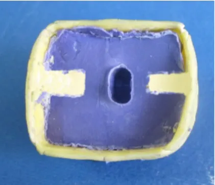
A layer of wax was then placed on the mounted tooth and two stops were created on the wax (Figures 2 and 3). A special tray was then fabri-cated and an impression was made using regular type polyvinyl siloxane (Zhermack, BadiaPolesine RO, Italy). This was repeated 20 times and the impressions were poured with epoxyresin (Exacto-Form Model Resin, Bredent, Senden, Germany) [6] and 20 resin dies with chamfer finish line were fabricated as such.
The chamfer finish line of the same tooth was then converted to a 90° shoulder with one-millimeter depth using a flat-end cylindrical diamond bur with one-millimeter diameter.
Care was taken not to change the depth of preparation when converting the chamfer finish line to shoulder (Figure 4).
Twenty polyvinyl siloxane impressions were made and 20 resin dies were fabricated. An impression was made of each epoxy resin die using polyvinyl siloxane and poured with type IV dental stone (GC Fujirock EP, GC America Inc., IL, USA).
Nail varnish was applied on stone dies except for one-millimeter band around the margin.
Forty resin dies and 40 stone dies were fabricated as such. IPS e.max crowns were fabricated on stone dies.
Figure 3. Regular polyvinyl siloxane impression
For preparation of IPS e.max Press crowns, the core was first waxed. The thickness of wax was 0.8mm, which was thicker than the core of
metal-ceramic restorations (thickness was
controlled with a wax gage).
Wax cores were sprued. The sprues for flasking of IPS empress were thicker than those for metal-ceramic restorations and connected to the wax pattern at a direct angle. A specific flask was used for flasking. The flask was heated up to 800°C to melt and eliminate the wax pattern. The ceramic block was entered into the sprue by an aluminum forceps and casting was performed in a furnace. After heating up to 920°C, the melted ceramic was slowly packed into the mold under vacuum. After packing, the surface of restoration was air-abraded and the sprue was cut.
The core was then adapted to the die [2] and immersed into Invex liquid (hydrofluoric acid base) for 10 to 30 minutes. Next, the frame was washed with water and dried. The frame was then sandblasted again with aluminum oxide particles at 1-2bar pressure and after drying, porcelain was applied using enamel and dentin porcelain powder (Ivoclar Vivadent) [2].
Crowns with Zir CAD cores were fabricated differently. The dies were scanned in CAD/CAM machine and the final thickness of cores was considered to be 0.5mm. After preparation by the machine, cores were sintered in a furnace for four hours at 1480°C. A layer of Zir Liner was applied on the prepared cores and after baking, porcelain was applied using enamel and dentin porcelain powder (Ivoclar Vivadent) [2].
Application of porcelain in all groups was performed by one technician. Thus, a total of 10 specimens with IPS e.max Zir CAD core and chamfer finish line, 10 specimens with IPS e.max Press core and chamfer finish line, 10 specimens with IPS e.max Zir CADcore and shoulder finish line and 10 specimens with IPS e.max Press core and shoulder finish line were fabricated.
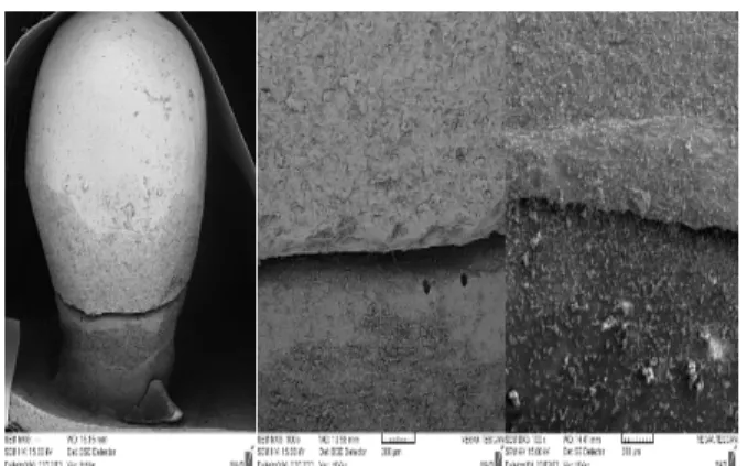
After fabrication of the crowns, their thicknesses were standardized using a gage [12] and their adaptation to resin dies was evaluated under a scanning electron microscope (Leo 1500VP, Germany) at ×100 magnification. Eventually 40 IPS e.max crowns with acceptable quality were obtained [13].
Crowns were then cemented using Panavia F21 resin cement. Excess cement was then removed and the specimens were stored in saline at room temperature for 24 hours [6]. (Figure 5)
Figure 1. Chamfer finish line
Figure 4. Shoulder finish line
Figure 2. Applying two layers of wax and creating stops before the fabrication of tray
Figure 5. Assessment of the marginal fit of restorations before cementation under a SEM
Fracture resistance was assessed in a universal testing machine (Gotech AI, 700LAC, Arizona, USA) (Figure 6). Load was applied by a stainless steel ball with 5mm diameter to the center of the occlusal surface parallel to the longitudinal axis of the tooth at a crosshead speed of 1mm/min. Load was applied starting from zero N and continued until fracture [6-8]. Fracture resistance data were automatically recorded by the device software. Specimens were also visually inspected for evaluation of the mode of failure. One-way ANOVA was used to compare the fracture resistance of groups.
Figure 6. Assessment of fracture resistance using a universal testing machine
Results
The mean fracture resistance was 1426N in the ZirCAD chamfer group, 1361.3N in the ZirCAD shoulder group, 1059.9 Nin the Press chamfer group and 1295.8N in the Press shoulder group. The load at fracture in each group is shown in Table 1 and the mean and standard deviation of
load at fracture in each group are shown in Table
2. One sample Kolmogorov-Smirnov test
confirmed normal distribution of data. One-way ANOVA was used to compare the fracture resistance of groups and found no significant difference in this regard (p=0.095).
Each specimen was visually inspected to determine the mode of failure. In the ZirCAD chamfer group, core and porcelain fracture occurred in three samples and the remainder fractures were exclusively in the porcelain. In the ZirCAD shoulder group, fracture of the core and porcelain occurred in two samples and the rest of the fractures were exclusively in the porcelain.
In Press chamfer group, all fractures occurred in the core and porcelain, except two cases with only porcelain fracture. In the shoulder Press group, all fractures occurred in the core and porcelain.
Discussion
This study aimed to compare the fracture resistance of all ceramic Press and Zir CAD crowns with chamfer and shoulder finish lines. The results showed that the mean fracture resistance was1426N in the chamfer Zir CAD, 1361. 3N in the shoulder Zir CAD, 1059.9N in the chamfer Press and 1295.8N in the shoulder Press group. The difference in this regard among groups was not significant.
Rammelsberg et al. reported that the fracture resistance of posterior Artglass crowns with chamfer finish line was higher compared to shoul-der finish line [14].
Webber et al. showed that the thickness of restoration wall did not affect the fracture resistance of Procera [5].
Cho et al. Reported that the fracture resistance of Ceromer/FRC crowns with chamfer finish line was higher compared to shoulder [10]. Lorio et al. demonstrated that the fracture resistance of sin-tered alumina cores fabricated by the CAD/CAM system with shoulder finish line was higher compared to chamfer [8].
Jalalian et al. stated that the fracture resistance of In Ceram core with chamfer finish line was higher than shoulder [6].
In another study, Jalalian et al. showed that the fracture resistance of zirconia cores with chamfer finish line was higher than shoulder [7].
According to the information provided in
According to the information provided in the catalogues of Ivoclar, the fracture resistance is 2.75 MPa for Press ingots and 6 MPa for ZirCAD ingots. Thus, the fracture resistance of ZirCAD ingots is approximately twice the rate for Press ingots. In the current study, the fracture resistance of Press and ZirCADwas almost the same. The reason may be that the porcelain fracture occurs sooner than core fracture. Also, core thickness is 0.8mm in the Press group and 0.5mm in the Zir-CAD group according to the instructions provided by Ivoclar. This difference in thickness can some-how explain the close fracture resistance of the two groups. Thus, it can be concluded that after appli-cation of porcelain, finish line design does not af-fect the fracture resistance of IPS e.max posterior single crowns.
The modulus of elasticity of core supporting materials affects the fracture resistance of core [15]. For this reason, epoxy resin dies were used in the current study, which are superior to brass dies [16]. Another difference in the clinical setting is in the bond between the adhesive cements and die. It is wise to believe that absence of hybrid layer at
the dentin-cement interface affects the
crown-diebiomechanical behavior. Since this factor was the same among groups, comparison of the groups was possible.
In 2002, Charles et al. evaluated the maximum clenching load. They compared the clenching load of 44 adults with posterior edentulism with maximum clenching load of 20 subjects with sound dentition. They estimated that the mean clenching load of 44 adults suffering from posterior edentulism was 462N (range 98 to 1031N); this value was 720N in the 20 healthy subjects (range 244-1243N) [17].
Since the fracture resistance in the four groups in our study was much higher than the load applied intra-orally, both types of cores and finish line designs can be successfully used in the clinical setting as an alternative to metal ceramic restorations. Since no significant difference was found in fracture resistance of shoulder and chamfer finish lines, both designs can be the catalogues of Ivoclar, the fracture resistance is 2.75 successfully used in the posterior teeth with all ceramic Press system.
Conclusion
Since both finish lines and core designs showed almost equal fracture resistance much higher than the load applied in the oral clinical setting, both finish line designs and cores can be successfully used in the clinical setting and have no superiority over one another for use in posterior single crowns. Table 1.Fracture resistance of specimens in the four groups (in N)
Table 2. The mean and standard deviation of load at fracture (fracture resistance) in the four groups
Group Mean fracture resistance (N) Standard deviation of fracture resistance (N)
ZirCAD chamfer 1426 335.16
ZirCAD shoulder 1361.3 386.76
Press chamfer 1059.9 113.96
Press shoulder 1295.8 413.26
ZirCAD chamfer (N) ZirCAD shoulder (N) Press chamfer (N) Press shoulder (N)
1603 1093 1049 1257
1256 1688 1066 880
1654 2020 1101 1020
1651 1551 988 1380
1602 1778 1146 1447
1530 1029 1010 1482
879 1040 1076 762
1075 1094 1296 2076
1936 1441 1011 1731
1074 879 856 923
References
1. Jung YS, Lee JW, Choi YJ, Ahn JS, Shin SW, Huh JB. A study on the in-vitro wear of the natural tooth structure by opposing zirconia or dental porcelain.J Adv Prosthodont.2010 Sept;2(3):111-5. 2. Rosenstiel SF, Land MF, Fujimoto J. Contemporary fixed prosthodontics. Mosby Incorporated; 2006, 774-790.
3. Barath VS, Faber FJ, Westland S, Niedermeier W. Spectrophotometric analysis of all-ceramic materials and their interaction with luting agents and different backgrounds. Advances in Dental Res. 2003 Dec; 17(1):55-60.
4. Komine F, Tomic M, Gerds T, Strub JR. Influence of different adhesive resin cements on the fracture strength of aluminum oxide ceramic posterior crowns. J Prosthet Dent. 2004 Oct; 92 (4):359-64.
5. Webber B, McDonald A, Knowles J. An in-vitro study of the compressive load at fracture of procera All ceramic crowns with varying thickness of veneer porcelain. J Prosthet Dent. 2003 Feb; 89(2): 154-60.
6. Jalalian E, Aletaha N. The effect of two marginal designs (chamfer and shoulder) on the fracture resistance of all ceramic restorations, Inceram: An in vitro study. J Prosthod Res. 2011 (April); 55(2):121-125.
7. Jalalian E, Atashkar B, Rostami R. The Effect of Preparation design on the fracture resistance of Zir - conia crown copings (Computer associated design/computer asso-ciated machine, CAD/CAM system. J Dent, Tehran University of Medical Sciences. 2011; 8(3):123-129.
8. Di Iorio D, Murmura G, Orsini G, Scarano A, Cupatis. Effect of margin design on the fracture resistance of procera All ceramic cores: An invitro stududy. J Contemp Dent Pract. 2008; 9(2):1-8.
9. De Jager N, Pallav P, Feilzer AJ. The influence of design parameters on the FEA-determined stress distribution in CAD/CAM produced All ceramic dental crown. Dent Mater. 2005 Mar; 21(3):242-51.
10. Cho L, Choi J, Jin Yi Y, Jin Park C. Effect of finish line variants on marginal accuracy and fracture strength of ceramic optimized polymer/ fiber-reinforced composite crowns. J Prosthet Dent. 2004 Jun;91(6):554-60.
11. Potiket N, Chiche G, Finger IM. In vitro fracture strength of teeth restored with different all-ceramic crown systems. J Prosthet Dent. 2004 Nov;92(5):491-5.
12. Amr S. Kassen, Osama Atta, Omar EI-Mowafy. Fatigue resistance and microleakage of CAD/CAM ceramic and composite molar crowns. J of Prosthod. 2012; 21(1):28-32.
13. Yeo I-S, Yang J-H, Lee J-B. In vitro marginal fit of three all-ceramic crown systems. J Prosthet Dent. 2003 Nov; 90(5):459-64.
14. Rammersberg P, Eickemeyer G, Pospiech P. Fracture resistance on posterior metal free polymer crowns. J Prosthet Dent. 2000 Sep; 84(3):303-308. 15. Scherrer SS, de Rijk KG. The fracture resistance of all ceramic crowns on supporting structure with different elastic moduli. Int J Prosthod. 1993 Sep-Oct;6(5):462-7.
16. Ayad MF. Effect of the crown preparation margin and die type on the marginal accuracy of Fiber-reinforced composite crowns. J Contemp Dent Pract. 2008 Feb; 9(2):9-16.
17. Gibbs CH, Anusavice KJ, Young HM, Jones JS, Esquivel-Upshaw JF. Maximum clenching force of patients with moderate loss of posterior tooth support: A pilot study. J Prosthet Dent. 2002 Nov; 88(5):498-502.