Page 1 of 6 Original Research
Read online:
Scan this QR code with your smart phone or mobile device to read online. Authors:
Richard J. Cormack1
Mollie C.M. Ferris2
Jason K. Wong2
Stefan Przybojewski2 Affiliations:
1RCA Diagnostics, Calgary,
Canada
2Foothills Medical Centre,
University of Calgary, Canada Corresponding author: Mollie Ferris,
doctormollie@gmail.com Dates:
Received: 11 Apr. 2016 Accepted: 25 Aug. 2016 Published: 23 Nov. 2016 How to cite this article: Cormack RJ, Ferris MCM, Wong JK, Przybojewski S. Splenic artery embolisation in the non-operative
management of blunt splenic trauma in adults. S Afr J Rad. 2016;20(1), a1014. http:// dx.doi.org/10.4102/sajr. v20i1.1014
Copyright:
© 2016. The Authors. Licensee: AOSIS. This work is licensed under the Creative Commons Attribution License.
Introduction
At our institution, haemodynamically stable patients with blunt splenic injury are managed non-operatively with the goal of preserving splenic function by avoiding splenectomy. In blunt splenic trauma, non-operative management (NOM) generally includes bed rest, limited oral intake and close clinical and biochemical observation. However, high failure rates, necessitating splenectomy, have been reported for observational NOM alone in patients with American Association for the Surgery of Trauma (AAST) grades III to V splenic injuries and those demonstrated to have vascular injuries.1,2,3 Splenic artery embolisation (SAE) has increasingly been utilised as an adjunct to NOM in order to improve splenic salvage rates.2,4,5,6,7
Patients with suspected splenic trauma who are candidates for NOM should have a contrast-enhanced computed tomography (CT) scan to evaluate and grade the extent of injury.4 CT can accurately assess the severity of splenic injury using the AAST grade, estimate the volume of haemoperitoneum, characterise vascular injuries and help direct appropriate therapy.3,8
The primary purpose of this study was to determine the splenic salvage rate in the NOM of blunt splenic injury. The secondary purpose was to evaluate the complications of SAE.
Research method and design
All patients admitted to our level I trauma centre, between January 2005 to April 2010, who underwent SAE for blunt splenic injury, were retrospectively reviewed. Patient demographics included age and sex. By study design, all patients included in the analysis required documentation of splenic injury on an admission CT scan. All pre-procedure CT scans were retrospectively reviewed by an interventional radiologist and a program-year 4 radiology resident for evidence of a vascular injury (i.e. arteriovenous fistula [AVF], pseudoaneurysm [PA], vessel truncation and active extravasation of intravenous [IV] contrast), estimation of the volume of haemoperitoneum and determination of AAST injury grade. Significant haemoperitoneum was defined as blood extending beyond the perisplenic recesses. If a post-procedural CT scan was obtained, it was reviewed for evidence of a complication, including splenic infarction, abscess formation, pleural effusion and persistent or new vascular injury.
Background: The purpose of this study was to evaluate the splenic salvage rate with
angioembolisation in the non-operative management (NOM) of blunt splenic injury.
Methods: We conducted a retrospective analysis of patients presenting to our Level I trauma
centre with computed tomography (CT)-confirmed splenic injury following blunt trauma and in whom angioembolisation was utilised in the algorithm of NOM. Data review included CT and angiography findings, embolisation technique and patient outcomes.
Results: Between January 2005 and April 2010, 60 patients with splenic injury following blunt
trauma underwent NOM, which included splenic artery embolisation (SAE). All patients included in the study required a preadmission. CT scan was used to document the American Association for the Surgery of Trauma (AAST) grade of splenic injury. The average injury grade was 3.0. The non-operative splenic salvage rate following SAE was 96.7% with statistically similar salvage rates achieved for grades II to IV injuries. The quantity of haemoperitoneum and the presence of a splenic vascular injury did not significantly affect the splenic salvage rate. The overall complication rate was 27%, of which 15% were minor and 13% were major.
Conclusion: SAE is a safe and effective treatment strategy in the NOM of blunt splenic injury.
The quantity of haemoperitoneum, the presence of vascular injury and embolisation technique did not significantly affect the splenic salvage rate.
Splenic artery embolisation in the non-operative
management of blunt splenic trauma in adults
Read online:
Scan this QR code with your smart phone or mobile device to read online.
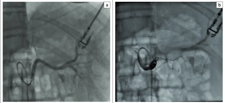
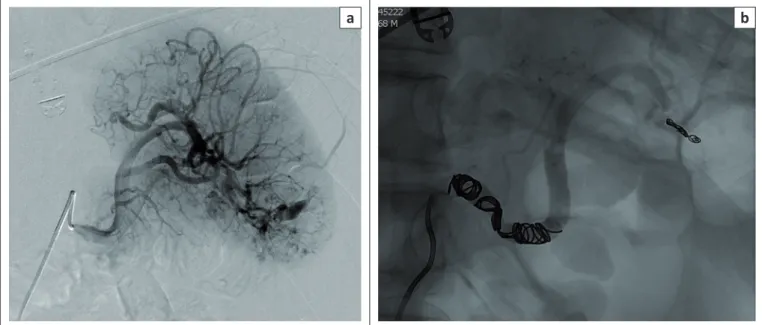
The splenic angiograms and procedural notes were reviewed for the findings that necessitated embolisation, the SAE technique and the embolic agent used. The SAE technique consisted of main SAE (Figure 1), selective distal branch vessel embolisation (Figure 2) or a combined main and distal SAE (Figure 3). The technique and embolic agents used for the SAE were at the discretion of the performing interventional radiologist.
The primary outcome of the study was splenic salvage, defined as the percentage of patients not requiring splenectomy. Splenic salvage rates were analysed, taking into consideration the AAST injury grade, quantity of haemoperitoneum and the presence of a vascular injury. Failure of NOM was defined as the need for abdominal
surgery during hospital admission regardless of the indication.
Post-procedure, patients were assessed clinically for evidence of complications. Follow-up CT examination was performed in patients with clinical deterioration at the discretion of the referring physician. Complications were stratified into major and minor categories based on the 2010 Society of Interventional Radiology Quality Improvement Guidelines for Percutaneous Transcatheter Embolisation.9 Minor complications included post-procedure pain and endovascular coil migration. Major complications included death, splenic infarct involving greater than 25% of the splenic parenchyma, post-procedural abscess formation, persistent or new vascular injury and left pleural effusion.
a
b
FIGURE 1: Fluoroscopic images demonstrating: (a) angiography of the main splenic artery and (b) coil embolisation of the proximal main splenic artery.
a
b
FIGURE 2: Fluoroscopic images demonstrating: (a) angiography of the main splenic artery with a splenic contusion and pseudoaneurysm formation and (b) coil embolisation of both the proximal main and distal (black arrow) splenic branches.
Descriptive statistics were performed and validated by a statistician at our institution.
Ethical considerations
Formal ethics approval was obtained from the Institutional Ethics Review Board.
Results
During the study period, there were 67 patients who underwent SAE for blunt splenic trauma. Seven patients did not have an available pre-procedure CT (these were performed at peripheral rural centres from which the patient was subsequently transferred) and thus were excluded from the data analysis. Therefore, a total of 60 patients were included in the study (46 males and 14 females) with a mean age of 36 years (ranging between 15 to 88 years).
On pre-procedural CT, there were 8 grade II, 44 grade III and 8 grade IV splenic injuries. In the study cohort, there were no patients with either an AAST splenic grade I injury or a grade V injury. Forty-three patients presented with a significant haemoperitoneum and 31 patients had evidence of a vascular injury (Table 1). Four patients had a PA involving one branch vessel and active extravasation of IV contrast from a separate branch vessel.
The chosen SAE technique was at the discretion of the performing interventional radiologist at the time of the procedure: 54 patients were treated with a proximal SAE (53 with coils and 1 with an Amplatzer plug) and 6 patients underwent combined selective distal and main SAE. Selective embolisation was performed either with coils alone or coils in combination with N-butyl cyanoacrylate glue. No exclusive selective distal embolisation was performed (Table 2).
Overall, the splenic salvage rate following SAE was 97%. In 16 patients with grade II and grade IV splenic injuries, there was a 100% splenic salvage rate. In 44 patients with a grade III injury, there was a 95% splenic salvage rate.
Of the 60 patients, 40 (67%) progressed to further assessment with a CT examination post-embolisation. Two patients failed NOM, one of whom required subsequent abdominal surgery for haemodynamic instability from multiple concurrent abdominal injuries and the other required splenectomy because of a splenic abscess. Both patients had grade III splenic injuries and were treated with proximal SAE. Both patients also had evidence of a vascular injury at presenting CT, one with active extravasation of IV contrast and the other with a PA (Figure 4). Both patients had significant haemoperitoneum at the time of presentation.
a
b
FIGURE 3: Fluoroscopic images demonstrating: (a) angiography of the main splenic artery showing pseudoaneurysm and arteriovenous fistula formation in the lower pole of the spleen and (b) combined distal and proximal coil embolisation.
TABLE 1: Splenic injury grades and vascular injuries.
Injury type Variable Number of patients (n)
AAST splenic injury grade I 0
II 8
III 44
IV 8
V 0
Vascular injury Haemoperitoneum 43
Pseudoaneurysm 15
Extravasation 18
AVF 2
AAST, American Association for the Surgery of Trauma; AVF, arteriovenous fistula.
TABLE 2: Embolisation technique.
Treatment Treatment detailsk Number of patients
Embolic agent Coils 58
Coils and N-butyl
cyanoacrylate glue 1
Amplatzer device 1
Splenic artery embolisation Main splenic artery 54 Main and selective artery 6
The overall complication rate of SAE per patient in our study was 27%, with 16 of the 60 patients having at least one complication. In total, there were 19 complications in 16 patients, with 14 patients having one complication each, one patient having two complications and one patient having three complications. Of the complications, 10 were major and nine were minor. The most common major complication was a left pleural effusion, occurring in four patients following SAE, one of whom required chest tube placement for symptomatic relief. The most common minor complication was distal migration of a main splenic artery coil, occurring in seven patients. The patient with three complications had a distal coil migration that was further complicated by a post-procedure splenic infarct and subsequent abscess formation (Figure 5). There was one death following SAE, but this was not directly related to the procedure but because of other injuries (Table 3).
Discussion
The spleen is the most frequently injured solid organ in blunt abdominal trauma.10 For many years, splenectomy was the preferred method of treatment for traumatic splenic injury;
however, because of the risk of fatal post-splenectomy sepsis and impaired resistance to infection with encapsulated organisms, many trauma physicians began seeking alternative management strategies.11 Over the past several decades, there has been a shift in the approach to blunt splenic injury, with NOM now preferred for haemodynamically stable patients.2,4,5,6,7 This generally consists of various observational strategies including bed rest, limiting oral intake, serial haemoglobin and/or haematocrit levels and regular physical examinations.6 In 2000, the multicentre Eastern Association for the Surgery of Trauma (EAST) trail, demonstrated an overall observational NOM failure rate of 10.8%.12 Significantly, higher failure rates were demonstrated with
a
b
FIGURE 4: Vascular injuries in two patients who failed NOM management with splenic artery embolisation: (a) Patient 1: contrast-enhanced axial CT shows active extravasation of IV contrast (white arrow) beyond the splenic margin and (b) Patient 2: fluoroscopic image from selective splenic artery angiogram shows splenic artery pseudoaneurysm (black arrow).
a
b
c
CT, computed tomography.
FIGURE 5: Complications included coil migration, splenic infarction and splenic abscess: (a) Fluoroscopic image shows distal migration of main splenic artery embolisation coil (arrow), (b) contrast-enhanced coronal CT shows non-enhancement of the superior pole of the spleen consistent with splenic infarct and (c) contrast-enhanced axial CT shows rim enhancing splenic abscess (circle).
TABLE 3: Outcomes of splenic artery embolisation.
Outcomes Complications Patients
n %
Minor complications Pain 2 3
Coil migration 7 12
Major complications Death 1 2
Infarct (>25%) 3 5
Abscess 1 2
Artery dissection 1 2
grades III–V splenic injuries and with vascular injuries. SAE as an adjunct therapy to observational NOM continues to gain favour as multiple studies have demonstrated improved non-operative splenic salvage rates, especially in those patients at risk of failing observational management.2,4,5,6,7,12 The use of SAE for blunt splenic injury was first described in 1995 by Sclafani et al.4 Since then, there have been several retrospective studies analysing the efficacy of transcatheter SAE with success rates ranging from 75% to 100%.2,4,5,6,7 This study further contributes to the existing literature that SAE is an effective management strategy for blunt splenic injury and can achieve excellent salvage rates, even for higher grade splenic injuries and vascular injuries.5,6 The overall splenic salvage rate was 96.7% (including a salvage rate of 96.2% for grades III and IV injuries), which is comparable with other published studies.2,4,5,6,7 In our study, there were no patients with grade V injuries.
Multiple studies have indicated that the presence of a splenic vascular injury increases the failure rate of NOM.2,6,13 In our study, 31 patients had evidence of a vascular injury on either their pre-procedure CT or on catheter angiography (Table 2). Both patients who failed NOM had evidence of a vascular injury: one had a PA and the other had active extravasation of IV contrast. Both patients were treated by main SAE. The quantity of haemoperitoneum was also assessed, being significant if blood extended beyond the splenic bed. Both patients who failed NOM had a significant quantity of haemoperitoneum on their pre-procedure CT. There was, however, no statistically significant correlation between the quantity of haemoperitoneum and the splenic salvage rate.
There remains debate in the literature regarding the optimal technique for SAE in the setting of trauma.6,10,13,14 Described approaches include main SAE, superselective embolisation of an injured distal splenic artery or a combined main and distal SAE. Embolisation of the proximal splenic artery allows for reconstitution of distal arterial flow via short gastric, gastroepiploic and pancreatic arteries.10,14 Proximal embolisation reduces the arterial pressure in the splenic parenchyma, allowing the spleen to heal and reducing the risk of re-bleeding.10 In cases of significant focal arterial injury demonstrated by angiography, a distal branch vessel embolisation is often preferred, excluding it from the circulation and reducing the risk of ongoing haemorrhage.14 A 2004 study by Haan et al. demonstrated statistically equivalent efficacy rates with either a main approach or a distal approach, but a statistically higher failure rate if a combined embolisation was performed.6 A 2011 systematic review by Schnüriger et al. was inconclusive in determining whether proximal or distal SAE should be performed to avoid significant re-bleeding.14 In our study, there was a 100% splenic salvage rate for patients undergoing main and distal SAE.
Complications after SAE were relatively frequent, occurring in 16 of the 60 patients (27%). The overall major complication
rate was 8 of 60 patients (13%). The 2010 Society of Interventional Radiology Quality Improvement Guidelines for Percutaneous Transcatheter Embolisation list overall major complication rates of 8% – 22%, with a suggested threshold of 15%.9 Other studies list overall complication rates of up to 38% and major complication rates ranging from 0% to 28.5%.6,7,14,15 The most common major complication in our study was the development of a post-procedural left pleural effusion seen in four patients, followed by partial splenic infarction in three patients. One patient died after SAE, but this was secondary to multiple other severe traumatic injuries rather than the sequelae of the embolisation procedure. One patient developed a large
splenic infarct (involving an estimated 50% of the
parenchyma) and a subsequent splenic abscess that was treated by splenectomy. Minor complications occurred in 9 of 60 patients (15%) and included post-procedure pain in two patients and clinically silent distal migration of intended proximal coils in seven patients. Of note is that detachable coils were not yet available during the study period. Although our complication rates are slightly high, they do still correlate with other studies.
Of the 60 patients in our study cohort, 16 patients (27%) failed initial observational NOM based on clinical deterioration or CT findings of vascular injury, who then went on to delayed (at least 48 h post-admission) but successful SAE. Of these patients, 12 had a grade III injury, 2 had a grade II injury and another 2 had a grade IV injury. This further corroborates the findings from the 2014 study by Miller et al. that all patients with grades III–V injuries should proceed directly to SAE to reduce NOM failure rates.5
Limitations of the study
Limitations of this study include that it is a single-centre retrospective analysis and no patients with grade V injuries were referred for SAE. Another limitation with respect to the assessment of post-procedure complications is that not every patient had a follow-up CT examination.
Conclusion
Our retrospective study contributes to the existing literature that SAE is an effective and safe therapy to improve splenic salvage rates in patients with high AAST grade splenic and vascular injuries because of blunt abdominal trauma.
Acknowledgements
We would like to thank Yukun Zhang, statistical analyst, for validation of the statistical methods and Justin Chun and Sherif Idris for assisting with chart review.
Competing interests
The authors declare that they have no financial or personal relationships which may have inappropriately influenced them in writing this article.
Authors’ contributions
J.K.W. and S.P. were the project leaders and responsible for project design. S.P., R.C. and M.C.M.F. collected data and were responsible for most of the manuscript creation and preparation. All authors were involved in manuscript editing.
References
1. Tinkoff G, Esposito TJ, Reed J, et al. American Association for the Surgery of Trauma organ injury scale I: Spleen, liver, and kidney, validation based on the National Trauma Data Bank. J Am Coll Surg. 2008;207:646–655. http://dx.doi. org/10.1016/j.jamcollsurg.2008.06.342
2. Sabe A, Calridge J, Rosenblum D, Lie K, Malangoni M. The effects of splenic artery embolisation on nonoperative management of blunt splenic injury: A 16-year experience. J Trauma. 2009;67:565–572. http://dx.doi.org/10.1097/TA.0b013e 3181b17010
3. Marmery H, Shanmuganathan K, Alexander M, Mirvis S. Optimization of selection for nonoperative management of blunt splenic injury: Comparison of MDCT grading systems. AJR Am J Roentgenol. 2007;189:1421–1427. http://dx.doi.org/ 10.2214/AJR.07.2152
4. Sclafani S, Shaftan G, Scalea T, et al. Nonoperative salvage of computed tomography-diagnosed splenic injuries: Utilization of angiography for triage and embolisation for hemostasis. J Trauma. 1995;39:818–825. http://dx.doi.org/10. 1097/00005373-199511000-00004
5. Miller PR, Chang MC, Hoth JJ, et al. Prospective trial of angiography and embolisation for all grade III to V blunt splenic injuries: Nonoperative management success rate in significantly improved. J Am Coll Surg. 2014;218:644–651. http:// dx.doi.org/10.1016/j.jamcollsurg.2014.01.040
6. Haan J, Biffl W, Knudson M, et al. Splenic embolisation revisited: A multicenter review. J Trauma. 2004;54:542–547. http://dx.doi.org/10.2214/AJR.07.2152 7. Wei B, Hemmila M, Arbabi S, Taheri P, Wahl W. Angioembolisation reduces
operative intervention for blunt splenic injury. J Trauma. 2008;64:1472–1477. http://dx.doi.org/10.1097/TA.0b013e318174e8cd
8. Thompson B, Munera F, Cohn S, et al. Novel computed tomography scan scoring system predicts the need for intervention after splenic injury. J Trauma. 2006;60:1083–1086. http://dx.doi.org/10.1097/01.ta.0000218251.67141.ef 9. Angle JF, Siddiqi NH, Wallace MJ, et al. Quality improvement guidelines for
percutaneous transcatheter embolisation: Society of Interventional Radiology Standards of Practice Committee. J Vasc Interv Radiol. 2010;21:1479–1486. http://dx.doi.org/10.1016/j.jvir.2010.06.014
10. Madoff D, Denys A, Wallace M, et al. Splenic artery interventions: Anatomy, indications, technical considerations, and potential complications. Radiographics. 2005;25:S191–S211. http://dx.doi.org/10.1148/rg.25si055504
11. Dasgupta N, Matsumoto A, Arslan B, Turba U, Sabri S, Angle J. Embolisation therapy for traumatic splenic lacerations. Cardiovasc Intervent Radiol. 2012;35:795–806. http://dx.doi.org/10.1007/s00270-011-0186-y
12. Peitzman AB, Heil B, Rivera L, et al. Blunt splenic injury in adults: Multi-institutional study of the Eastern Association for the Surgery of Trauma. J Trauma. 2000;49: 177–189. http://dx.doi.org/10.1097/00005373-200008000-00002
13. Imbrogno BF, Ray CE. Splenic artery embolisation in blunt trauma. Semin Intervent Radiol. 2012; 29: 147–149. http://dx.doi.org/10.1055/s-0032-1312577 14. Schnüriger B, Inaba K, Konstantinidis A, Lustenberger T, Chan LS, Demetriades D.
Outcomes of proximal versus distal splenic artery embolisation after trauma: A systematic review and meta-analysis. J Trauma. 2011;70:252–260. http://dx.doi. org/10.1097/TA.0b013e3181f2a92e
15. Ekeh AP, Khalaf S, Ilyas S, Kauffman S, Walusimbi M, McCarthy MC. Complications arising from splenic artery embolisation: A review of an 11-year experience. Am J Surg. 2013;205:250–254. http://dx.doi.org/10.1016/j.amjsurg. 2013.01.003