Manisha Sharma et al JMSCR Volume 07 Issue 05 May 2019 Page 898
Original Article
Paramedian Epidural with Midline Spinal in the Same Intervertebral Space:
An Alternative Technique for Combined Spinal and Epidural Anaesthesia
Authors
Manisha Sharma
1, Manish Anand
2, Nand Kishore
3, Waquas Ahmed
41,4
Department of Anaesthesia, Indira Gandhi Institute of Medical Sciences, Patna, Bihar
2
Consultant, Heart Hospital, Kankerbagh, Patna, Bihar
3
Consultant, Department of Surgery, Medipark Hospital, Patna, Bihar *Corresponding Author
Manish Anand
Consultant, Heart Hospital, Kankerbagh, Patna, Bihar, India
Abstract
Background & Aim: The combined spinal epidural technique (CSE) involves subarachnoid blockade and epidural catheter placement in procedure. Aim was to compare two different approaches of CSE, paramedian epidural with midline Subarachnoid block, that is Single space dual needle technique (SDT) with the Single space technique (SST).
Materials and Methods: The study was randomised, comparative &prospective. A total of 80 patients were divided in Group I SST & Group II SDT. Inclusion criteria: ASA grade I/II, undergoing Hysterectomy etc. Exclusion criteria: Patient refusal, allergy, coagulopathy, IHD, local infection etc. Group I- Needle through needle technique. Group II-Epidural in paramedian position with midline spinal. Parameters observed were technique performance time, time to surgical readiness, in Epidural block: attempts for space localisation, accidental dural puncture. For epidural catheter: attempts for insertion, presence of blood / CSF, paraesthesia, inability to push test dose. While Subarachnoid block; attempts, appreciation of dural puncture, reflux of CSF < 5 seconds.
Results: Both were comparable in technique performance time & time to surgical readiness. Parameters relating to epidural block were comparable.The appreciation of dura in SAB in group I was 26 and group II was 39(p 0.0002) which was statistically significant. The incidence of reflux of CSF within 5 seconds in the group I was 33 and group II was 40 which was statistically significant (p 0.0056).
Conclusion: Paramedian epidural with midline spinal at the same space is an acceptable alternative to the Single Space technique.
Keywords: Paramedian, Epidural, Spinal, Midline.
Introduction
Neuraxial anaesthesia is the term for central blocks involving the spinal, epidural, and caudal spaces. Corning published studies documenting success with neuraxial blocks in 18851. Most operations
below the neck can be performed under neuraxial anaesthesia. Neuraxial blocks reduces incidence of venous thrombosis, pulmonary embolism; minimizing transfusion requirements and respiratory compromise following thoracic and upper abdominal surgery. A decreased stress response
www.jmscr.igmpublication.org Index Copernicus Value: 79.54
ISSN (e)-2347-176x ISSN (p) 2455-0450
Manisha Sharma et al JMSCR Volume 07 Issue 05 May 2019 Page 899
have positive cardiac benefits, reducing perioperative and postoperative ischemia.
The combined spinal–epidural technique (CSE) involves subarachnoid blockade and epidural catheter placement during the same procedure. The combination of two different routes of anaesthesia administration improves effectiveness and reduces side effects (Stevens and Edwards, 1999)2. Subarachnoid block (SAB) provides fast and reliable segmental anaesthesia with minimal risk for toxicity, while epidural anaesthesia provides perioperative anaesthesia, followed by excellent analgesia in the postoperative period.3,4 CSE anaesthesia reduces the problems, such as unpredictable level of blockade after SAB, and problems of missed segments, incomplete motor block ,that can occur with epidural anaesthesia3. Ability to perform CSE through single inter-vertebral space has made SST (Single space technique) a popular technique. Despite this advantage SST suffers from complications, technical problems and there is the cost factor. Migration of epidural catheter in subarachnoid space have been reported5 leading to extensive block6. Delayed respiratory depression due to drug entering into subarachnoid space through migrated catheter has also been claimed7. Metallic flecks getting deposited in the epidural space while using needle through needle technique have raised concern8. Meningitis9, knotting of catheter5, inadvertent dural puncture with the wide bore Tuohy10needle are additional problems with Needle through needle technique.
There is risk of damage to the epidural catheter or spinal needle if SAB is carried out after epidural catheterization4.Introducing a spinal needle with an epidural catheter in place could allow the spinal needle to strike the epidural catheter leading to spinal needle damage11 or catheter fracture12. In an attempt to find out the best CSE technique, we conducted this study, wherein epidural was performed by paramedian approach and SAB in midline, both at the same inter-vertebral space that is Single space dual needle technique (SDT) and
this was compared with the Single space (Needle through needle NTN) technique (SST).
Materials & Methods
The study was randomised, comparative & prospective. It was time bound & hospital based.
Sample size calculation
N=
Where 1.96 = Z value for 95% significance level, 0.84 =conventional multiplier for power 80%,
=squared pooled deviation=23.75, M1= 16.4
min, M2=19.5 min; N= 40 in each group.
All data so obtained was meticulously documented and statistically analysed. Quantitative data was analysed using Percentage, Mean, Standard deviation, unpaired t test. Qualitative data was analysed by chi square test.
Approval from institutional ethical committee was taken. Procedure was explained to patients and informed written consent was taken .Patients were allocated in either groups using computer generated random numbers.
Group I: Single space (needle through needle NTN) technique (SST) n=40.
Group II: Single space dual needle (Paramedian epidural with midline spinal) technique (SDT) n=40.
Inclusion criteria: ASA grade I/II, Female patients between 40 to 65 years, height 145-165 cms, Weight 45-70 kgs, patients undergoing elective Hysterectomy.
Exclusion criteria: Patient refusal, Patient with known allergy to local Anaesthetics, coagulopathy, platelet count <75,000/mm3, IHD, valvular heart disease, local infection, spinal deformity, raised intracranial tension, neurological disease.
Manisha Sharma et al JMSCR Volume 07 Issue 05 May 2019 Page 900
Group I: SST
In Group I, CSE kit was used. In sitting position, after painting and draping, L2-L3/ L3-L4 vertebral inter-space was locally infiltrated with inj. Xylocaine 2% 2ml. Epidural space was located in midline using 16 G Tuohy’s needle. The spinal needle (26 G, pencil point, 117mm) was introduced through epidural needle and dural puncture was appreciated. After free flow of CSF, 2.5 ml of 0.5% hyperbaric Bupivacaine was injected intrathecally. Thereafter, epidural catheter 20 G was introduced and catheter was fixed leaving 9cm inside the epidural space in the cephalad direction.
Group II: SDT
In Group II,Epidural set and spinal needle (26 G, pencil point, 95mm) was used.At L2-L3/ L3-L4 inter-space point of entry for epidural needle was at 1.5cm lateral to the caudal part of spinous process of corresponding vertebra. After local infiltration, the touhy needle 16 G pointing in cephalad direction was directed towards midline. After confirming space, needle was left in the space. Spinal needle was introduced in the midline in the same space and dural puncture was appreciated. After free flow of CSF, 2.5 ml 0.5% hyperbaric Bupivacaine was injected intrathecally. Thereafter, epidural catheter 20 G was introduced through the epidural needle and catheter was fixed leaving 9cm inside epidural space in the cephalad direction.
Surgery was allowed initially under Sub arachnoid block. Test dose of inj. Xylocaine with adrenaline 2%, 3ml was given in epidural. If any blood or CSF was aspirated in the catheter or positive response to test dose observed, then no top up was given and surgery was carried under SAB only.
When Spinal anaesthesia level started weaning off, (regression of two segment sensory level, tested by pin prick) Epidural top up was given intraoperatively, using increments of 4 ml of 0.5% Bupivacaine otherwise 8 ml of 0.25% Bupivacaine with 25µgfentanyl in the postoperative period. A maximum of three attempts was tried for each procedure.
Inj. Atropine 0.6mg was given if heart rate was< 50/min. Any relevant fall in blood pressure i.e more
than 20% fall from the baseline treated with intravenous fluids followed by vasopressors. Inj. Mephenteramine 6mg with repeated doses was given if MAP was < 65mmHg.
Parameters observed were
1. Technique performance time (T1): Time taken after painting and draping to the time when the patient was made supine was noted as T1.
2. Time to surgical readiness (T2): Time taken after painting and draping to the achievement of sensory level of T 6 was
noted as T2.
3. While performing the epidural block following was recorded:
a) Number of attempts for space localisation b) Accidental Dural puncture.
4. While passing epidural catheter parameters seen were:
a) Number of attempts for insertion of catheter
b) Presence of blood or CSF in the catheter c) Paraesthesia during the catheter insertion d) Inability to push test dose (due to kinking, blockade, malposition and coiling)
e) Failure to extend analgesia after epidural top-up.
5. While performing Subarachnoid block; a record of the following was made:
a) Number of attempts taken for SAB, b) Appreciation of puncture of dura,
c) Reflux of CSF < 5 seconds
Manisha Sharma et al JMSCR Volume 07 Issue 05 May 2019 Page 901
Results
Figure 1: Demographic profile of patients
The observed demographic data signifies that the patients in both groups were comparable and of similar profile.
Table 1: Technique performance time (T1) and time to surgical readiness (T2)
Time Group I (SST)
[+ SD]
Group II (SDT) [+ SD]
P value
T1 (minutes) 05:09 + 01:02 05:40 + 01:09 0.568
T2 (minutes) 11:36 + 01:43 12:05 + 01:29 0.177
The time taken is comparable in both the groups with no significant difference.
Table 2: Characteristics of Epidural Block
Characteristic of Epidural Block Group I (SST) Group II (SDT) P Value Average number of attempts at
epidural space localisation[+ SD]
1.175 + 0.501 1.375 + 0.628 0.119
Accidental puncture of Dura [%] 2 [5%] 1 [2.5%] 0.556
The average number of attempts required at epidural space localisation in group I was 1.175 + 0.501 and for group II it was 1.375 + 0.628 which is slightly more in group II but the difference is statistically insignificant with p-value 0.119.
The incidence of accidental puncture of Dura in group I is 2 out of 40 [5%] and in group II is 1 out of 40 [2.5%] with p-value 0.556 indicating no significant difference.
Table 3: Parameters of Epidural catheter insertion
Parameters of Epidural catheter insertion
Group I (SST) [n= 38]
Group II (SDT) [n=39]
P value
Average number of attempts required [+ SD]
1.158 + 0.37 1.077 + 0.27 0.277
Presence of Blood/CSF in the Epidural catheter [%]
2 [5.26%] 0 [0.0%] 0.147
Incidence of Paraesthesia [%] 7 [18.42%] 5 [12.82%] 0.498
Inability to push test dose [%] 2 [5.26%] 1 [2.56%] 0.54
Failure to extend level after top-up [%]
3 [7.89%] 1 [2.56%] 0.292
The following parameters were statistically insignificant and comparable in both the groups.
40
52.95 56
151.01
40
53.05 55.95
153.5
0 20 40 60 80 100 120 140 160 180
N (sample size) Average age (years) Average weight (kg) Average height (cm)
Group I (SST)
Manisha Sharma et al JMSCR Volume 07 Issue 05 May 2019 Page 902
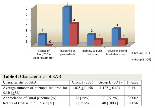
Figure 2: Comparison of various complications in Epidural catheter insertion
Table 4: Characteristics of SAB
Characteristic of SAB Group I (SST) Group II (SDT) P value
Average number of attempts required for SAB [+SD]
1.025 + 0.158 1.125 + 0.404 0.151
Appreciation of Dural puncture [%] 26 [65%] 39 [97.5%] 0.0002
Reflux of CSF within 5 sec [%] 33[82.5%] 40 [100%] 0.0056
The average number of attempts required for subarachnoid block is statistically insignificant in both the groups. Incidence of appreciation of dura is statistically significant (p 0.0002). The incidence of reflux of CSF from spinal needle within 5 seconds is statistically significant (p 0.0056).
Discussion
Though Single space technique is most widely used, it carries drawbacks like metallic particle toxicity, 3 interpretation of test dose, warning paraesthesia, epidural catheter placement in subarachnoid space, catheter coiling, kinking, knotting5, delayed respiratory depression due to epidural drug passing into subarachnoid space7 etc. Also longer spinal needles are required which delays CSF reflux due to resistance of longer needle. Special CSE kits are needed which is costly and not easily available. Studies conducted have shown that in paramedian epidural, the angulation of the epidural needle causes less accidental dural puncture13,14, less paraesthesia, lesser bloody tap and also more cephalad direction of the epidural catheter due to lesser kinking, coiling and knotting5. It does not require any CSE kit and is cost effective.
The demographic profile was comparable in both the groups as shown in Figure 1. The time taken for anaesthetic technique performance (T1) in group I was 05:09 + 01:02 minutes as compared to 05:40 + 01:09minutes for group II. The time for surgical readiness (T2), for achievement of analgesia up to T6 spinal level, in group I was 11:36 + 01:43 minutes and 12:05 +01:29 minutes for group II. Both the differences were statistically insignificant as shown in Table 1. Deepti Saigal et al15 in 2013, compared three groups SST, SDT and DST (double segment technique- epidural one space above spinal in the midline). Time for performance of anaesthetic technique (T1) was 12.18±6.092 min, 13.41±2.848 min, 11.63±3.243 min in SST, SDT, DST respectively (P=0.268) and time to surgical readiness (T2) was 17.64±5.877 min, 18.28±3.624 min, 16.87±3.137 min respectively (P=0.462). In their study comparative time was higher than our study probably because we had excluded time taken for painting and draping in our study.
The average number of attempts required at epidural space localisation in group I was 1.175 + 0.501 and for group II, it was 1.375 + 0.628 which is slightly more in group II but the difference is statistically
0 1 2 3 4 5 6 7
Pesence of blood/CSF in Epidural catheter
Incidence of paraesthesia
Inability to push test dose
Failure to extend level after top-up
2
7
2
3
0
4
1 1
Group I (SST)
Manisha Sharma et al JMSCR Volume 07 Issue 05 May 2019 Page 903
insignificant (p-value 0.119). The incidence of accidental puncture of dura in group I was two out of 40 [5%] and in group II is 1 out of 40 [2.5%] with p-value 0.556 indicating no significant difference in both groups as shown in Table 2. In such cases epidural block was abandoned. In a cadaveric study by Blomberg, RG13in 1988, found that there is presumably lower risk of accidental dural puncture using paramedian approach.
The average number of attempts required for epidural catheter insertion in group I was 1.158 + 0.37 and for group II was 1.077 + 0.27 with p-value of 0.277 which is statistically insignificant as shown in Table 3. However there is slight increase in average number of attempts for epidural catheter insertion in SST group indicating towards difficulty in inserting the epidural catheter in that group. In a study by Takahashi et al epidural catheterization was not possible in two cases of SST (n=169)16. There was no case with difficulty in advancement of the epidural catheter in SDT group. The influence of the dorsomedian connective tissue band can explain the difficulty in traversing of the midline epidural catheters13.In a study Blomberg13and colleagues found less resistance to catheter insertion due to steeper angle of entry of paramedian epidural needle into epidural space, facilitating catheter insertion. In an epiduroscopic cadaver study, it was demonstrated that when catheter is advanced in the midline there is considerable dural tenting and course of the catheter is unpredictable due to strands of connective tissue restricting movement of dura mater13. Leeda M et al17 in 2005, studied epidural catheter insertion via midline and paramedian approach and found that catheter insertion was faster using the paramedian approach. Incidence of blood or CSF in the epidural catheter in group I was 2 out of 38 [5.26%] as compared to 0 in case of group II. The difference in incidence was found to be insignificant with p-value of 0.147 as shown in figure 2. Use of midline approach to epidural catheterization in SST is more likely to encounter the epidural venous plexus. Deepti Saigal et al15, found lower incidence of blood in the epidural catheter in Paramedian epidural with midline spinal
group. The incidence of paraesthesia while advancement of epidural catheter were comparable with p-value of 0.498 which is insignificant. Deepti Saigal et al15, found lower incidence of paraesthesia in paramedian epidural with midline spinal group. Leeda M et al17 in 2005, found a higher incidence of paraesthesias in the midline group (33% vs. 6.7%) compared to paramedian epidural. Blomberg and colleagues13 reported an incidence of 36% with the midline approach as opposed to 4% with the paramedian approach.
The risk of epidural catheter penetrating the duramater through the hole made by spinal needle is a major concern. However, there was no such case in our as well as other studies18.Epiduroscopy studies have concluded that it is impossible to force epidural catheter through the hole made in dura by a fine spinal needle.6
Lack of appreciation of dural puncture was significant (35%, P value 0.0002) in group I and was 2.5% in SDT group. Paech and Evans could not feel the dural puncture in 6-12% cases while performing CSE by NTN technique19.Lack of dural puncture appreciation may lead to failure of SAB in Needle through needle technique3.
Reflux of CSF <5 seconds was seen in 33 patients in group I and all 40 cases in group II as shown in Table 4 . There was a significantly lower incidence of cases with instant reflux of CSF in SST group (82.5%) as compared with 100% incidence in SDT group (P value 0.0056). The speed of reflux of CSF primarily depends on the gauge of spinal needle used. However, the spinal needle used in SST was longer (117 mm) than the ones used in SDT (95 mm). The delay in reflux of CSF in SST can probably be attributed to the length of spinal needle, which increases the resistance and hence diminishing the speed of flow of CSF20.
Manisha Sharma et al JMSCR Volume 07 Issue 05 May 2019 Page 904
significant result was found in Group II in terms of appreciation of dural puncture and reflux of CSF within 5 seconds while performing subarachnoid block.
Conclusion
Based on the results of this study, we conclude that Paramedian epidural with midline spinal at the same space (SDT) is an acceptable alternative to the Single Space (Needle through Needle) technique (SST). The paramedian epidural with midline spinal is a lesser used technique and needs to be popularised in developing countries like ours where CSE kits may not be readily available and also not affordable at large.
References
1. Corning, JL. Spinal anaesthesia and local medication of the cord. N Y Med J. 1885; Vol. 42: 483.
2. Stevens DS, and W. T. Edwards. Management of pain in intensive care settings. Surgical Clinics of North America. 1999; v. 79, no. 2, p. 371-386.
3. Cook, T. M. Combined spinal-epidural techniques. Anaesthesia. 2000; 55(1): 42-64. 4. Rawal, N., B. Holmstrom, J. A. Crowhurst, and Z. A. Van. The combined spinalepidural technique. Anesthesiol. Clin. North America. 2000; 18(2): 267-295.
5. Norris M, Grieco W et al. Complications of labour analgesia: Epidural versus combined spinal epidural techniques. Anesthesia Analgesia. 1994; 79: 529-37.
6. Holmstrom B, Rawal N, et al. Risk of catheter migration during combined spinal epidural block: Percutaneous epiduroscopy study. AnaesthAnalg. 1995; 80: 747-753. 7. Eldor J, Guedj P, Levine S. Delayed
respiratory arrest in combined spinal-epidural anesthesia: Case report. RegAnesth. 1994 Nov-Dec; 19(6): 418-22.
8. Editor J. Metallic fragments and the combined spinal epidural technique. Br J Anesth. 1992; 69: 663.
9. Cascio M, Heath G. Meningitis following a combined spinal epidural technique in a labouring term parturient. Can J Anaesth. 1996 Apr; 43(4): 399-402.
10.Morgan BM, Kadim MY. Mobile regional analgesia in labour. British Journal of Obstetrics and Gynaecology. 1994; 101: 839 841.
11.Levin A, Segal S, Datta S. Does combined spinal–epidural analgesia alter the incidence of paraesthesia during epidural catheter insertion? Anesthesia and Analgesia. 1998; 86: 445–51.
12.Kestin IG. Spinal anaesthesia in obstetrics. British Journal of Anaesthesia. 1991; 67: 663.
13.Blomberg RG. Technical advantages of the paramedian approach for lumbar epidural puncture and catheter introduction: A study using epiduroscopy in autopsy patients. Anaesthesia. 1988; 43: 837–43.
14.Satoki Inoue, Masahiko Kawaguchi, Hitoshi Furuya. Cephalad Angulation of Epidural Needle Insertion May Be an Important Factor for Safe Epidural Space Approach: a Mathematical Model. Rev Bras Anestesiol. Clinical information. 2011; 61: 6: 764-769. 15. D. and Wason R. Paramedian epidural with
midline spinal in the same intervertebral space: An alternative technique for combined spinal and epidural anaesthesia Indian Journal of Anaesthesiology. 2013; 57(4): 364-370.
16.Takahashi R, Yamada K, Yoshiyama T, Nitta S, Hamatani K. Comparison of double-segment technique with single-space technique for cesarean section using combined spinal epidural anesthesia. Masui. 1999; 48: 57-61.
Manisha Sharma et al JMSCR Volume 07 Issue 05 May 2019 Page 905
18.Casati A, D´Ambrosio A, Negri PE, Fanelli G, Tagariello V, Tarantino F. A clinical comparison between needle-through-needle and double segment-techniques for combined spinal and epidural anaesthesia. RegAnesth Pain Med. 1998; Jul-Aug; 23(4): 390-4.
19.Paech MJ, Evans SF. Prospective clinical evaluation of two combined spinal-epidural kits. Anaesth Intensive Care. 1995; 23: 600-4.