Dr Prakhar Katta et al JMSCR Volume 06 Issue 07 July 2018 Page 551
Use of 1.3 mm Low Profile Titanium Miniplates for the Treatment of
Mandibular Fractures
Authors
Dr Prakhar Katta
1, Dr Vikas Singh
2*, Dr Bindu Bhardwaj
3, Dr Ruchika Tiwari
4,
Gaurang Thanvi
51
3rd Year P.G., Dept. of Oral & Maxillofacial Surgery, Mahatma Gandhi Dental College, Jaipur, Rajasthan 2
Professor, Dept. of Oral & Maxillofacial Surgery, Mahatma Gandhi Dental College, Jaipur, Rajasthan 3
Professor & Head, Dept. of Oral & Maxillofacial Surgery, Mahatma Gandhi Dental College, Jaipur, Rajasthan
4
Professor, Dept. of Oral & Maxillofacial Surgery, Mahatma Gandhi Dental College, Jaipur, Rajasthan 5
2nd Year Resident, Dept. of Oral & Maxillofacial Surgery, Mahatma Gandhi Dental College, Jaipur, Rajasthan
*Corresponding Author
Dr Vikas Singh
Professor, Dept. of Oral & Maxillofacial Surgery, Mahatma Gandhi Dental College, Jaipur, Rajasthan, India
Abstract
Background: The aim of mandibular fracture treatment is the restoration of anatomic form and function, with particular care to reestablish the occlusion.
Material & Methods: A observational study done on the selected cases (total 20) were treated by open reduction and fixation with two 1.3mm low profile titanium miniplates in twenty cases. The other associated fractures of the mandible, maxilla or condyle which required open reduction and fixation were treated as per routine using titanium miniplates and screws.
Results: Our study showed that the all 20 patients were healthy adult males with their ages ranging from 18 to 53 year, the mean age was (28.95). Out of 20 patients in 13 (65%) patients the aetiology was road traffic accident (R.T.A.), in 4 (20%) patients the history was fall and in three (15%) patients the history was assault. The Rt. Parasymphysis fracture is commonest in the mandible. Screw of 1.3 mm can be used in mandibular fracture satisfactorily and also 6 mm length of screw can be used.
Conclusion: We concluded that the plates due to their thinness and flexibility don’t require adaptation. They will adapt to the mandible by itself while tightening of the screws.
Keywords: Titanium screw, Fractures, Fixation, Parasymphysis fracture.
Introduction
In a developing country like India, with increasing urbanization, and rapid influx of high speed automobiles, poor road conditions and over population, the road traffic accidents are scaling
heights and the incidence of traumatic injuries to the maxillofacial skeleton are increasing alarmingly. Statistics shows that the maxillofacial injuries make up to 48% of all forms of injuries of which road traffic accidents and assaults
www.jmscr.igmpublication.org Impact Factor (SJIF): 6.379
Dr Prakhar Katta et al JMSCR Volume 06 Issue 07 July 2018 Page 552 contribute to 34%. The major cause of mandibular
fractures was motor vehicle accidents (52.5%) and altercations were the next most frequent cause (18.7%).
The aim of mandibular fracture treatment is the restoration of anatomic form and function, with particular care to reestablish the occlusion. The management of trauma has evolved greatly over the years from supportive bandages, splints, circum mandibular wiring, extra oral pins, and semi rigid fixation with transosseous wiring to rigid fixation. Kruger Eberhard and W Schilli, in (1982)1 and more lately reverted back to semi rigid fixation with miniplates. It was after the Second World War that the treatment modality has changed from closed reduction to open reduction and direct fixation using transosseous wiring, bone plates and screws. Amongst the various treatment modalities available for fractures of the jaws, rigid fixation was preferred over semi rigid fixation
The current focus is towards the use of miniplates with monocortical screws in the treatment of mandibular fractures. Although many studies have shown two miniplates to be more stable than a single miniplate. The titanium plates and screws have gained worldwide acceptance due to its properties like high tensile strength and low modulus of elasticity
The use of low profile titanium miniplates also offers good rigidity, comfort and ease of application despite there reduced thickness. Since these plates are thin, it is not necessary to bend them. These plates coapt to bone by tightening the screws. The bulky plates result in subjective discomfort such as thermal sensitivity. The 2.0 mm and 1.5mm titanium miniplates are thick as compared to 1.3mm titanium miniplates, and are palpable though the thin skin. In this study, we wish to establish their efficacy in treating mandibular fractures.
Material & Methods
Amongst the cases reported to the Department of Oral and Maxillofacial Surgery, at Mahatma
Gandhi Dental College, Jaipur, Rajasthan. Twenty cases that had sustained fractures in the mandibular region were selected.
The selected cases were treated by open reduction and fixation with two 1.3mm low profile titanium miniplates in twenty cases. The other associated fractures of the mandible, maxilla or condyle which required open reduction and fixation were treated as per routine using titanium miniplates and screws.
Criteria for Patient Selection
The criteria governing the selection were as follows: -
1. Patients should be available for periodic review. 2. Fractures with/without displacement requiring or indicative of open reduction.
3. Those fractures showing early signs of infection were also included in the study.
4. Malunited cases were not included.
5. Patients with bone loss, intraoral or extraoral wounds incapable of immediate and complete closure following reduction of fracture or those with pathological abnormalities of bone were not selected.
Surgical Technique
The patient was taken to the operation theatre. After intubation, antibiotic ointment was put in both the eyes and sterile gauze pads were placed over the eyes, a throat pack was placed in all patients. The patients were scrubbed and draped as per routine. Surgical approach for all the fractures was either through the intraoral or existing laceration.
Intra-Oral Approach
Dr Prakhar Katta et al JMSCR Volume 06 Issue 07 July 2018 Page 553 Adequate exposure of the fractured segments was
obtained. The segments were manipulated with periosteal elevator or reduction forceps and satisfactorily reduced. Intra oral occlusion was achieved with Erich arch bar and intermaxillary fixation was done. The bone plates were adapted and held with plate holding forceps to make bur holes for the screws. The fractured segments were then fixed using two six-holed titanium miniplates with gap after three holes.
Fixation with two six holed titanium miniplates was achieved with two plates over the fracture line at a distance of 0.5 cm over the buccal cortex. Monocortical self-tapping screws of 6 mm were used. Care was taken to avoid the roots of the teeth and the inferior alveolar canal.
Once adequate fixation was achieved the area was irrigated with Betadine and saline and after adequate hemostasis the wound was closed in layers with 4-0 catgut/vicryl, adhesive pressure bandage was given extraorally and intermaxillary fixation was released after 10 days.
Results
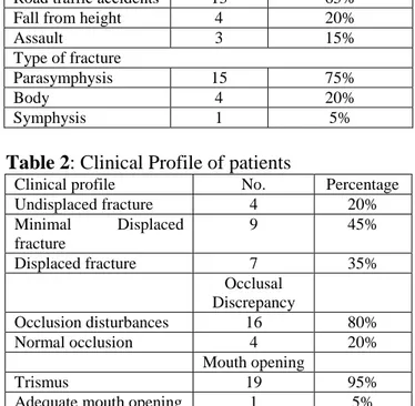
Our study showed that the all 20 patients were healthy adult males with their ages ranging from 18 to 53 year, the mean age was (28.95). Out of 20 patients in 13 (65%) patients the aetiology was road traffic accident (R.T.A.), in 4 (20%) patients the history was fall and in three (15%) patients the history was assault. In ten (50%) patients, the fracture site was right parasymphysis, in five (25%) patients the fracture site was left parasymphysis, in two (10%) patients the fracture site was right body of the mandible, in two (10%) patients the fracture site was left body of the mandible, and in one (5%) patient the fracture site was symphysis (table 1).
In four (20%) patients fractures were undisplaced. In nine (45%) patients the fracture was minimally displaced. In seven (35%) patients, the fracture was displaced, 16 (80%) patients showed disturbance of occlusion and four (20%) patients showed normal occlusion. Trismus was present in 19(95%) patients and one (5%) patient had
adequate mouth opening (table 2). Radiologically healing of the fractures was evaluated at the intervals of 1st week, 1st month, 3rd month and 6th month. None of the case had delayed healing, non-union. Adequate amount of bone healing was seen by end of 6 months.
Table 1: Profile of patients
Profile of patients No. Percentage Mean age 28.95 years Range= 18-53 yrs Cause of injury
Road traffic accidents 13 65%
Fall from height 4 20%
Assault 3 15%
Type of fracture
Parasymphysis 15 75%
Body 4 20%
Symphysis 1 5%
Table 2: Clinical Profile of patients
Clinical profile No. Percentage
Undisplaced fracture 4 20%
Minimal Displaced fracture
9 45%
Displaced fracture 7 35%
Occlusal Discrepancy
Occlusion disturbances 16 80%
Normal occlusion 4 20%
Mouth opening
Trismus 19 95%
Adequate mouth opening 1 5%
Discussion
Dr Prakhar Katta et al JMSCR Volume 06 Issue 07 July 2018 Page 554 In the management of midface fractures,
1.5-1.3mm titanium mini/microplates are routinely used. During dynamic movements of speech and mastication, the direction and magnitude of forces are similarly transmitted to maxilla and mandible. Hence, 1.3mm low profile titanium microplates can provide similar stability when used in treatment of mandibular fractures.
The current study was undertaken to treat mandibular fractures with 1.3mm low profile titanium miniplates. Twenty cases of mandibular fractures were treated with open reduction and internal fixation using thin low profile titanium miniplates and 1.3mm self-threading screws. To overcome the effects of tension and compression which occur at the fracture site during loading as well, to overcome the possible complication of plate fracture because of its thinness and malleability, two microplates fixation was considered more effective than one. Three screws of 1.3mm self threading in nature were used on either side of the fractured segments.
Duration of intermaxillary fixation ranged from 5-10 days. Patients were evaluated for complications during a follow up period till six months for adequate jaw opening, occlusion, infection, wound dehiscence and radiological assessment of bone healing.
Postoperatively adequate mouth opening was achieved in all the patients with normal occlusion. One case (5%) showed postoperative infection of wound dehiscence, which was managed by daily irrigation.
The results in our study are comparatively better than other studies of Passeri et al., (1993)4 who reported 17% of postoperative infection whereas, Kim et al5,6 reported post operative infection of 7.9%.
However, we advocated 5-10 days of postoperative IMF. Histologically during fracture healing primary callus formation takes place about 10 days and subsequently there is enough stability as stated by B.G. Miller (1990)7 Thus the advantage of using 1.3mm titanium miniplates are, thinness of plates, ease of application, easy
adaptation, it cannot be palpated through the skin, minimum incision is required and are comparable to the Champy’s system.
However, the sample size in this study is only 20 cases. These plates and screws are indigenously developed. To establish the efficacy of this system, the components of titanium plates, their histological response in the soft tissue and a larger sample is necessary.
Conclusion
We concluded that the plates due to their thinness and flexibility don’t require adaptation. They will adapt to the mandible by itself while tightening of the screws. Minimum pressure is required during tightening of the screws as excessive pressure can lead to breaking of screw head.
References
1. Kruger E., Schilli W., “Oral and maxillofacial traumatology” 1982, Chicago, Quintessence Publishing Co., page 181.
2. Fonseca Raymond J. and Robert V. Walter 1997 “Oral and Maxillofacial Trauma”, Pennsylvania, W.B. Saunders Company, 2nd Edition Vol 1: 474-478 J. Oral Maxillofac. Surg., 52 : 1032-1036.
3. J.L. Williams Rowe and Williams, “Maxillofacial Injuries”, London, Churchill Livingstone 1994, 2nd Edition: Vol 2,: 644-645.
4. Passeri L. A., E. Ellis III, F. Sinn. “Complications of non rigid fixation of mandibular angle fractures”. J. Oral Maxillofac. Surg.,1993; 51 : 382-384. 5. Young-kyun-kim, Hwan-ho yeo,
Seung-cheul lim. “Tissue response to titanium plates – A transmitted electron microscopic study” J. Oral Maxillofac. Surg., 1997; 55:322-326
Dr Prakhar Katta et al JMSCR Volume 06 Issue 07 July 2018 Page 555 Reconstructive Surgery,2001; Vol.108.
No.1, 38-43.