Case Report
A rare case of hydatid cyst of the interventricular septum
Monila Patel, Rafe Khan, Ruchir Dave, Jyoti Vora, Sneha Shah, Shreyans D. Singhvi*,
Preksha T. Singh
INTRODUCTION
A Hydatid disease or Echinococcosis is a zoonotic disease caused by the larvae (met cestode) of the cestode species of the genus Echinococcus like E. granulosus, E. multilocularis, E. vogeli or E. oligarthus.1 According to
study in 2009, it is estimated that the worldwide incidence of cystic echinococcosis is about 100,000-300,000 cases annually.1
Humans acquire primary cystic echinococcus by ingestion of E. granulosus eggs excreted by infected carnivores usually canines. Humans are not the definite host of the disease. There are several intermediate hosts of Echinococcus. They get infected from the parasite by ingesting the eggs of the parasite and in the viscera of these hosts the eggs develop into the larval stages. Humans are accidental hosts of the disease. The infection may be acquired by contact with infected definitive hosts, egg-containing feces, or egg-contaminated plants or soil followed by their ingestion.2
Human echinococcosis occurs in 4 forms- cystic echinococcosis, alveolar echinococcosis, polycystic echinococcosis and unicystic echinococcosis, but the there are two forms which are more common in humans, these are-cystic echinococcosis or hydatidosis and alveolar echinococcosis.
The disease can be prevented by programs like deworming of canines, which are the definite host of the disease.
The disease is known to occur in all continents except Antarctica and in 100 countries worldwide.3 Hydatid
disease is endemic in India.4 For India, the highest
prevalence has been reported from the erstwhile state of Andhra Pradesh, Tamil Nadu and Saurasthra region of Gujarat.4
This case report deals with a rare occurrence of hydatid cyst in the Interventricular Septum. Cardiac involvement in case of hydatid cyst ranges from 0.5-2%.5
Department of Internal Medicine, Sardar Vallabhbhai Patel Institute of Medical Sciences and Research, Ahmedabad, Gujarat, India
Received: 17 February 2020
Accepted: 20 March 2020
*Correspondence:
Dr. Shreyans D. Singhvi,
E-mail: shreyanssinghvi4@gmail.com
Copyright: © the author(s), publisher and licensee Medip Academy. This is an open-access article distributed under the terms of the Creative Commons Attribution Non-Commercial License, which permits unrestricted non-commercial use, distribution, and reproduction in any medium, provided the original work is properly cited.
ABSTRACT
A Hydatid disease or Echinococcosis is a zoonotic disease caused by the larvae (metacestode) of the cestode species of the genus Echinococcus. Humans are the accidental hosts of the diseases; they usually acquire it from canines; which are the definite host. It can present with systemic cyst, while cardiac manifestation of the disease is rare, due to contractile property of the heart’s muscle fiber which provide resistance. In this case report, the patient is diagnosed with hydatid cyst in the inter ventricular septum; it’s diagnosis and its successful treatment with surgery and albendazole. As, inter ventricular septum hydatid cyst occurs in only 0.5-2% cases, it’s a unique case and its successful treatment and diagnosis can help the physicians in the future to treat a similar case as this.
Keywords: Cardiac, Cyst, Hydatid, Interventricular, Septum
CASE REPORT
A 36-year-old female patient presented to the OPD of the department of medicine of a tertiary care hospital of Ahmedabad with chief complaints of occasional chest pain and dyspnoea on accustomed exertion since the past 1 month.
The general physical examination was performed. She was well alert and oriented to time, place and person. Her vitals were taken which recorded a pulse rate of 96 beats per minute, in the right radial artery. She was afebrile and her Blood pressure recorded was 100/60mmHg and a saturation of 98% on room air with bilateral normal breath sounds.
Furthermore, cardiac examination was performed during which- Cardiac auscultation revealed an ejection systolic murmur in left parasternal region.
Her laboratory investigations are as follows-
Hemoglobin (hb): 9.8mg/dl (normal range:12.0-15.5 mg/dl),
Total leucocyte count: 15,400/mm3 (normal range-3600-11,200/mm3),
Erythrocyte sedimentation rate (ESR): 24mm/hr (normal: <20 mm/hr),
Platelets count- 2.4 lakh/cumm (normal range :1.7 lakh/cumm- 3.0 lakh/cumm),
Blood Potassium - 4.5 mmol/L,
Serum Creatinine- 0.58 mg/dl.
Radiological examination was performed -Chest X ray with Postero-anterior View (PA View) - which did not give any significant results.
Further, an Electrocardiography (ECG) investigation was performed which revealed a sinus Rhythm.
Later, 2d Echocardiography was performed for further evaluation, which revealed- Left ventricular ejection fraction - 66%, No regional wall abnormalities (RWMA) at rest, trivial Mitral Regurgitation, trivial Aortic regurgitation and trivial tricuspid regurgitation.
It also revealed an Inter-ventricular Septal Cyst with Mid Cavitary Obstruction.
Also, the Left ventricular Size appeared normal with fair Left ventricular function with reduced Left ventricular compliance.
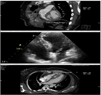
The next investigation performed was a Multislice Computed Tomography Angiogram (MSCT) which provided the following results- Profound evidence of large, rounded, well-defined, thin- walled, minimally enhancing, cystic lesion involving inter ventricular septum- projecting towards both right and left ventricles
at mid- cavity level with external compression over ventricular cavities (left ventricle more than right ventricle)- This suggested the probable diagnosis of hydatid cyst of approximate size 48.3 x 36.5 mm in axial plane with craniocaudal extent of 44.2 mm (Figure 1).
Figure 1: Cardiac multislice computed tomography (MSCT) - pre-operatively.
The patient was started on oral Albendazole and was referred to the CTVS department for surgical excision of the cyst. The cyst was excised successfully leaving behind a pseudo aneurysm.
The diagnosis of hydatid cyst disease was confirmed by histopathological examination following the operation. The patient was discharged on oral Albendazole following an uneventful postoperative stay.
After the successful surgery - 2d Echocardiography was performed which had the following results- Left Ventricular Ejection fraction - 60%, No regional wall abnormalities at rest. There was still presence of Mild Mitral Regurgitation, Trivial Aortic Regurgitation and Mild Tricuspid Regurgitation.
There was presence of a freely mobile structure attached to mid septal capsule. There was absence of any effusion.
Figure 2: Multislice computed tomography (MSCT) done post- operatively.
DISCUSSION
Hydatid cyst can affect various systemic organs and therefore, it can present a variety of signs and symptoms. This was a particularly rare manifestation of hydatid cardiac disease which in itself is an uncommon entity accounting for 0.5-2% of cases of cystic Echinococcus disease.5 The most affected areas of cardiac involvement
are the left ventricle (60-70%) and right ventricle (10%), while inter-ventricular septum, pericardium, and atria are the least affected.5
The clinical symptoms of cardiac hydatid cyst disease depend on various factors which include- the localization, size, age, the number of the cysts and the extent of calcification. In multiple cases of the disease, signs and symptoms may develop resulting from compression of the nearby cardiac structures such as - coronary artery, inter cardiac chamber or the conductive system of the heart. Electrocardiographic changes have been reported also, such as myocardial infarction, arrhythmias, bundle branch conduction disturbances, and sudden cardiac death.6,7
Cardiac hydatid disease, though rare, can be fatal with complications like cardiac tamponade and disseminated disease. While, it’s also given that the patients can remain asymptomatic during throughout; It is estimated that up to 10% of the patients remain asymptomatic for many years, although they are under the continuous threat of rupture. Mostly, the patients asymptomatic during the early states, diagnosis is most important during this stage of the disease to prevent dissemination, a careful clinical examination is crucial in early detection of the disease, Since It has been reported that up to 20% of fatal cases present with sudden death having previously shown no signs or symptoms of cardiac echinococcosis.7
In this case, the hydatid cyst was formed in the inter-ventricular system. According to a research study, involvement of the inter-ventricular septum occurs only in 4% of the cardiac cases.8 Alternatively, sometimes a
cardiac hydatid cyst may simulate mitral, pulmonary, and tricuspid valvular diseases.9
There are various clinical manifestations that can present to the hospital, they vary according to the cyst site, size, number and are due to related complications. Symptoms are nonspecific and include atypical chest pain, breath shortness, asthenia, and palpitations.10 Chest pain may be
due to compression of the small coronaries mimicking coronary artery disease and sometimes causing ischemic changes in the electrocardiogram.
Patients with a cardiac hydatid cyst usually have symptoms after its rupture. Sudden death caused by anaphylactic shock may occur after the rupture of a cyst.11 The rupture may be silent, and metastatic
echinococcosis of various organs may be a lateand often solely due to the presence of an underlying cardiac involvement.12 Rupture into the pericardium may result in
pericardial effusion, cardiac tamponade, and, in rare cases, constrictive pericarditis.13,14 Rupture in the
right-side chamber may lead to pulmonary emboli and rupture in the left side chamber may lead to systemic emboli; these can be grave situations for the patient.
Pulmonary embolism resulting from a hydatid cyst may cause pulmonary hypertension that is chronic, subacute, or acutely fatal.15 Several of thesepatients appear to
follow a path of continued pulmonary hypertension with acute attacks of acute pulmonary embolism. Pulmonary emboli are caused by vesicles or daughter cysts and by scolices.16
The rupture of a left-sided hydatid cyst may lead to form a cerebral embolus. As a contrast to primary cysts of central nervous system; these cysts are found in multiple numbers. Also, secondary multiple hydatid cysts of the central nervous system caused by cardiac embolization are infertile.17
The marked incidence of catastrophic complications of cardiac hydatid cyst emphasizes the need for early diagnosis.18 currently, the following tests have lost their
value due to their false positive results- Casoni’s intradermal test, the Weinberg reaction, and a peripheral blood eosinophil count. A definite diagnosis based on electrocardiography and chest radiography is not possible and adequate. Since the rupture of cyst can happen during Cardiac catheterization which can lead to an anaphylactic shock, its dangers outweigh its benefits. Furthermore, angiography cannot visualize a small intramural cyst and ruptured or degenerated cysts. Two-dimensional echocardiography allows the differentiation of a cyst from a solid mass.18 In addition, computed tomography
Two-dimensional echocardiography allows the differentiation of a cyst from a solid mass.20 Furthermore,
computed tomography can also be used, as it was used in this case.
A trans-thoracic echocardiogram can prove invaluable in the diagnosis of cystic lesions and structural anomalies after which a Trans-oesophageal echocardiography and further imaging can be done if required.
Surgical management with oral albendazole therapy remains the mainstay of treatment. The main principle of surgical treatment is to empty the cyst, remove daughter cysts and the germinative membrane, excise the pericyst, and then obliterate the residual cavity with sutures (capitonnage). The use of local scolicidal solution such as hypertonic saline solution is obligatory after cysto-pericystectomy in order to minimize the risk of dispersion of cystic content.21
CONCLUSION
As, Hydatid cyst occuring in the inter-ventricular septum is an extremely rare phenomenon, it can be easily misdiagosed and wrongly treated, therefore; this case report is of extreme value as it has provided the method of diagnosis as well as successful treatment of the hydatid cyst in this rare case.
ACKNOWLEDGEMENTS
Authors would like to thank the patient to give us the consent to present the case. Authors would also wish to thank the Department of medicine, Sardar Vallabhbhai Patel Institute of Medical Science and research for guiding and helping us in the quick diagnosis and treatment of the patient.
Funding: No funding sources Conflict of interest: None declared Ethical approval: Not required
REFERENCES
1. Kern P, Bardonnet K, Renner E, Auer H, Pawlowski Z, Ammann RW et al. European Echinococcosis Registry. European echinococcosis registry: human alveolar echinococcosis, Europe, 1982-2000. Emerg Infect Dis. 2003;9(3):343-9.
2. Larrieu EJ, Costa MT, del Carpio M, Moguillansky S, Bianchi G, Yadon ZE. A case-control study of the risk factors for cystic echinococcosis among the children of Rio Negro province, Argentina. Ann Trop Med Parasitol. 2002;96:43-52.
3. Eckert J, Deplazes P . Biological, epidemiological, and clinical aspects of echinococcosis: A zoonosis of Increasing Concern. Clin Microbiol Rev. 2004;17:107-35.
4. Tiwary AK, Tiwary RN. Hydatid disease in Chotanagpur region of South Bihar. Indian J Surg. 1988;50:14-8.
5. Perez-Gomez F, Duran H, Tamames S, Perrote JL, Blanes A. Cardiac echinococcosis: clinical picture and complications. Br Heart J. 1973;35(12):1326-31.
6. Vestri A, Nigri A, Massi L, Reale A. Electrocardiographic picture of myocardial infarct during echinococcosis of the heart. Boll Soc Ital Cardiol. 1972;17:752-4.
7. Miralles A, Bracamonte L, Pavie A, Bors V, Rabago G, Gandjbakhch I et al. Cardiac echinococ- cosis surgical treatment and results. J Thorac Cardiovasc Surg. 1994;107:184-90.
8. Dursun M, Terzibasioglu E, Yilmaz R, Cekrezi B, Olgar S, Nisli K, et al. Cardiac hydatid disease: CT and MRI findings. AJR Am J Roentgenol 2008;190(1):226-32
9. Hazan E, Leblanc J, Robıllard M, Mathey J. Hydatid cyst of the right ventricle revealed by an acute complication: emergency exeresis with prosthetic replacement of the tricuspid valve. Chirurgie. 1970;96:257-60.
10. Charfeddine S, Mallek S, Gueldiche M, Triki F, Jmâa HB, Frikha I, et al. A huge cardiac hydatid cyst: An unusual cause of chest pain revealing multivisceral hydatidosis in a young woman. J Saudi Heart Assoc. 2015;27(4):286-91.
11. Madariaga I, Fuente A, Lezaun R. Cardiac echinococ- cosis and systemic embolism: report of a case. Thorac Cardiovasc Surg. 1984;32:57-9. 12. Di Bello R, Menendez H. Intracardiac rupture of
hydatid cyst of the heart: a study based on three personal observations and 101 cases in the world literature. Circulation. 1963;27:366-74.
13. Di Bello R, Abo JC, Borges L. Hydatid constrictive peri- carditis: a new case and review of the literature. J Thorac Cardiovasc Surg. 1970;59:530-2. 14. Halliday JH, Jose AD, Nicks R. Constrictive pericarditis fol- lowing rupture of a ventricular hydatid cyst. Br Heart J. 1963;25:821.
15. Bayezid Ö, Öcal A, Is ̧ık Ö, Okay T, Yakut C. A case of cardiac hydatid cyst localized on the interventricular septum and caus- ing pulmonary emboli. J Cardiovasc Surg. 1991;32:324-6.
16. Gilsanz V, Campo C, Cue R, Estella J, Estrada RV, Perez-oteiza C, et al. Recurrent pulmonary embolism due to hydatid disease of heart: study of 3 cases, one with intermittent tricuspid valve obstruction (atrial pseudomyxoma) Br Heart J. 1977;39:553-8.
17. Turgut M, Benli K, Eryılmaz M. Secondary multiple intracranial hydatid cysts caused by intracerebral embolism of cardiac echinococcosis: an exceptional case of hydatidosis. J Neurosurg. 1997;86:714-8. 18. Malouf J, Saksouk FA, Alam S, Rizk GK, Dagher I.
19. Oliver JM, Sotillo JF, Domínguez FJ, López de Sá E, Calvo L, Salvador A, et al. Two-dimension- al echocardiographic features of echinococcosis of the heart and great vessels. Circulation. 1988;78:327-37.
20. Farooki ZQ, Adelman S, Green EW. Echocardiographic dif- ferentiation of a cystic and a solid tumor of the heart. Am J Cardiol. 1977;39:107-11.
21. Salih OK, Celik SK, Topcuoğlu MS, Kisacikoğlu B, Tokcan A. Surgical treatment of hydatid cysts of the
heart: a report of 3 cases and a review of the literature. Can J Surg. 1998;41(4):321-7.