Dr Prasun Sagar et al JMSCR Volume 07 Issue 11 November 2019 Page 232
Clinical and Laboratory Evaluation of Patients with Fever with
Thrombocytopenia
Authors
Dr Prasun Sagar
1, Dr Hardik Patel
2, Dr Sandeep Rai
31,2
Junior Resident, Department of General Medicine 3
Professor and Head of Unit, Department of General Medicine
Introduction
Thrombocytopenia is a disorder in which there are too few platelets in the blood. Platelets are small, disk-shaped cellular structures in the bloodstream that help the blood to clot. Thus thrombocytopenia is often characterized by excessive bleeding, including epistaxis and easy bruising. Thrombocytopenia can be diagnosed by a routine blood test. Thrombocytopenia arises for one of three reasons, the bone marrow may not produce enough platelets, too many platelets may be destroyed in the spleen, and thrombocytopenia can be caused by a variety of conditions.
Acute fever with thrombocytopenia (platelet count less than 150,000) is a common clinical problem in our medical wards. Febrile thrombocytopenia is the thrombocytopenia associated with fever. Diseases which commonly present with fever and thrombocytopenia are malaria, leptospirosis, rickettsial infections, septicemia, typhoid, borreliosis, arbovirus such as dengue or yellow fever, rodent-borne viruses such as Hanta and Lassa fever, human immunodeficiency virus (HIV), visceral leishmaniasis and TTP-HUS.1
Thrombocytopenia can occur as a complication of diseases such as leukemia or HIV infection. In some cases too many platelets are trapped and
stored in the spleen leaving too few platelets in circulation. Treatment of thrombocytopenia depends on the type and cause of platelet deficiency. Thrombocytopenia in fever being a prognostic factor can predict the cause and thus helps in early diagnosis and treatment of the same, preventing further fatal outcome associated with it such as intracerebral bleed, hemorrhage into vital organs, shock and finally leading to death.
Majority of studies on adult with thrombocytopenia have focused on specific etiology or associated with some symptom like fever. On the other hand, Bhalara et al has taken different etiologies in consideration for one month duration study on adults during rainy season where mosquito transmitted diseases were the commonest cause.2
Though patients can initially present with simple fever in due course it can lead to unpredictable outcomes including death at times therefore the aim of the study is to analyze the etiological factors of thrombocytopenia, as early diagnosis and timely intervention prevents adverse outcomes and saves life.
Aims and Objectives
To study clinical and laboratory evaluation of patients with fever with thrombocytopenia
http://jmscr.igmpublication.org/home/ ISSN (e)-2347-176x ISSN (p) 2455-0450
Dr Prasun Sagar et al JMSCR Volume 07 Issue 11 November 2019 Page 233 attending M.G.M Hospital Kamothe, Navi
Mumbai
Material and Methods
This prospective observational study was conducted on 100 patients of fever with thrombocytopenia attending to M.G.M Hospital Kamothe, Navi Mumbai during the period from November 2016 to October 2018. Patients presenting with fever, temperature of more than 100° F, and thrombocytopenia i.e. platelet count of < 1.5 lakhs were enrolled in the study. Patients with known inherited causes for thrombocytopenia, patients on drugs causing thrombocytopenia, known Autoimmune causes for thrombocytopenia, HIV infection, leukemia’s and myelodysplastic syndromes and other blood dyscrasias were excluded.
Methodology
Patients presenting to department of medicine in OPD or Emergency ward with complaints of Fever, and on investigation found to be having thrombocytopenia were screened for this study and those fulfilling Inclusion criteria were enrolled in the study after obtaining informed consent.
Patients were selected with the Simple random sampling method. Patients taken up for the study, and their details were entered in semi-structured proformas which included sociodemographic details, clinical history of patients, general and systemic examination, and Investigations. Further follow of patients in regard to their clinical prognosis and Investigations including platelet count were taken on regular interval, and depending on their clinical profile. Meanwhile, simultaneous records of diagnosis, treatment, prognosis were made as usual. Records were collected for 100 patients and then analyzed statistically through SPSS.
Statistical Analysis
All the collected data was entered in Microsoft Excel sheet and then transferred to SPSS software ver. 17 for analysis. Qualitative data was
presented as frequency and percentages and analyzed using chi-square test. P-value < 0.05 was taken as level of significance
Results
Table no 1Demographic profile
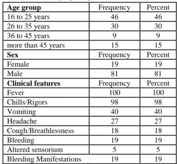
Age group Frequency Percent
16 to 25 years 46 46
26 to 35 years 30 30
36 to 45 years 9 9
more than 45 years 15 15
Sex Frequency Percent
Female 19 19
Male 81 81
Clinical features Frequency Percent
Fever 100 100
Chills/Rigors 98 98
Vomiting 40 40
Headache 27 27
Cough/Breathlessness 18 18
Bleeding 19 19
Altered sensorium 5 5
Bleeding Manifestations 19 19
The most common age group amongst study population was 16 to 25 years (46%) followed by 26 to 35 years (30%), more than 45 years (15%) and 36 to 45 years (9%). There was male predominance (81%) amongst study population as compared to female (19%).
Fever (100%) was the most common clinical features amongst study population followed by chills/rigors (98%), vomiting (40%) and headache (27%). Bleeding manifestations was present in 19 % of study population. Splenomegaly and hepatomegaly was present in 7 % and 13 % of study population respectively.
Vivax Malaria (26 %) was the most common Peripheral smear findings amongst study population followed by Falciparum malaria (1%) and mixed malaria (1%). NS1 positive (47 %) was the most common diagnostic test for Dengue amongst study population followed by IgG positive (6%) and IgG &IgM (1%).
Leptospirosis and Widal test was positive in 13% and 2% of study population respectively.
Dr Prasun Sagar et al JMSCR Volume 07 Issue 11 November 2019 Page 234 Most of the study population had platelet count of
50000-1lakh/cumm (39%) followed by 1-1.5 lakh/cumm (37 %) and less than 50,000/cumm (24%).
Acinetobacterial sp., E.Coli, Klebsiella and S. typhi was found in 1%, 1%, 1% and 1% of study population on blood culture findings respectively. On USG Abdomen/Pelvis, hepatomegaly (17%) was the most common findings amongst study population followed by hepatosplenomegaly (15%) and splenomegaly (10%). On Bone marrow findings, Erythroid hyperplasia was observed in 1% of study population.
Dengue (53%) was the most common cause of thrombocytopenia amongst study population followed by Malaria (27%), Leptospirosis (13%), Septicemia (4%), Enteric fever (2%) and Dimorphic Anemia (1%)
Blood Product Transfusion was given in 16% of study population. Less than 10 days of hospital stay was observed in 94% of the study population while More than 10 days of hospital stay was observed in 6% of study population Eighty nine
percent of the study population were alive while death was occurred in 11% of study population Deranged prothrombin time was observed most commonly in dengue (45.8%) followed by malaria (37.5%), leptospirosis (6.3%), septicemia (4.2%) and enteric fever (4.2%) and the difference was statistically significant (P value – 0.04).
Deranged INR was observed most commonly in dengue (44.4%) followed by malaria (27.8%), leptospirosis (13.9%), septicemia (8.3%) and enteric fever (5.6%) and the difference was statistically insignificant (P value – 0.181).
Blood product transfusion requirement was observed most commonly in dengue (43.8 %) followed by leptospirosis (25.0%), septicemia (18.8%) and malaria (12.5%) and the difference were statistically significant (P value – 0.01). Death requirement was occurred most commonly in dengue (36.40%) followed by leptospirosis (36.40 %) and septicemia (27.30 %) and the difference was statistically significant (P value – 0.0001)
Table no 2 Comparison of Final diagnosis with Prothrombin time amongst study population
Prothrombin time
Total
Deranged Normal
Diagnosis Dengue Count 22 31 53
% 45.8% 59.6% 53.0%
% 2.1% 0.0% 1.0%
Enteric fever Count 3 0 3
% 6.3% 0.0% 2.0%
Leptospirosis Count 3 10 13
% 6.3% 19.2% 13.0%
Malaria Count 18 9 27
% 37.5% 17.3% 27.0%
Septicemia Count 2 2 4
% 4.2% 3.8% 4.0%
Total Count 48 52 100
% 100% 100.0% 100.0%
As seen in the above table, deranged Prothrombin time was observed most commonly in Dengue (45.8%) followed by Malaria (37.5%) ,
Dr Prasun Sagar et al JMSCR Volume 07 Issue 11 November 2019 Page 235
Table no 3 Comparison of Final diagnosis with INR amongst study population
INR
Total Deranged Normal
Diagnosis Dengue Count 16 37 53
% 44.4% 57.8% 53.0%
Dimorphic Anemia Count 0 1 1
% 0.0% 1.6% 1.0%
Enteric fever Count 2 0 2
% 5.6% 0.0% 2.0%
Leptospirosis Count 5 8 13
% 13.9% 12.5% 13.0%
Malaria Count 10 17 27
% 27.8% 26.6% 27.0%
Septicemia Count 3 1 4
% 8.3% 1.6% 4.0%
Total Count 36 64 100
% 100% 100.0% 100.0%
As seen in the above table, deranged INR was observed most commonly in dengue (44.4%) followed by malaria (27.8%), leptospirosis
(13.9%), septicemia (8.3%) and enteric fever (5.6%) and the difference was statistically insignificant (P value – 0.181).
Table no 4 Comparison of Final diagnosis with Blood Product Transfusion amongst study population
Blood Product Transfusion
Total
No Yes
Diagnosis Dengue Count 46 7 53
% 54.8% 43.8% 53.0%
Dimorphic Anemia Count 1 0 1
% 1.2% 0.0% 1.0%
Enteric fever Count 2 0 2
% 2.4% 0.0% 2.0%
Leptospirosis Count 9 4 13
% 10.7% 25.0% 13.0%
Malaria Count 25 2 27
% 29.8% 12.5% 27.0%
Septicemia Count 1 3 4
% 1.2% 18.8% 4.0%
Total Count 84 16 100
% 100.0% 100.0% 100.0%
As seen in the above table, Blood product transfusion requirement was observed most commonly in dengue (43.8%) followed by
leptospirosis (25.0%), septicemia (18.8%) and malaria (12.5%) and the difference was statistically significant (P value – 0.01).
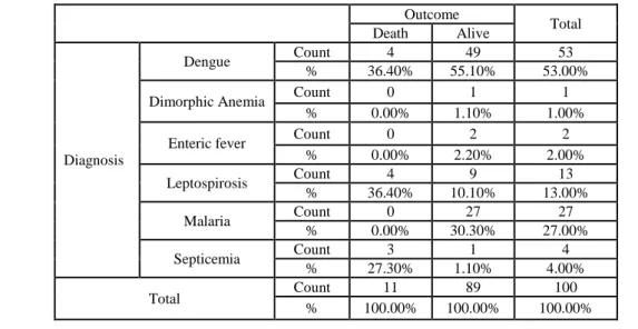
Table no 5 Comparison of Final diagnosis with Outcome amongst study population
Outcome Total
Death Alive
Diagnosis
Dengue Count 4 49 53
% 36.40% 55.10% 53.00%
Dimorphic Anemia Count 0 1 1
% 0.00% 1.10% 1.00%
Enteric fever Count 0 2 2
% 0.00% 2.20% 2.00%
Leptospirosis Count 4 9 13
% 36.40% 10.10% 13.00%
Malaria Count 0 27 27
% 0.00% 30.30% 27.00%
Septicemia Count 3 1 4
% 27.30% 1.10% 4.00%
Total Count 11 89 100
Dr Prasun Sagar et al JMSCR Volume 07 Issue 11 November 2019 Page 236 As seen in the above table, death occurred most
commonly in dengue (36.40%) and leptospirosis (36.40%) each and septicemia (27.30%) and the difference was statistically significant ( p value – 0.0001). The common causes of death were dengue hemorrhagic fever leptospirosis with ARDS and septicemia with shock.
Discussion
Thrombocytopenia (TCP) refers to a reduction in platelet count below 1.5lakh/microlitre.3 It is the commonest platelet abnormality encountered in clinical practice with variable clinical expression. With the widespread use of automated cell counters clinicians in any field may encounter TCP. The symptomatology may vary greatly and the underlying cause may be either inconsequential or life threatening. 4 In a tropical country like India infectious causes predominate and are usually associated with fever.5 TCP may give a clue to presence of infections like malaria, dengue, leptospirosis and viral infections.
Febrile thrombocytopenia is a distinct clinical entity, commonly encountered in infectious diseases. A number of infections such as malaria, dengue fever, scrub typhus, leptospirosis, chickungunya, enteric fever, bacterial and fungal sepsis as well as certain other viral infections result in thrombocytopenia. The varied etiological profile and unpredictable clinical outcome often poses a diagnostic as well as therapeutic challenge to clinicians.
Age group
In the present study, 16 to 25 years (46%) was the most common age group amongst study population followed by 26 to 35 years (30%), more than 45 years (15%) and 36 to 45 years (9%). This findings is in agreement with the study conducted by Yasmeen Khatib et al., in which there were 68 cases (22.67%) seen in the 21-30 year age group followed by 58 patients in the 31-40 year age group.(19.33%).6 This findings is also in agreement with the study conducted by Smita Masamatti et al ., in which the commonest age group affected was between 18 to 36 years
(48.27%), the reason for this increased incidence in males and younger age group in the given locality has been attributed to the prolonged outdoor activities and increased chances of exposure to mosquitoes and also majority of the women being homemaker.7 In another study by Shah et al maximum cases of thrombocytopenia were also found in the 21-30 years age group and male preponderance was seen as in our study.8
Sex
In the present study, there was male predominance (81%) amongst study population as compared to female (19%). This findings is in agreement with the study conducted by Yasmeen Khatib et al., in which there were 176(58.67%) males and 124(41.33%) females. 6 This findings is also in agreement with the study conducted by Bhalara, et al in which out of 412 cases (236 male and 176 female patients).2 According to another study by Badvi A. J. et al, male to female ratio was 64:36 and 77% of patients were in age group under 10 years.9 Similar sex distribution was seen in certain local and international studies.10-12
Clinical Features
In the present study, Fever (100%) was the most common Clinical features amongst study population followed by Chills/Rigors (98%), Vomiting (40%) and Headache (27%). This findings is in agreement with the study conducted by Yasmeen Khatib et al., in which The main presenting features in patients with TCP were fever 205 cases (68.3%).6 This findings is also in agreement with the study conducted by Bhalara, et al in which fever associated with thrombocytopenia was found in 327 (79.3%).2 In another study by Ahmed S et al study,16 frequently noted clinical features included fever (97%), vomiting (68%), abdominal pain (68%) and rashes (65%). Gastrointestinal bleeding (61%) and epistaxis (26%) were commonest haemorrhagic manifestations.11
Dr Prasun Sagar et al JMSCR Volume 07 Issue 11 November 2019 Page 237 with bleeding tendencies was seen in 70 cases
(23.3%).6 Nair et al 4 have reported bleeding manifestations in 41.3% of their cases while Shah et al have reported them in 30% of their cases.8 Signs of bleeding were reported in 24% children and in 23% adults by Kuhne T et al.10
Examination findings
In the present study, Splenomegaly and Hepatomegaly was present in 7 % and 13 % of study population respectively. This findings is in agreement with the study conducted by Yasmeen Khatib et al., in which hepatomegaly 48 cases (16%), splenomegaly 46 cases (15.3%).16
Peripheral smear findings
In the present study, Vivax Malaria (26 %) was the most common Peripheral smear findings amongst study population followed by Falciparum Malaria (1%) and Mixed malaria (1%).This findings is in agreement with the study conducted by Guruprasada Shetty et al study,30 Plasmodium vivax found in 66%, Plasmodium falciparum in 16% and mixed in 18% of cases.13 This findings is in agreement with the study conducted by Naveen Kulkarni et al., vivax malaria, falciparum and mixed infections were noted in 53.2%, 26.6% and 19 % cases respectively.14
Platelet count
In the present study, most of the study population had Platelet count of 50000-1lakh/cumm (39%) followed by 1-1.5 lakh/cumm (37 %) and less than 50,000/cumm (24%).This findings is in agreement with the study conducted by Bhalara, et al in which out of 109 patients, 62 (56.8%) had platelet count between 50,000 and 1,00,000, followed by 28 (25.7%) patients who had count between 20,000 and 50,000. 2 This findings is in agreement with the study conducted by Yadav et al., most of the study population had platelet distribution 50,001-1, 00,000/cumm was seen in 53.8% cases.15 Similar results (56.8%) were obtained in Nair PS et al study for this platelet distribution range.4
Final diagnosis
In the present study, Dengue (53%) was the most common cause of thrombocytopenia amongst
study population followed by Malaria (27%), Leptospirosis (13%), Septicemia (4%), and Enteric fever (3%). This findings is in agreement with the study conducted by Bhalara, et al in which the most common etiology responsible for newly diagnosed thrombocytopenia in adult patients was found to be dengue/dengue-like fever (28.6%).2 This findings are also seen in the study conducted by Smita Masamatti et al., in which the commonest etiology for fever with thrombocytopenia was Dengue (48.28%), followed by septicemia (19.83%), typhoid (15.52%). It was more common among males (64.82%) than in females (35.18%).7 In another study by Badvi A. J. et al study, enteric fever contributed to about 5% of cases of febrile thrombocytopenia which is similar to the present study.9 Dengue and malaria were the common causes due to the higher prevalence of these infections during the study period (October), which may be the reason for variation between different studies. The pathogenesis of thrombocytopenia in dengue fever is not clearly understood. Increased peripheral destruction of antibody coated platelets is strongly suspected as the possible mechanism. Other modes include acute bone marrow suppression leading to amegakaryocytic condition, mild DIC like presentation and enhanced platelet destruction by the reticuloendothelial system.16
Morbidity
In the present study, Blood Product Transfusion requirement was observed most commonly in Dengue (43.8 %) followed by Leptospirosis (25.0%), septicemia (18.8%) and Malaria (12.5%) and the difference was statistically significant (P value – 0.01).
Blood Product Transfusion was given in 16% of study population. In a study by Anubha Sharma blood product transfusion was given in 38% of cases.17
Dr Prasun Sagar et al JMSCR Volume 07 Issue 11 November 2019 Page 238
Final outcome
In the present study, 89% of the study populations were Alive while death was occurred in 11% of study population. In the study by Lohitashwa et al septicemia accounted for 78% and dengue for 22% of mortality.18 Kumar et al study on fever with thrombocytopenia found 9.47% mortality where 83.33% were due to septicemia with multiorgan dysfunction and 16.67% were due to complicated malaria.19
Coagulation profile vs Final diagnosis
In the present study, deranged Prothrombin time was observed most commonly in Dengue (45.8%) followed by Malaria (37.5%), Leptospirosis (6.3%), septicemia (4.2%) and Enteric fever (6.3%) and the difference was statistically significant ( P value – 0.04). similarly in the study by Tejas N. Modi et al., Viremia 241(61.32%) constitutes the commonest a etiology of febrile thrombocytopenia of which 220(55.98%) were of dengue fever and 21(5.34%)viral infection other than dengue, followed by Malaria 102(25.95%), Septicemia 21(5.34%), Megaloblastic anemia 11(2.79%), Hematological malignancy 11(2.79%) and Enteric fever 6(1.58%). 20
In the present study, deranged INR was observed most commonly in dengue (44.4%) followed by malaria (27.8%), leptospirosis (13.9%), septicemia (8.3%) and enteric fever (5.6%) and the difference was statistically insignificant (P value – 0.181). Many studies have shown an association of dengue with TCP which if severe can lead to bleeding tendency.21-23 Leptospirosis can also present with TCP.24, 25
Outcome vs Final diagnosis
In the present study, Death was occurred most commonly in Dengue (36.40%) followed by Leptospirosis (36.40 %) and septicemia (27.30 %) and the difference was statistically significant (P value -0.0001). The common causes of death were Dengue Hemorrhagic Fever Leptospirosis with ARDS, septicemia with shock
Conclusion
Dengue is the most common diagnosis made in adult patients who present with fever and are detected to have thrombocytopenia at admission followed by malaria and leptospirosis. Most of the study population had Platelet count of 50000-1lakh/cumm followed by 1-1.5 lakh/cumm and less than 50,000/cmm. In malaria, P. vivax was most common followed by P. falciparum infection and then mixed infection. Nineteen percent of patients with thrombocytopenia tend to have bleeding manifestations, most common being the bleeding from gums. Dengue was the most common cause of thrombocytopenia followed by Malaria, Leptospirosis and Septicemia. Blood Product Transfusion was needed in 16% of the patients.
References
1. Lee GR, Foerster J, Lukens J, Paraskevas F, Greer JP, Rodgers GM. Shirley Parker Levine - Miscellaneous causes of thrombocytopenia. In: Wintrobe’s Clinical Haematology. Vol. 2, 10th edition, Lipincott Williams: Philadelphia 1999; 1623-1629.
2. Bhalara SK, Shah S, Goswami H, Gonsai RN. Clinical and etiological profile of thrombocytopenia in adults: A tertiary-care hospital based crosssectional study. Int J Med Sci Public Health. 2015;4:7-10. 3. Platelet and Blood vessel disorders. In:
Richard E Behrman, Robert M Kliegman, Hal B Jensen, eds. Nelson Textbook of Pediatrics, 17th Edition. Saunders, 2004: 1770 – 77.
4. Sekhon SS,Roy Vivek.Thrombocytopenia in adults: A practical approach to evaluation and management. Southern medical journal.2006;vol99(5) 491-498. 5. Nair PS, Jain A, Khanduri U, Kumar V. A
Dr Prasun Sagar et al JMSCR Volume 07 Issue 11 November 2019 Page 239 6. Yasmeen Khatib, Dr. Vaishali Jain , Dr.
Richa Patel, One year study of thrombocytopenia in a peripheral hospital of Mumbai, 2016; Volume 6:Issue 4: PP. 26-30
7. Smita Surendra. Masamatti et al., Laboratory and Etiological Profile of Febrile Thrombocytopenia Cases, National Journal of Laboratory Medicine. 2016 Jul, Vol-5(3): PO44-PO48
8. Shah HR, Vaghani BD, Gohel P,Virani BK. Clinical profile review of patients with thrombocytopenia:A study of 100 cases at a tertiary care centre. Int J Cur Res Rev.2015;vol 7 (6):33-37.
9. Jawed Ahmed Badvi, Bahawaluddin Jamro, Aftab Ahmed Soomro, Shankar lal, Saifullah Jamro. An experience of thrombocytopenia in children at tertiary care hospitals sukkur and larkana MC. 2012;19:23-26.
10.Kuhne T, Berchtold W, Michaels LA, Wu R, Donato H et al. Newly diagnosed immune thrombocytopenia in children and adults: a comparative prospective observational registry of the International Cooperative Immune Thrombocytopenia Study group. Haematologica J. 2011;96:1831-1837.
11.Ansari S, Khoharo HK, Abro A, Akhund IA, Qureshi F. Thrombocytopenia in plasmodium malaria. J Ayub Med Coll Abbottabad. 2009;21:145-147.
12.Kibria SG, Islam MDU, Chowdhury AJ, Ali MY, Haque MR, Mustanzid SM, Ali SY. Prevalence of hematological disorder; A bone marrow study of 177 cases in a private hospital at Faridpur. Faridpur Med Coll J. 2010;5:11-13.
13.Guruprasada Shetty et al., Thrombocytopenia in children with malaria–A study from coastal Karnataka, India, Asian Pacific Journal of Tropical Disease Volume 2, Issue 2, April 2012, Pages 107-109
14.Naveen Kulkarni et al / A clinical study of febrile thrombocytopenia at a Tertiary Care Hospital in North Karnataka , IJBR (2017) 08 (01)
15.Vishal Yadav, Abhishek Singhai, Study of febrile thrombocytopenia in Malwa region of India Asian Journal of Medical Sciences | Sep-Oct 2017 | Vol 8 | Issue 5:83-87
16.Stasi R, Provan D. Management of immune thrombocytopenic purpura in adults. Mayo Clin Proc. 2004;79:504-22. 17.Anubha Sharma et al., A prospective
observational study of thrombocytopenia in high risk neonates in a tertiary care teaching hospital, Sri Lanka Journal of Child Health, 2015: 44(4): 213-219
18.Lohitashwa SB, Vishwanath BM, Srinivas G. A study of clinical and lab. Profile of fever with thrombocytopenia. Abstract free Paper Oral Presentation – APICON,
2008. Availableat:
http://www.japi.org/march_2009/oral_pres entation
19.Kumar P, Chandra K. A Clinical study of febrile thrombocytopenia: A hospital-based retrospective study. Indian Journal of Clinical Practice. 2014;24(10):952-957 20.Modi T et al: Clinical Profile of Febrile
Thrombocytopenia , Journal of Research in Medical and Dental Science ,2016; Vol. 4 Issue 2
21.Anuradha S, Singh N P, Rizvi S N, Agarwal S K, Gur R, Mathur M D. The 1996 outbreak of dengue hemorrhagic fever in Delhi, India. Southeast Asian J Trop Med Public Health 1998 Sep; 29(3): 503 – 6.
22.Sharma S, Sharma S K, Mohan A, Wadhwa J, Dar L, Thakur S, et al. Dengue/ DHF. Clinical profile of DHF in adults during 1996. Dengue Bulletin 22: 1 – 6.
Dr Prasun Sagar et al JMSCR Volume 07 Issue 11 November 2019 Page 240 diagnostic, prognostic and therapeutic role
in DHF/ DSS in children. Ind J Hematol & Blood Transf. 2000; Vol. 18, No. 1: 13 – 15.
24.Sharma Jayashree, Suryavanshi Moushumi. Thrombocytopenia in leptospirosis and role of platelet transfusion. Asian J Transf Sci 2007 July; 2(1): 52 – 55.