www.msjonline.org pISSN 2320-6071 | eISSN 2320-6012
Original Research Article
Relationship of thyroid function with body fat, waist circumference,
sub-cutaneous and visceral adipose tissue in eu-thyroid female subjects
Sanhita Mukherjee
1*, Debasis Deoghuria
2, Diptakanti Mukhopadhyay
3INTRODUCTION
The relationship between thyroid function and body weight in euthyroid individuals has been given a great medical concern. Evidence suggests that slight variations in thyroid function, even as indicated by tests that are within laboratory reference ranges, contribute to the development of regional obesity and the tendency to gain weight.1,2 Higher serum TSH concentrations are
consistently found in obese children and adults compared with normal-weight individuals.3-6 Several previous
studies have reported associations between thyroid function parameters such as TSH and thyroid hormone levels, within the normal range, and changes in BMI and fat distribution.7-10 Few studies reported that central fat
accumulation, as assessed by waist circumference measurement, was positively and independently associated with TSH and free triiodothyronine (fT3)
ABSTRACT
Background: The relationship between thyroid function, body weight and central obesity in euthyroid individuals has been given a great medical concern. Central abdominal adiposity can be divided into adipose tissue present in subcutaneous areas (SCAT) and visceral areas (VAT). The type of adiposity may be implicated in the associations of thyroid function parameters with obesity. The aim of the present study was to evaluate thyroid function and its possible relationship with BMI, body fat percent, waist circumference, SCAT and VAT in eu-thyroid female subjects.
Methods: Randomly selected eu-thyroid female subjects were included in this study. Their BMI was estimated by metric method, body fat percent was measured by Harpenden skin fold caliper. Ultrasound (US) was used to assess regional adiposity. Serum TSH was measured from fasting serum sample by Enzyme-linked-immuno-sorbant (ELISA) Assay. Data was analysed by SPSS (version 18).
Results: A positive linear association was found between serum TSH level and BMI, body fat percent, waist circumference and VAT. (r=0.342, p <0.002, r=0.628, p <0.00, r=0.289, p <0.009, r=0.375, p <0.000). No significant association was found with SCAT and TSH. There is gradual increase in serum TSH values among three BMI groups i.e. normal weight (BMI <23 kg/m2), over-weight (BMI 23.00-24.99 kg/m2), and obese (BMI >25 kg/m2) females.
Body fat percentage, BMI and VAT were found to be significant independent predictors of TSH.
Conclusions: Selective reduction of visceral fat may induce greater beneficial effects on the parameters of thyroid hormones than subcutaneous fat reduction.
Keywords: Body fat, TSH, SCAT, VAT
1Department ofPhysiology, Calcutta National Medical College, Kolkata, West Bengal, India 2Department of Radiodiagnosis, B.S. Medical College, Bankura, West Bengal, India
3Department of Community Medicine, College of Medicine and Sagar Dutta Hospital, Kamarhati, Kolkata, West
Bengal, India
Received: 27 October 2018
Accepted: 29 January 2019
*Correspondence:
Dr. Sanhita Mukherjee, E-mail: drsanhita@gmail.com
Copyright: © the author(s), publisher and licensee Medip Academy. This is an open-access article distributed under the terms of the Creative Commons Attribution Non-Commercial License, which permits unrestricted non-commercial use, distribution, and reproduction in any medium, provided the original work is properly cited.
levels in overweight and obese euthyroid women.10
However, these results have not been confirmed by all investigators; Manji et al. reported no associations of TSH or fT4 levels with BMI in euthyroid individuals.11
Obese individuals vary in their body fat distribution. There are differences between adipose tissues present in subcutaneous areas (SCAT) and visceral adipose tissue (VAT) present in the abdominal cavity.12 These include
anatomical, cellular, molecular, physiological, clinical and prognostic differences. Anatomically, VAT is present mainly in the mesentery and omentum, and drains directly through the portal circulation to the liver. VAT compared with SCAT is more cellular, vascular, innervated and contains a larger number of inflammatory and immune cells, lesser preadipocyte differentiating capacity and a greater percentage of large adipocytes.12
It is well known that adipose tissue and especially visceral fat is implicated in the pathogenesis of the metabolic syndrome (MS). Subjects with increased central fat distribution are at higher risk for the development of cardiometabolic disease.13 Visceral and
subcutaneous fat (SF) expresses thyroid hormone receptors as well as TSH receptors that may also directly influence various functions of adipose cells.14-18
However, it is not known whether the type of adiposity could be implicated in the associations of thyroid function parameters with obesity.
The aim of the present study was to evaluate thyroid function and its possible relationship with BMI, body fat percent, waist circumference, SCAT and VAT in eu-thyroid female subjects.
METHODS
This descriptive cross-sectional study was carried out at B. S. Medical College, Bankura, West Bengal, over a period of one year (January 2014-July 2015). Institutional ethical committee clearance and informed consent from all the subjects were obtained.
All the eu-thyroid female subjects attending Biochemistry Department for testing their thyroid profile during the study period were included in this study provided they have fulfilled the following inclusion criteria. Inclusion criteria for the subjects were: no significant clinical abnormalities on physical examination, no history of intake of lipid-lowering, hypoglycemic, antihypertensive drugs, no history of cardiovascular or respiratory disease, normal ECG (Electrocardiogram), normal fasting blood sugar level (<6-10m mol/l), normal systolic blood pressure (<130mmHg), normal diastolic blood pressure (<85mmHg) for at least three measurement.
A total of 103 eu-thyroid female subjects (i.e. TSH level within 0.4µIU/ml to 3µIU/ml) 19 who fulfilled the study criteria were included in the study. Body Mass Index and Body fat percent were measured in these subjects.
Body Mass Index was estimated by metric method.20 The
standing height of the subjects was measured with the same stadiometer, without footwear; to the nearest centimetre. Weight was measured, which was the nearest to 0.1kg, with the subjects in the standing position, before lunch, with light clothes and without footwear, by using a standardized weighing scale.21
Table 1:Body density equations (Linear regression equations; Durnin and Wormersley).
Body Density= C [M (log10 Sum of all four skin folds)]
Male 17-19 yrs
20-29 yrs
30-39 yrs
40-49 yrs
50+ yrs
C 1.1620 1.1631 1.1422 1.1620 1.1715
M 0.0630 0.0632 0.0544 0.0700 0.0779
Female 16-19 yrs
20-29 yrs
30-39 yrs
40-49 yrs
50+ yrs
C 1.1549 1.1599 1.1423 1.1333 1.1339
M 0.0678 0.0717 0.0632 0.0612 0.0645
Fat %= [(4.95/Body Density)-4.5]*100
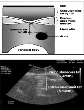
Figure 1: Abdominal adipose tissue landmark. (A): anatomical landmark of abdominal adipose tissue,
(B): illustration of abdominal adipose tissue and anatomical landmarks used for ultrasound
measurement.24
weight, subjects with a BMI value between 23 to 24.99 (kg/m2) was classified as the overweight group and those
who have a BMI value more than 25 (kg/m2) was
classified as obese.21,22 Waist circumference was
measured at the midpoint between the lower margin of the least palpable rib and the top of the iliac crest, using a stretch resistant tape.22
Harpenden skin fold caliper was used to measure percent of body fat. The four-site system was used in female subjects. Site 1-biceps, site 2- triceps, site 3- sub-scapular, site 4- supra-iliac. To calculate % Body fat linear regression equations of Durnin and Wormersley was used.23
Authors used ultrasound (US), a reliable low-cost method, to assess regional adiposity. All ultrasonographic procedures were performed by the same examiner using a 3.5-MHz probe located 1 cm from the umbilicus. Two US measurements of intraabdominal (“visceral”) and
subcutaneous fat were taken. US-determined
subcutaneous fat was defined as the distance between the skin and external face of the rectus abdominis muscle, and visceral fat was defined as the distance between the internal face of the same muscle and the anterior wall of the aorta (Figure 1).24 The intra examination coefficient
of variation for US was 0.8%.
Serum TSH was estimated by Enzyme-linked-immuno-sorbant assay (ELISA). Five ml of cord blood samples were collected from the peripheral veins in sterile test tubes. The serum separated by centrifugation was used for quantitative estimation of TSH, total T3, total T4 by a micro plate immuno-enzymatic assay (Ranbaxy). The intra and inter-assay coefficient of variation (CV) for TSH estimation were 4.33% and 7.5% respectively and the sensitivity limit was 0.078 μ IU/ml. That for total T3 was 5.73% and 6.7% respectively with the sensitivity limit 0.04 ng/ml and total T4 were 4.7% and 5.4% respectively with the sensitivity limit 0.4 μg/dl.
The collected data was analyzed with SPSS (version 18) statistical package. Pearson’s correlation test was used to see the association between TSH, BMI, Body fat Percent, WC, Subcutaneous and visceral fat.
The study population were further subdivided into three groups normal weight (BMI<23 kg/m2), over-weight
(BMI 23.00-24.99 kg/m2) and obese (BMI>25 kg/m2) in
accordance with their BMI. Analysis of Variance (ANOVA) Test among the above three groups was done to see the changes of TSH level with increasing body mass index.
As TSH, BMI, body fat could be affected by a third factor, which according to the observation could be age, author included age as independent variable in multiple regression analysis.
Multiple linear regression analysis was done to show the relation between TSH level, body fat percentage, BMI, SCAT, VAT and age of the study participants.
RESULTS
A total of 103 eu-thyroid female subjects were included in the study. Their mean age was 56 years (Range 30-82). A significant positive association was observed between serum TSH and BMI (r=0.342, p<0.002), Body fat percent (r=0.628, p <0.000), WC (r=0.289, p <0.009),VAT (r=0.375, p <0.000) in eu-thyroid female subjects (Table 2) but no significant association was found with SCAT (r=0.105, p=0.316).
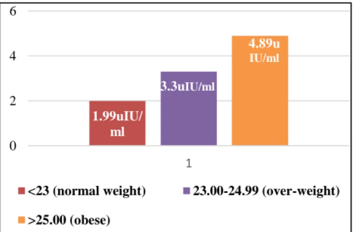
Figure 2: Serum TSH values in normal weight, over-weight and obese eu-thyroid females.
There are significant (p <0.003) inter-group variations among BMI and serum TSH concentration (Table 3). Serum TSH values gradually increases among three BMI groups i.e. normal weight (BMI <23 kg/m2; TSH Mean
±SD: 1.99±1.49), over-weight (BMI 23.00-24.99 kg/m2;
TSH Mean ±SD: 3.30±2.78), and obese (BMI >25 kg/m2;
TSH Mean ±SD: 3.28 ±2.29) females (Figure 2).
Table 2:The association between BMI, body fat percent, waist circumference, sub-cutaneous Adipose tissue, visceral adipose tissue and serum TSH in eu-thyroid female subjects.
BMI (kg/m2) % Body fat WC (cm) SCAT (cm) VAT (cm)
TSH (µ IU/ml)
Pearson correlation (r) 0.342*** 0.628*** 0.289** 0.105 0.375**
Sig. (2 tailed) <0.002 0.000 .009 0.316 0.000
n 103 103 93 93 93
**. Correlation is significant at the 0.01 level (2-tailed).
1.99uIU/ ml
3.3uIU/ml
4.89u IU/ml
0 2 4 6
1
<23 (normal weight) 23.00-24.99 (over-weight)
In order to control the effect of a third factor, e.g. age, between the association of serum TSH and BMI, age was
included as an independent variable in multiple regression analysis.
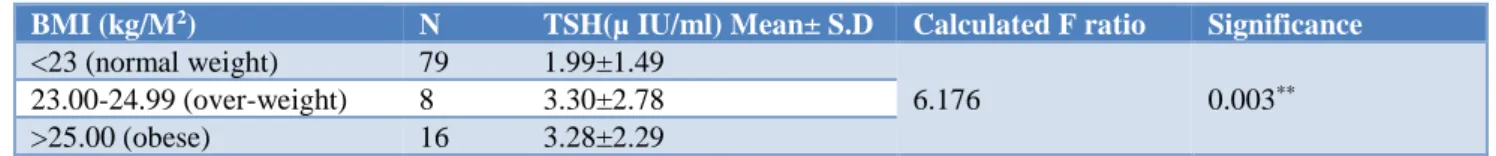
Table 3: Serum TSH concentration in normal weight, over-weight and obese eu-thyroid females.
BMI (kg/M2) N TSH(µ IU/ml) Mean± S.D Calculated F ratio Significance
<23 (normal weight) 79 1.99±1.49
6.176 0.003**
23.00-24.99 (over-weight) 8 3.30±2.78
>25.00 (obese) 16 3.28±2.29
Table 4:Multiple linear regression showing relation between TSH level, body fat percentage and age of the
study participants.
Variable Unstandardized coefficient Sig.
B Std. Error
Constant -0.512 0.798 0.523
B. Fat% 0.113 0.039 0.005
Age in yrs -0.003 0.019 0.879
Dependent variable: TSH (Uiu/ml); TSH = - 0.512 + 0.113 b.fat% - 0.003 Age in yrs, body fat percentage is a significant predictor of TSH but age is not.
It shows Body fat percentage and BMI were significant independent predictors of TSH, but age was not. (TSH = - 0.512 + 0.113 b. fat % - 0.003 age in yrs) (Table 4). Abdominal fat was further subdivided into SCAT and VAT. Multiple linear regression analysis was done to show the relation between TSH level, body fat percentage, BMI, SCAT, VAT and age of the study
participants. Linear regression model shows VAT is positive and independent predictor of TSH (TSH = 0.481 - 0.039 (subcutaneous fat) + 0.742 (Visceral fat). Inclusion of age did not show significant R2 change, and so, was not included in the final model (Table 6).
Table 5:Multiple linear regression showing relation between TSH level, body mass index and age of the
study participants.
Variable Unstandardized coefficient Sig.
B Std. Error
Constant -1.045 0.989 0.293
BMI 0.128 0.047 0.008
Age in yrs 0.025 0.015 0.103
Dependent variable: TSH (Uiu/ml); TSH = -1.045 + 0.128 BMI + 0.025 Age in yrs BMI is a significant predictor of TSH but age is not. Upper two tables demonstrated model fit R-square change and Sum of squares in regression showed that body fat % is a better predictor.
Table 6:Linear Regression model showing relation between TSH level, SCAT and VAT of the study participants.
Variable Unstandardized coefficients t Sig. 95.0% Confidence Interval for B
B Std. Error Lower bound Upper bound
Constant 0.481 0.573 0.838 0.404 -0.659 1.620
Subcut. Fat (cm) -0.039 0.342 -0.115 0.908 -0.718 0.639
Visceral fat (cm) 0.742 0.201 3.685 0.000 0.342 1.143
TSH = 0.481 - 0.039 (subcutaneous fat) + 0.742 (visceral fat)* Inclusion of age did not show significant R2 change, and so, was not included in the final model.
DISCUSSION
In the present study, author have found that although in the normal range, serum TSH is positively associated with BMI and body fat percent in eu-thyroid female subjects. This finding is in agreement with the study of Nyrnes A et al, who showed serum TSH within the normal range to be significantly and positively associated with BMI in non-smoking men and women.25,26 Knudsen
N et al, also showed small differences in thyroid function
may be important for Body Mass Index and the occurrence of obesity in the population.8 Although Manji
et al. reported no associations of TSH or fT4 levels with BMI in euthyroid individuals.11
that showed no association regarding BMI categories and plasma TSH levels in their euthyroid cohort, overweight, obese, morbid obese, and normal weight subjects.27 But
they mentioned that absence of correlation between plasma TSH levels and obesity in their study does not mean that TSH and thyroid hormones have no effect on adiposity.
In order to control the effect of a third factor, e.g. age, between the association of serum TSH and BMI, author included age as an independent variable in multiple regression analysis. Authors found body fat percentage and BMI were significant independent predictors of TSH but age is not. This is in accordance with Nyrnes A et al, who reported age is an unlikely explanation for the association between TSH and BMI.26
To have a better idea of association between serum TSH and regional adiposity markers author further subdivided abdominal fat into SCAT and VAT and measured it by US which has proved to be a suitable non-invasive and reliable tool for quantifying abdominal fat as useful as CT.28 Present study revealed one interesting finding that
TSH is significantly and positively associated with VAT but no significant association was found with SCAT and TSH. Furthermore, VAT is positive and independent predictor of TSH. Very few previous studies focused on this topic and one of such study even contradict this report as they did not find any significant association between Visceral Fat and fT4 or TSH levels.29 But in the
subgroup of overweight and obese subjects they found the inverse association of Subcutaneous Fat layer with fT4 levels.
It is now well known that VAT is associated with increase cardiovascular risk.30 SCAT does not pose the
same health danger as the VAT and visceral fat expresses thyroid hormone receptors as well as TSH receptors that may also directly influence various functions of adipose cells.14-18
All forms of weight loss affect visceral fat more than subcutaneous fat.30 Hence Selective reduction of visceral
fat may induce greater beneficial effects on the parameters of thyroid hormones than subcutaneous fat reduction.
The present study has few limitations. Present study is a cross-sectional study. A longitudinal study with more sample size will give better idea of the relationship between thyroid profile and regional adiposity markers.
CONCLUSION
There is significant positive association of TSH with BMI, Body fat percent, WC and VAT in eu-thyroid female subjects. No significant association was found between TSH and SCAT. Hence selective reduction of visceral fat may induce greater beneficial effects on the
parameters of thyroid hormones than subcutaneous fat reduction.
Funding: No funding sources Conflict of interest: None declared
Ethical approval: The study was approved by the Institutional Ethics Committee
REFERENCES
1. Knudsen N, Laurberg P, Rasmussen LB, Bülow I, Perrild H, Ovesen L, Jørgensen T. Small differences in thyroid function may be important for body mass index and the occurrence of obesity in the population. J Clin Endocrinol Metabol. 2005 Jul 1;90(7):4019-24.
2. Fox CS, Pencina MJ, D’Agostino RB, Murabito JM, Seely EW, Pearce EN, et al. Relations of thyroid function to body weight: cross-sectional and longitudinal observations in a community-based
sample. Arch Internal Med. 2008 Mar
24;168(6):587-92.
3. Reinehr T, de Sousa G, Andler W.
Hyperthyrotropinemia in obese children is reversible after weight loss and is not related to lipids. J Clin Endocrinol Metabol. 2006 Aug 1;91(8):3088-91.
4. Reinehr T, Isa A, De Sousa G, Dieffenbach R, Andler W. Thyroid hormones and their relation to weight status. Hormone Res Paediat. 2008;70(1):51-7.
5. Rotondi M, Leporati P, La Manna A, Pirali B, Mondello T, Fonte R, et al. Raised serum TSH levels in patients with morbid obesity: is it enough to diagnose subclinical hypothyroidism?. Eur J Endocrinol. 2009 Mar 1;160(3):403-8.
6. Shintani M, Nishimura H, Akamizu T, Yonemitsu S, Masuzaki H, Ogawa Y, et al. Thyrotropin decreses leptin production in rat adipocytes. Metabolism. 1999 Dec 1;48(12):1570-4.
7. Mukherjee S. Relationship between Body Mass Index (BMI), body fat percent and serum TSH level in eu-thyroid female subjects. IOSR J Dental Med Sci. 2018;17(9):1-4.
8. Knudsen N, Laurberg P, Rasmussen LB, Bülow I, Perrild H, Ovesen L, et al. Small differences in thyroid function may be important for body mass index and the occurrence of obesity in the population. J Clin Endocrinol Metabol. 2005 Jul 1;90(7):4019-24.
9. Fox CS, Pencina MJ, D’Agostino RB, Murabito JM, Seely EW, Pearce EN, et al. Relations of thyroid function to body weight: cross-sectional and longitudinal observations in a community-based
sample. Arch Internal Med. 2008 Mar
24;168(6):587-92.
resistance, metabolic parameters and blood pressure in overweight and obese women. Clin Endocrinol. 2007 Aug 1;67(2):265-9.
11. Manji N, Boelaert K, Sheppard MC, Holder RL, Gough SC, Franklyn JA. Lack of association between serum TSH or free T4 and body mass index in euthyroid subjects. Clin Endocrinol. 2006 Feb 1;64(2):125-8.
12. Ibrahim MM. Subcutaneous and visceral adipose tissue: structural and functional differences. Obesity Rev. 2010 Jan 1;11(1):11-8.
13. Chan JM, Rimm EB, Colditz GA, Stampfer MJ, Willett WC. Obesity, fat distribution, and weight gain as risk factors for clinical diabetes in men. Diab Care. 1994 Sep 1;17(9):961-9.
14. Obregon MJ. Thyroid hormone and adipocyte differentiation. Thyroid. 2008 Feb 1;18(2):185-95. 15. Villicev CM, Freitas FR, Aoki MS, Taffarel C,
Scanlan TS, Moriscot AS, et al. Thyroid hormone receptor β-specific agonist GC-1 increases energy expenditure and prevents fat-mass accumulation in rats. J Endocrinol. 2007 Apr 1;193(1):21-9.
16. Viguerie N, Millet L, Avizou S, Vidal H, Larrouy D, Langin D. Regulation of human adipocyte gene expression by thyroid hormone. J Clin Endocrinol Metab. 2002 Feb 1;87(2):630-4.
17. Ortega FJ, Moreno‐Navarrete JM, Ribas V, Esteve E, Rodriguez‐Hermosa JI, Ruiz B, et al. Subcutaneous Fat Shows Higher Thyroid Hormone Receptor‐α1 Gene Expression Than Omental Fat. Obesity. 2009 Dec;17(12):2134-41.
18. Antunes TT, Gagnon A, Chen B, Pacini F, Smith TJ, Sorisky A. Interleukin-6 release from human abdominal adipose cells is regulated by thyroid-stimulating hormone: effect of adipocyte differentiation and anatomic depot. Am J Physiol Endocrinol Metabol. 2006 Jun;290(6):E1140-4. 19. Use of thyroid function tests: guidelines
development group (2006-06-01). .UK Guidelines for the Use of Thyroid Function Tests (pdf).
Available at:
http://www.btf-thyroid.org/images/documents/tft_guideline_final_v ersion_july_2006.pdf. Accessed on 2 February 2012.
20. Eknoyan G, Quetelet A. (1796-1874) the average man and indices of obesity". Nephrology Dialysis Transplantation. 2007;23(1):47-51.
21. Obesity and overweight - World Health
Organization. Available at:
https://www.who.int/news-room/fact-sheets/detail/obesity-and-overweight.
22. National Center for Health Statistics (NCHS). National Health and Nutrition Examination Survey Data: Anthropometry Procedures Manual. 12/15/06,
Available at:
http://www.cdc.gov/nchs/data/nhanes/nhanes_01_02 /body_measures_year_3.pdf.
23. Tanner JM, Whitehouse RH. Revised standards for triceps and subscapular skinfolds in British children. Arch Dis Childhood. 1975 Feb 1;50(2):142-5. 24. Armellini F, Zamboni M, Rigo L, Todesco T,
Bosello O, Bergamo‐Andreis IA, et al. The contribution of sonography to the measurement of intra‐abdominal fat. J Clin Ultrasound. 1990 Sep;18(7):563-7.
25. Després JP, Lamarche B. Effects of diet and physical activity on adiposity and body fat distribution: Implications for the prevention of cardiovascular disease. Nutr Res Rev. 1993 Jan;6(1):137-59.
26. Nyrnes A, Jorde R, Sundsfjord J. Serum TSH is positively associated with BMI. Int J Obes. 2006 Jan;30(1):100.
27. Bakiner O, Bozkirli E, Cavlak G, Ozsahin K, Ertorer E. Are plasma thyroid-stimulating hormone levels associated with degree of obesity and metabolic syndrome in euthyroid obese patients? A Turkish cohort study. ISRN Endocrinology. 2014 Jan 2;2014.
28. Berker D, Koparal S, Isik S, Pasaoglu L, Aydin Y, Erol K, et al. Compatibility of different methods for the measurement of visceral fat in different body mass index strata. Diagnostic and interventional radiology. 2010 Jun 1;16(2):99.
29. Alevizaki M, Saltiki K, Voidonikola P, Mantzou E, Papamichael C, Stamatelopoulos K. Free thyroxine is an independent predictor of subcutaneous fat in euthyroid individuals. Eur J Endocrinol. 2009 Sep 1;161(3):459-65.
30. Armellini F, Zamboni M, Rigo L, Bergamo-Andries IA, Robbi R, De Marchi M, et al. Sonography detection of small intra abdominal fat variation. Int J Obes. 1991; 15: 847-52.
![Bis[hydrotris(4 chloro 3,5 dimethylpyrazolyl)borato]nickel(II)](data:image/gif;base64,R0lGODlhAQABAIAAAP///wAAACH5BAEAAAAALAAAAAABAAEAAAICRAEAOw==)